• Localized nodular parenchyma with fibrous septa and stellate central scar
Septa contain thick-walled vessels and mononuclear inflammatory infiltrate
• Ductular reaction at junction between septa and parenchyma
Ancillary Tests
• Glutamine synthetase: Characteristic map-like pattern, with sparing of areas around scar and fibrous septa
• Serum amyloid A is typically negative; focal staining in few case
• C-reactive protein staining typically restricted to periseptal areas
Top Differential Diagnoses
• Hepatocellular adenoma
• Cirrhosis
• Hepatocellular carcinoma
• Nodular regenerative hyperplasia
Ultrasound Findings Oblique transabdominal ultrasound shows a hypoechoic central scar in the center of an isoechoic mass (FNH) . The scar may show vascular calcification but the lesion itself rarely calcifies.
Nodular Architecture Low-power photomicrograph of focal nodular hyperplasia shows the nodular hepatic parenchyma separated by fibrous septa. This appearance can mimic biliary cirrhosis.
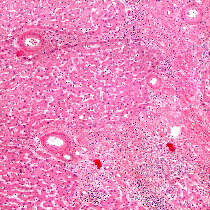
Aberrant Arterioles Thick-walled arteries are typically seen at the periphery of the fibrous septa in FNH. Less commonly, the aberrant arterioles can be seen in the parenchyma.
Ductular Reaction FNH typically shows a marked ductular reaction in the fibrous septa. By definition, normal interlobular bile ducts and normal portal tract are absent.
TERMINOLOGY
Abbreviations
• Focal nodular hyperplasia (FNH)
Synonyms
• Focal cirrhosis
Definitions
• Benign tumor-like lesion caused by hyperplastic response to localized vascular abnormality
• Most lesions formerly labeled as telangiectatic FNH are thought to be inflammatory hepatocellular adenomas

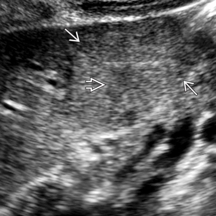
 in the center of an isoechoic mass (FNH)
  . The scar may show vascular calcification but the lesion itself rarely calcifies.
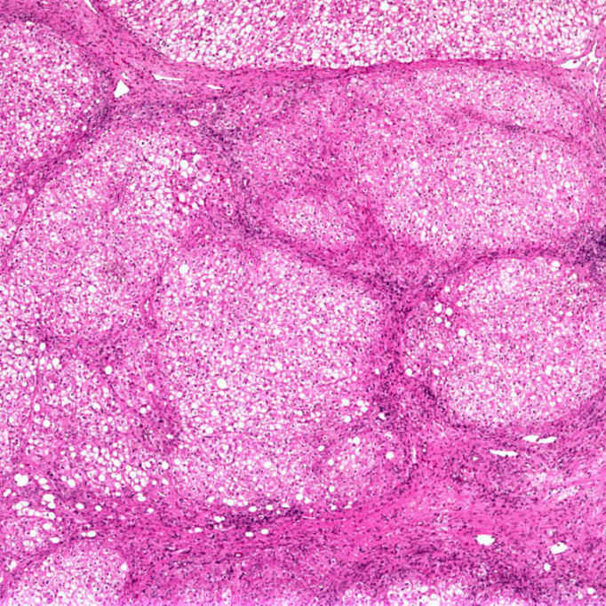
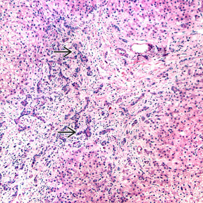
 in the fibrous septa. By definition, normal interlobular bile ducts and normal portal tract are absent.


