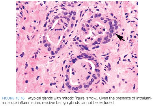
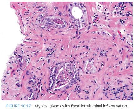
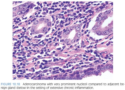
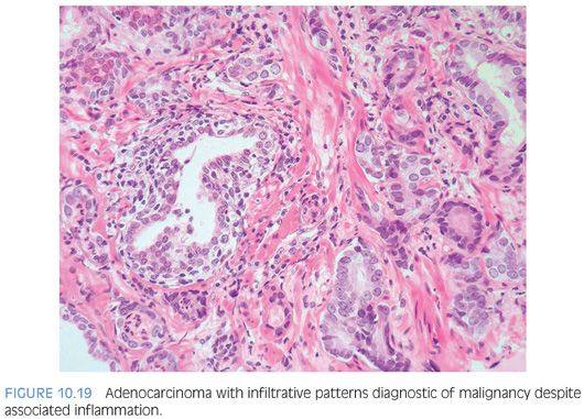
In the presence of inflammation, one must be cautious in diagnosing cancer (Figs. 10.16 and 10.17). Rarely, one can establish a diagnosis of cancer associated with inflammation (Fig. 10.18). Figure 10.19 demonstrates a focus of crowded small glands infiltrating in between larger benign glands associated with extensive inflammation. This focus is diagnostic of cancer because the pattern of numerous small glands in between larger benign glands cannot be attributed to inflammation. Furthermore, the degree of cytologic atypia present in the small atypical glands is significantly greater than the adjacent benign glands even though both are exposed to the same inflammatory milieu.
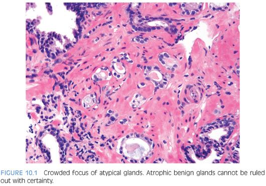
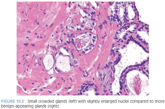
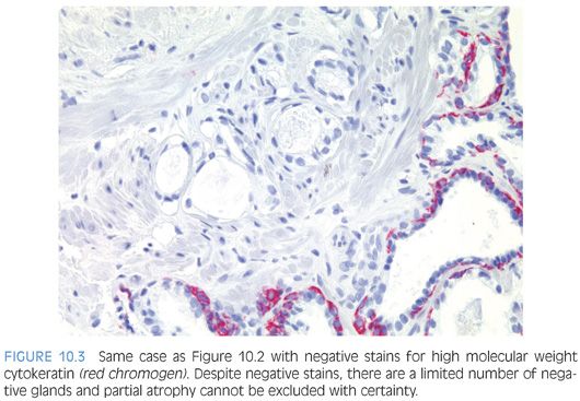
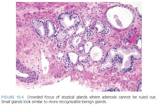
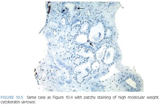
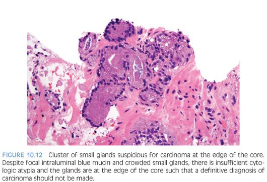
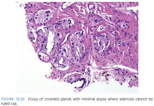
In some cases where the number of atypical glands are so few or the glands have no other atypical features other than that they are crowded, adenosis cannot be excluded (Fig. 10.20). Even if the glands are negative for basal cell markers in a small focus, the lesion could still be adenosis because basal cell stains can be very patchy in adenosis. In a small focus of atypical glands on prostate biopsy, negative staining for basal cell markers should not necessarily lead to a definitive malignant diagnosis in all cases, because almost half of these biopsies on follow-up sampling are benign.24
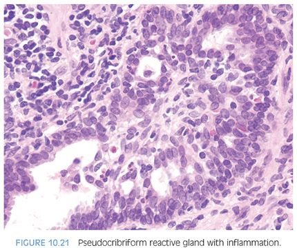
In the setting of an inflamed prostate, one should also be cautious in the evaluation of isolated glands with abnormal architecture. Although rarely carcinomas may be inflamed, inflammation tends to preferentially localize away from malignant glands. In areas of intense chronic inflammation, prostatic acini appear atrophic with a high nuclear to cytoplasmic ratio. These basophilic glands may show some architectural abnormalities such as pseudocribriform formation with budding off of little glands (Fig. 10.21). Streaming of basophilic epithelium in areas of intense chronic inflammation resembles transitional cell metaplasia. The finding of occasional large nucleoli is not uncommon in areas of intense acute or chronic inflammation. The distinction of these inflammatory atypias from carcinoma first relies on the recognition that the atypical glands are located in an area of intense inflammation. In addition, the glands have a very basophilic appearance in contrast to the usual gland-forming prostatic adenocarcinomas that have abundant, often pale-staining cytoplasm. The high nuclear to cytoplasmic ratio seen in inflamed glands is predominantly seen in only the more poorly differentiated prostatic carcinomas that lack good gland formation. Careful examination of these basophilic glands will also demonstrate the finding of a basal cell layer in most instances.
Stay updated, free articles. Join our Telegram channel

Full access? Get Clinical Tree


