Fibromatosis

 4-6% of Caucasian adults > 50 years of age


• Deep fibromatosis
image 2.40-4.43 new cases per 100,000 persons/year

• Recurrences common for both superficial and deep fibromatoses, but deaths only from deep fibromatoses

• Increased incidence in familial adenomatous polyposis


Microscopic


• Sweeping fascicles of myofibroblasts

• Smooth nuclear membranes and small nucleoli in most cells

• Occasional cells with stellate cytoplasmic contours

• Occasional foci with storiform pattern similar to nodular fasciitis

• Some cases show keloid-like collagen

• Small but conspicuous vessels

• Gaping, thin-walled vessels with perivascular sclerosis often feature of mesenteric fibromatosis


Ancillary Tests


• β-catenin (+) (nuclear), especially in deep lesions, SMA(+); desmin (-), keratins (-), CD117(+/-)


Top Differential Diagnoses


• Scar

• Tenosynovial giant cell tumor (giant cell tumor of tendon sheath, pigmented villonodular tenosynovitis)

• Leiomyoma and leiomyosarcoma

• Clear cell sarcoma

• Epithelioid sarcoma

• Low-grade fibromyxoid sarcoma

image
Fibromatosis, Clinical Image
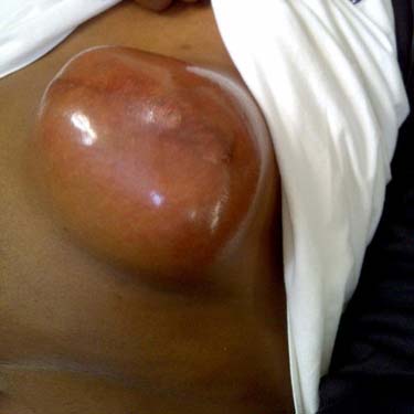
This fibromatosis is unusually superficial and arose in the chest wall of a young adult. These tumors are typically deep (deep fascia or within muscle).


image
Fibromatosis, Low Magnification
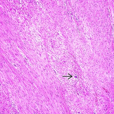
This image of a deep-type fibromatosis shows sweeping fascicles of spindle cells separated by regularly spaced collagen. Even at this magnification, small vessels are readily apparent image.

image
Fibromatosis, Gross Specimen
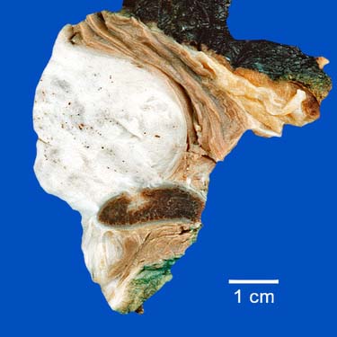
This gross photograph shows a large, deep fibromatosis of the shoulder that has eroded into the scapula. Superficial portions of such lesions can be encountered on skin biopsies.

image
Fibromatosis, Neck
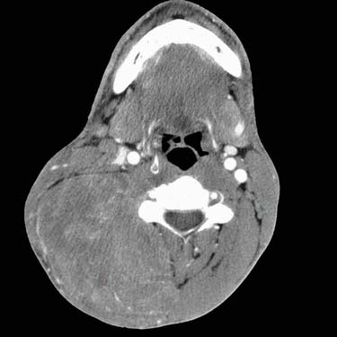
This radiologic image shows a large fibromatosis of the head and neck in an elderly alcoholic man. Although more arise in the shoulder girdles (or in the abdominal wall in young women), the head and neck is a common location for fibromatoses.


TERMINOLOGY


Synonyms


• Deep fibromatosis: Aggressive fibromatosis, desmoid tumor

• Superficial fibromatoses: Palmar fibromatosis (Dupuytren contracture, Dupuytren disease), plantar fibromatosis (Ledderhose disease), penile fibromatosis (Peyronie disease), knuckle pads


Definitions


• Palmar fibromatosis: Nodular myofibroblastic proliferation of volar surface of hand that is prone to local persistence but does not metastasize

• Plantar fibromatosis: Nodular myofibroblastic proliferation of plantar surface of foot that is prone to local persistence but does not metastasize

• Peyronie disease: Penile fibrous lesion causing various deformities; initially pain with erection, erectile dysfunction

• Knuckle pads: Well-circumscribed thickening of skin over metacarpophalangeal and, more commonly, proximal interphalangeal joints

• Deep fibromatosis: Myofibroblastic proliferation of deep soft tissues with infiltrative growth pattern; prone to local recurrences but does not metastasize


CLINICAL ISSUES


Epidemiology


• Incidence
image Palmar fibromatosis

– 4-6% of Caucasian adults > 50 years of age; reports of up to 75% of Celtic male patients

– Uncommon in nonwhites; marked male predominance

image Plantar fibromatosis

– 1-2 per 100,000 persons per year (Northern Europe)

– Most patients 30-50 years of age; slight male predominance

image Penile fibromatosis

– ~ 3.5% of white men > 50 years of age

image Deep fibromatosis

– 2.40-4.43 new cases per 100,000 persons per year (Scandinavian data)

image Knuckle pads (rare)


Presentation


• Superficial fibromatoses present as nodular lesions on palms, soles, knuckles, or penis
image Variable tenderness

• Deep fibromatoses present as firm large masses, typically with intramuscular component
image Relationship to age and gender
– In children and older adults, no gender predominance: Lesions of shoulders, chest wall, back, thigh, head, and neck

– In women in childbearing years: Abdominal wall
image May also arise in shoulder girdles, chest wall, back, thigh, head, and neck

image Usually clinically concerning for sarcomas based on large size, deep location

• Familial adenomatous polyposis-associated lesions
image Risk of fibromatoses is 2.56/1,000 person/year; comparative risk is 852x that of general population

image Patients have numerous colon adenomas and must undergo colectomy to forestall development of colorectal carcinoma

• Occasionally associated with scar (“cicatricial fibromatosis”)


Treatment


• Superficial fibromatoses treated by excision
• Nonsurgical treatments for penile lesions: Verapamil, colchicine, Potaba, L-carnitine, and liposomal superoxide dismutase

• Deep fibromatoses treated by wide excision; for unresectable lesions, radiation, chemotherapy, hormone therapy

image Clear surgical margins are key to successful outcome

Only gold members can continue reading. Log In or Register to continue

Stay updated, free articles. Join our Telegram channel

Apr 24, 2017 | Posted by in PATHOLOGY & LABORATORY MEDICINE | Comments Off on Fibromatosis

Full access? Get Clinical Tree

Get Clinical Tree app for offline access