Nuclear staining with CAMTA1 immunohistochemistry


Top Differential Diagnoses
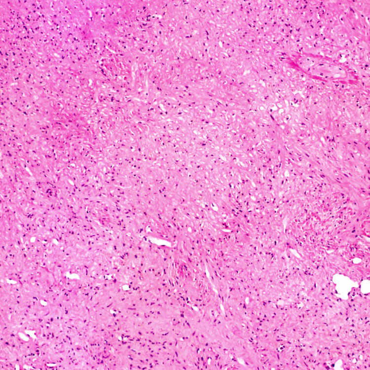
Central portion is typically hypocellular with loosely arranged spindle cells in a fibromyxoid or sclerotic stroma. The findings can simulate a scar or sclerosed hemangioma.
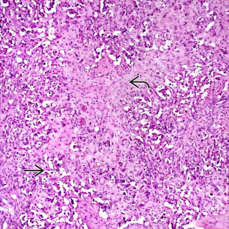
Poorly formed vascular lumina
 and infiltrating tumor cells in a fibromyxoid stroma
  can mimic cholangiocarcinoma.
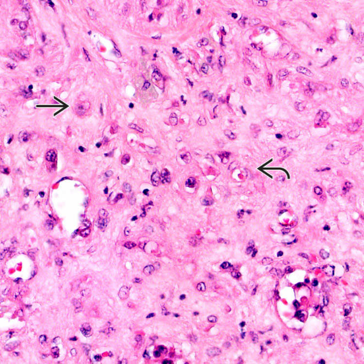
Cellular area with epithelioid tumor cells
 in a fibromyxoid stroma demonstrates mild nuclear atypia with no mitoses. Vascular differentiation is seen in the form of intracytoplasmic lumina with red blood cells
  .
.
 and extension along the sinusoids in adjacent liver.
CLINICAL ISSUES
Presentation


Treatment
• Primary treatment is hepatic resection


Stay updated, free articles. Join our Telegram channel
Full access? Get Clinical Tree
