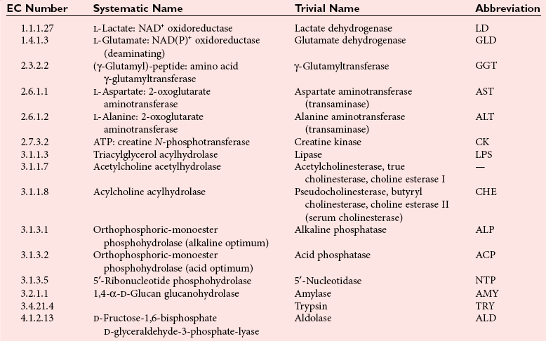
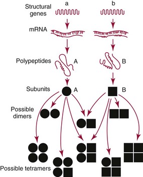
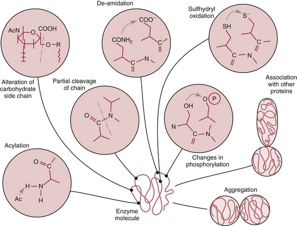
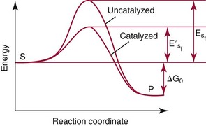
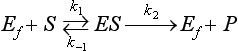
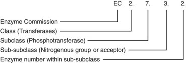
Enzyme and Rate Analyses

Chapter 15


Enzyme and Rate Analyses


Enzymes are proteins with catalytic properties; clinical enzymology is the application of the science of enzymes to the diagnosis and treatment of disease. The principles of clinical enzymology will be introduced and discussed in this chapter, as will information on how enzymes are measured and how they are used as analytical reagents in various types of rate analysis. Individual topics include basic principles, enzyme kinetics and analytical enzymology, and rate analyses.


Basic Principles


This section begins with a presentation of enzyme nomenclature, which is followed by a discussion of enzymes as proteins and catalysts.


Enzyme Nomenclature


Historically, individual enzymes were identified using the name of the substrate or group upon which they act and then adding the suffix –ase. For example, the enzyme hydrolyzing urea was urease. Later, the type of reaction involved was also identified, as in carbonic anhydrase, D-amino acid oxidase, and succinate dehydrogenase. In addition, some enzymes had been given empirical names such as trypsin, diastase, ptyalin, pepsin, and emulsin.


Because this combination of trivial common names and semisystematic names was found to be inadequate, in 1955 the International Union of Biochemistry (IUB) appointed an Enzyme Commission (EC) to study the problem of enzyme nomenclature. Its subsequent recommendations, with periodic updating, provide a rational and practical basis for identifying all enzymes now known and enzymes that will be discovered in the future (http://www.chem.qmw.ac.uk/iubmb/enzyme/accessed May 6, 2011).11


With the IUB system, a systematic and trivial name is provided for each enzyme. The systematic name describes the nature of the reaction catalyzed and is associated with a unique numeric code. The trivial or practical name, which may be identical to the systematic name but is often a simplification of it, is suitable for everyday use. The unique numeric designation for each enzyme consists of four numbers, separated by periods (e.g., 2.2.8.11), and is prefixed by the letters EC, denoting Enzyme Commission. The first number defines the class to which the enzyme belongs. All enzymes are assigned to one of six classes, characterized by the type of reaction they catalyze: (1) oxidoreductases, (2) transferases, (3) hydrolases, (4) lyases, (5) isomerases, and (6) ligases. The next two numbers indicate the subclass and the sub-subclass to which the enzyme is assigned. For example, these may differentiate the amino-transferring subclass from the phosphate-transferring category, or the ethanol acceptor sub-subclass from that accepting acyl groups. The last number is the specific serial number given to each enzyme within its sub-subclass.


To illustrate how this system is used to name an enzyme, consider the enzyme creatine kinase, which catalyzes the following reaction:


image


Its system number is


image


Table 15-1 lists some selected enzymes of clinical interest, identified by trivial, abbreviated, and systematic names and by their code numbers.


Although it is not recommended by the EC, it is a common and convenient practice to use capital letter abbreviations for the names of certain enzymes, such as ALT (formerly GPT) for alanine aminotransferase. Other examples are AST for aspartate aminotransferase, LD for lactate dehydrogenase, and CK for creatine kinase (see Table 15-1).


Enzymes as Proteins


All enzyme molecules possess the primary, secondary, and tertiary structural characteristics of proteins (see Chapter 21). In addition, most enzymes exhibit quaternary structure. The primary structure, the linear sequence of amino acids linked through their α-carboxyl and α-amino groups by peptide bonds, is specific for each type of enzyme molecule. Each polypeptide chain is coiled up into three-dimensional secondary and tertiary structure. Secondary structure refers to the conformation of limited segments of the polypeptide chain, namely α-helices, β-pleated sheets, random coils, and β-turns. The arrangement of secondary structural elements and amino acid side chain interactions that define the three-dimensional structure of the folded protein is referred to as its tertiary structure. In many cases, biological activity, such as the catalytic activity of enzymes, requires two or more folded polypeptide chains (subunits) to associate to form a functional molecule. The arrangement of these subunits defines the quaternary structure. The subunits may be copies of the same polypeptide chain (e.g., the MM isoenzyme of creatine kinase, the H4 isoenzyme of lactate dehydrogenase), or they may represent distinct polypeptides.


The application of physical methods, such as x-ray crystallography and multidimensional nuclear magnetic resonance (NMR), has provided structural insights upon which enzyme mechanisms have been built. Furthermore, the tools of molecular biology, such as molecular cloning, have enabled the purification and characterization of enzymes that previously were available only in minute amounts. Molecular biology also enables the manipulation of the amino acid sequence of enzymes, and site-directed mutagenesis (substituting one amino acid residue for another) and deletion mutagenesis (eliminating sections of the primary structure) have enabled the identification of chemical groups that participate in ligand binding and in specific chemical steps during catalysis.


In general, no feature of primary structures, such as repetition of particular amino acid sequences, is common to all enzyme molecules. However, considerable homologies of sequence are found between enzymes that appear to share a common evolutionary origin, such as the proteases, trypsin and chymotrypsin, and similarities of sequence are even more marked among the members of a family of isoenzymes. The amino acid sequence in the immediate neighborhood of the active center of the enzyme (discussed later) is often closely similar in enzymes of related function (e.g., the serine proteases are so called because they all have this amino acid in the active center).


Enzyme molecules differ in the proportion of secondary structures—such as α-helices—they contain, although no enzyme molecule so far studied approaches the large proportion of α-helices found in myoglobin and hemoglobin. The tertiary structures of different types of enzyme molecules are as individually characteristic as their primary structures; nevertheless, some common features exist. Enzyme molecules are roughly globular in overall shape, with a preponderance of polar amino acid side chains on the outside of the molecule and nonpolar side chains in the interior. The ionizable residues in contact with the surrounding medium are responsible for many of the properties of the enzyme molecules in solution, such as their migration in an electric field and their solubility. Covalent disulfide bridges may link different parts of the polypeptide chains in some enzyme molecules, but the three-dimensional structure is mainly stabilized by the large number of hydrophobic interactions that are formed between the nonpolar side chains in the interior of the molecule.


The biological activity of a protein molecule depends generally on the integrity of its structure, and any disruption of its structure is accompanied by loss of activity, a process known as denaturation. If the process of denaturation is minimal, it may be reversed with the recovery of enzyme activity upon removal of the denaturing agent. However, prolonged or severe denaturing conditions result in an irreversible loss of activity. Denaturing conditions include elevated temperatures, extremes of pH, and chemical addition. Heat inactivation of most enzymes takes place at an appreciable rate at room temperature and in most cases becomes almost instantaneous above 60 °C. The polymerases are an exception and retain activity at temperatures as high as 90 °C—a property that has been made use of in the polymerase chain reaction (see Chapter 17). Low temperatures are used to preserve enzyme activity, especially in aqueous solutions, such as serum (see Chapter 22). Extremes of pH also cause unfolding of enzyme molecular structures and, except for a few exceptions, should be avoided when enzyme samples are preserved. Addition of chemicals, such as urea and detergents, disrupts hydrogen bonds and hydrophobic interactions so that exposure of enzymes to strong solutions of these reagents results in inactivation.


Specificity and the Active Center


With the exception of enzymes such as proteases, nucleases, and amylases, which act on macromolecular substrates, enzyme molecules are considerably larger than the molecules of their substrates. Consideration of the structure of an enzyme’s active site and its relationship to the structures of the enzyme’s substrate(s) in its ground and transition states is necessary to understand the rate enhancement and specificity of the chemical reactions performed by the enzyme. The active site of an enzyme will vary between enzymes but in general,5


1. The active site of an enzyme is relatively small compared with the total volume of the enzyme molecule because its structure may involve less than 5% of the total amino acids in the molecule.


2. The active sites of enzymes are three-dimensional structures that are formed as a result of the overall tertiary structure of the protein. This results when the amino acids and cofactors in the active site of an enzyme are spatially structured in an exact, three-dimensional relationship with respect to one another and the structure of the substrate molecule.


3. Typically, the attraction between the molecules of the enzyme and its substrate molecules is noncovalent binding. Physical forces used in this type of binding include hydrogen bonding, electrostatic and hydrophobic interactions, and van der Waals forces.


4. Active sites of enzymes typically occur in clefts and crevices in the protein. This excludes bulk solvent and reduces the catalytic activity of the enzyme.


5. The specificity of substrate binding is a function of the exact special arrangement of atoms in the enzyme active site that complements the structure of the substrate molecule.


Isoenzymes and Other Multiple Forms of Enzymes


An isoenzyme is defined as “one of a group of related enzymes catalyzing the same reaction but having different molecular structures and characterized by varying physical, biochemical, and immunologic properties.” However, the IUB recommends that use of the term isoenzyme be restricted to forms that originate at the level of the genes that encode the structures of the enzyme proteins in question.4


Isoenzymes may occur within a single organ or even within a single type of cell. The forms can be distinguished on the basis of differences in various physical properties, such as electrophoretic mobility and resistance to chemical or thermal inactivation. They often have significant quantifiable differences in catalytic properties, but all forms of a particular enzyme retain the ability to catalyze its characteristic reaction.


The existence of multiple forms of enzymes in human tissue has important implications in the study of human disease. The presence in different organs of isoenzymes with distinctive properties helps in our understanding of organ-specific patterns of metabolism, but genetically determined variations in enzyme structure between individuals account for such characteristics as differences in sensitivity to drugs and differences in metabolism, which manifest themselves as hereditary metabolic diseases. For diagnostic enzymology, the existence of multiple forms of enzymes, whether due to genetic or nongenetic causes, provides opportunities to increase the diagnostic specificity and sensitivity of enzyme assays in body fluid samples.


Similar to other proteins, enzymes usually elicit the production of antibodies when they are injected into animals of a species other than those in which they originate. Even small structural differences between closely similar molecules, such as the members of a family of isoenzymes, are often sufficient to render them antigenically distinct, allowing antibodies to be produced specific to a single type of molecule. The availability of enzyme-specific antisera opens up a wide range of methods in enzyme analysis, some of which—such as immunoassay—do not depend on the catalytic activity of the enzyme molecules. The availability of immunochemical methods has been particularly important in the analysis of isoenzyme mixtures. Many commercial immunoassays now use monoclonal antibodies to increase specificity.


Genetic Origins of Enzyme Variants


True isoenzymes result from the existence of more than one gene locus coding for the structure of the enzyme protein. Many human enzymes (perhaps more than one third) are known to be determined by more than one structural gene locus. Genes at the different loci have undergone differential modifications during the course of evolution, so that the enzyme proteins coded by them no longer have identical structures, although they are recognizably similar; in other words, they are isoenzymes.


The multiple genes that determine a particular group of isoenzymes are not necessarily closely linked on one chromosome. For example, the structural genes that code for human salivary and pancreatic amylases both are located on chromosome 1, whereas the genes that code for cytoplasmic and mitochondrial malate dehydrogenase are carried on chromosomes 2 and 7, respectively. Among the enzymes of clinical importance that exist as isoenzymes because of the presence of multiple-gene loci are lactate dehydrogenase, creatine kinase, α-amylase, and some forms of alkaline phosphatase.


An enzyme can exist in molecular forms that differ from one individual to another because of the existence of alternative alleles that are inherited according to mendelian laws. These give rise to gene products with the same function, and isoenzymes that result from the existence of allelic genes are termed allozymes. The proportion of human gene loci subject to allelic variation is considerable, and the probability that individual human beings will differ to some degree in their isoenzyme patterns is correspondingly high.


The number of allelic variants and the frequency with which particular variants occur within the population vary considerably from one enzyme to another. For example, mutations at either of the two principal loci that determine human lactate dehydrogenase are extremely rare, but a high incidence of mutant alleles occurs at the single locus that determines the structure of placental alkaline phosphatase. More than 400 mutations in the glucose-6-phosphate dehydrogenase gene have now been identified on the X chromosome [up-to-date genetic information on this and other enzymes can be obtained from the Online Mendelian Inheritance in Man (OMIM) database at http://www.ncbi.nlm.nih.gov/Omim/searchomim.html/ accessed May 6, 2011]. Some of these alleles are extremely rare, whereas others occur with appreciable frequency in particular populations or geographical locations. When isoenzymes, because of variation at a single locus, occur with appreciable frequency in a human population, the population is said to be polymorphic with respect to the isoenzymes in question.


Another category of multiple molecular forms can arise when enzymes are oligomeric and consist of molecules made up of subunits. The association of different types of subunits in various combinations gives rise to a range of active enzyme molecules. When the subunits are derived from different structural genes—multiple loci or multiple alleles—the hybrid molecules so formed are called hybrid isoenzymes. The ability to form hybrid isoenzymes is evidence of considerable structural similarities between the different subunits. Hybrid isoenzymes can be formed in vitro, but they are also formed in vivo in cells in which different types of constituent subunits are present in the same subcellular compartment.


The number of different hybrid isoenzymes that can be formed from two nonidentical protomers depends on the number of subunits in the complete enzyme molecule. For a dimeric enzyme, one mixed dimer (hybrid isoenzyme) can be formed. If the enzyme is a tetramer, three heteropolymeric isoenzymes may be formed (Figure 15-1). Examples of hybrid isoenzymes include the mixed MB dimer of creatine kinase and the three hybrid isoenzymes—LD-2, LD-3, and LD-4—of lactate dehydrogenase.


Nongenetic Causes of Multiple Forms of Enzymes


Post-translational modifications of enzyme molecules give rise to multiple forms known as isoforms (Figure 15-2).


Modifications of residues in the polypeptide chains of enzyme molecules takes place in living cells to yield multiple forms. For example, removal of amide groups accounts for some of the heterogeneity of amylase and carbonic anhydrase (each of these enzymes also exists as a true isoenzyme). Modification can also take place as a result of extraction procedures. Many erythrocyte enzymes, including adenosine deaminase, acid phosphatase, and some forms of phosphoglucomutase, contain sulfhydryl groups that are susceptible to oxidation. In hemolysates, oxidation may be brought about by the action of oxidized glutathione, although in intact cells, this compound is present in its reduced form. Thus, variant enzyme molecules with altered molecular charge may be generated.


Serum isoforms of creatine kinase are formed as part of the normal clearance process of the cell. Human myocardial and skeletal muscle tissues have the CK-MM and CK-MB isoenzymes, which are modified upon release into the circulation. This modification is due to sequential removal of the C-terminal amino acid, lysine, by the action of carboxypeptidase (see Chapter 22 for additional details).


Modifications affecting nonprotein components of enzyme molecules may also lead to molecular heterogeneity. Many enzymes are glycoproteins, and variations in carbohydrate side chains are a common cause of nonhomogeneity of preparations of these enzymes. Some carbohydrate moieties, notably N-acetylneuraminic acid (sialic acid), are strongly ionized and consequently have a profound effect on some properties of enzyme molecules.1 For example, removal of terminal sialic acid groups from human liver and/or bone alkaline phosphatase with neuraminidase greatly reduces the electrophoretic heterogeneity of the enzyme.


Aggregation of enzyme molecules with each other or with nonenzymatic proteins may give rise to multiple forms that can be separated by techniques that depend on differences in molecular size. For example, four catalytically active cholinesterase components with molecular weights ranging from about 80,000 to 340,000 are found in most sera, with the heaviest component, C4, contributing most of the enzyme activity. Other enzyme forms are also occasionally present, but it appears that the principal serum cholinesterase fractions can be attributed to different states of aggregation of a single monomer.


A specific form of interaction between enzymatic and nonenzymatic proteins is the cause of unusual enzyme components noted when some samples of human plasma are fractionated by electrophoresis or chromatography. These components are the result of the combination of apparently normal enzyme or isoenzyme molecules with plasma immunoglobulins. The enzyme–protein complexes (macrocomplexes) thus formed may themselves be heterogeneous. Since the identification of macroamylase, the first such enzyme–immunoglobulin complex to be identified, similar complexes have been observed involving lactate dehydrogenase, creatine kinase, alkaline phosphatase, and other enzymes.


A single polypeptide chain in theory exists in an infinite number of different conformations. However, one specific conformation generally appears to be the most stable for any given sequence of amino acids, and this conformation is assumed by the chain as it is synthesized within the cell. Thus, the primary structure of the polypeptide chain also determines its three-dimensional secondary and tertiary structures. It is conceivable that in some cases, several alternative conformations (“conformers”) of a single chain that are almost equally stablel may be present, and therefore these alternative forms may coexist. This possibility was first suggested to account for the heterogeneity noted in preparations of the cytoplasmic and mitochondrial isoenzymes of malate dehydrogenase and has also been proposed as an explanation for the multiple electrophoretic zones of erythrocyte acid phosphatase. However, no multiple-enzyme forms have been shown unequivocally to be due to conformational isomerism.


Distribution of Isoenzymes and Other Multiple Forms of Enzymes


The existence of multiple-gene loci and the isoenzymes derived from them has presumably conferred an evolutionary advantage on the species and has thus become part of its normal metabolic pattern. Some of these adaptations are related to the division of function between and within different types of specialized cells and tissues. Thus, the distribution of isoenzymes is not uniform throughout the body, and wide variations in the activities of different isoenzymes are found at the organ, cellular, and subcellular levels. Tissue-specific differences are also found in the distributions of some multiple forms of enzymes that are not due to the existence of multiple-gene loci. The tissue-specific distribution of isoenzymes and other multiple forms of enzymes provides the basis for organ-specific diagnosis through isoenzyme measurements.


Certain gene loci may be expressed almost exclusively in a single tissue, perhaps at a particular stage in development. In addition to the two gene loci that determine the two most common subunits of lactate dehydrogenase, a third locus is active only in mature testes. It determines the structure of a third type of subunit, X or C, which makes up a specific isoenzyme, LD-X or LDC, found only in testes. The isoenzyme of ALP that occurs in the human placenta is the product of a single structural gene locus, which is distinct from the loci that specify the structures of other forms of ALP, and the product of the placental phosphatase locus is normally detectable only in the placenta.


A particularly striking example of the local expression of multiple-gene loci is provided by distinct isoenzymes that occur exclusively in specific subcellular organelles. Differences between mitochondrial isoenzymes and their functionally analogous counterparts in the cytoplasm have been demonstrated in several cases (e.g., for aspartate aminotransferase and malate dehydrogenase).


Changes in Isoenzyme Distribution During Development and Disease


The patterns of several sets of isoenzymes change during normal development in tissues from many species. For example, during the embryonic development of skeletal muscle, the proportions of the electrophoretically more cathodal isoenzymes—both LD and CK—progressively increase in this tissue until approximately the sixth month of intrauterine life, when the pattern resembles that of differentiated muscle.


The liver also shows characteristic changes in the patterns of several isoenzymes during embryogenesis. In early fetal development, three aldolase isoenzymes—A, B, and C—together with various hybrid tetramers, can be detected in extracts of liver. However, at birth as in the adult liver, aldolase B is the predominant isoenzyme. Striking changes in the distribution of isoenzymes of alcohol dehydrogenase also occur in human liver during prenatal development.


Changes in isoenzyme patterns during development result from changes in the relative activities of gene loci within developing cells of a particular type (e.g., muscle cells). Other alterations in the balance of isoenzymes within the whole organism may derive from changes in the number or activity of cells that contain large amounts of a characteristic isoenzyme. An example of this is the increased number and activity of the osteoblasts, which are responsible for mineralization of the skeleton between the early postnatal period and the beginning of the third decade of life. An excess of ALP from active osteoblasts enters the circulation, where its presence can be recognized by its characteristic properties, and where it elevates the total serum ALP activity of young people to above that of skeletally mature adults. An ALP from the liver also contributes to the total activity of this enzyme in the plasma of healthy people, and the amount of this isoenzyme in plasma shows a small, progressive increase with age. The reason for the latter age-dependent change is not known, but it may result from increased synthesis of the isoenzyme by hepatocytes in response to continuing exposure to inducing factors.


Certain diseases, such as the progressive muscular dystrophies, appear to involve failure of the affected tissues to mature normally or to maintain a normal state. Cancer cells show progressive loss of the structure and metabolism of the healthy cells from which they arise. Therefore, the pattern of isoenzymes of mature, differentiated tissue may be lost or modified if normal differentiation is arrested or reversed, and many examples of isoenzyme changes accompanying such processes have been reported.


The distributions of isoenzymes of aldolase, LD, and CK in the muscles of patients with progressive muscular dystrophy have been found to be similar to those in the earlier stages of development of fetal muscle. Isoenzyme abnormalities in dystrophic muscle have been interpreted as failure to reach or maintain a normal degree of differentiation. Isoenzyme patterns seen in regenerating tissues may also show some tendency to approach fetal distributions. This tendency may result from relaxation or modification of control systems in rapidly dividing cells and may account for some of the isoenzyme changes noted (e.g., in muscle in acute polymyositis).


Reemergence of fetal patterns of isoenzyme distribution is a feature of malignant transformation in many tissues. This phenomenon was first studied extensively in the case of lactate dehydrogenase isoenzymes. Malignant tumors in general show a significant shift in the balance of isoenzymes toward electrophoretically more cathodal forms such as LD-4 and LD-5. The decline in activity of the LD-1 and LD-2 isoenzymes results in patterns that are reminiscent of those occurring in embryonic tissues. Tumors of prostate, cervix, breast, brain, stomach, colon, rectum, bronchus, and lymph nodes are among those that show this transformation. In contrast, comparatively benign gliomas show a relative increase in anionic isoenzymes. A relative increase in the proportion of cathodal isoenzymes of LD has also been observed in tissue adjacent to malignant tumors (e.g., the colon), although the cells in these regions are morphologically normal.


Differences in Properties Between Multiple Forms of Enzymes


Structural differences between the multiple forms of an enzyme give rise to greater or lesser differences in physicochemical properties, such as electrophoretic mobility, resistance to inactivation, and solubility, or in catalytic characteristics, such as the ratio of reaction with substrate analogs or response to inhibitors. Methods of isoenzyme analysis have therefore been designed to investigate a wide range of catalytic and structural properties of enzyme molecules.9 However, it is usually possible to make only limited deductions about the nature of the underlying structural differences between isoenzymes that are responsible for the dissimilar properties. Equally, the changes in catalytic and other properties that may result from specific structural alterations in enzyme molecules are difficult to predict from current theoretical knowledge of the relationship between structure and function of proteins.22


Techniques of molecular biology, such as gene cloning and sequencing, have revolutionized the investigation of the primary structures of isoenzymes. Differences in primary structures between isoenzymes, whether derived from multiple-gene loci or from different alleles, are now known to exist in a growing number of cases. Furthermore, many questions have been answered about whether multiple-enzyme forms represented true (genetically determined) isoenzymes or arose from post-translational modification.


Isoenzymes caused by the existence of multiple-gene loci usually differ quantitatively in catalytic properties. These differences may be manifested in such characteristics as molecular activity, Km values for substrate(s), sensitivity to various inhibitors, and relative rates of activity with substrate analogs (when the specificity of the isoenzymes allows the substrate to be varied), underscoring the biological importance of isoenzymatic variation. In contrast, multiple-enzyme forms that arise by such post-translational modifications as aggregation usually have similar catalytic properties.


Multilocus isoenzymes also usually differ in terms of antigenic specificity, although these differences may be less pronounced among isoenzymes that have emerged relatively recently in evolutionary history and are closely related in structure. Immunological cross-reaction is not uncommon among multilocus isoenzymes. Multiple-enzyme forms caused by postsynthetic modification frequently have common antigenic determinants. Isoenzymes derived from allelic genes (allozymes) are often antigenically similar, even to the extent that they may cross-react with antisera to the common isoenzyme even when a mutation has abolished enzyme activity altogether. The capacity for detecting differences between antigenically similar isoenzyme molecules depends on the extent of monoclonal antibody specificity.


Differences in resistance to denaturation (e.g., by heat, concentrated urea solutions, detergents) are commonly found between true isoenzymes, whether these are the products of multiple loci or multiple alleles. Other multiple forms of enzymes often do not differ or differ only slightly in this respect. The most commonly exploited difference between isoenzymes is the difference in net molecular charge that results from the altered amino acid compositions of the molecules; this forms the basis of separation by zone electrophoresis, ion-exchange chromatography, or isoelectric focusing. Separation methods that depend on differences in molecular size, such as gel filtration, do not distinguish between the small size differences that often exist between true isoenzyme molecules but are important in the detection of multiple forms that involve aggregation or association of enzyme molecules with other proteins.


Enzymes as Catalysts


A catalyst is a substance that modifies and increases the rate of a particular chemical reaction without being consumed or permanently altered; enzymes are protein catalysts of biological origin. Metabolism is a coordinated series of chemical reactions that occur within a living cell to provide energy and accomplish biosynthesis. The process can be regarded as an integrated series of enzymatic reactions, and some diseases as a derangement of the normal pattern of metabolism. Apart from these fundamental considerations, it is the remarkable properties of enzymes that make them such sensitive indicators of pathologic change.


Because of their remarkable catalytic activity, a given number of enzyme molecules convert an enormous number of substrate molecules to products within a short time. This property is used to measure increased amounts of enzymes in the bloodstream, although the amount of enzyme protein released from damaged cells is small compared with the total quantity of nonenzymatic proteins in blood. Thus a change in the quantity of a particular enzyme is recognized by its characteristic effect on a given chemical reaction.


Units for Expressing Enzyme Activity


When enzymes are measured by their catalytic activities, the results of such determinations are expressed in terms of the concentration of the number of activity units present in a convenient volume or mass of specimen. The unit of activity is the measure of the rate at which the reaction proceeds (e.g., the quantity of substrate consumed or product formed in a chosen unit of time). In clinical enzymology, the activity of an enzyme is generally reported in terms of unit of volume, such as activity per 100 mL or per liter of serum or per 1.0 mL of packed erythrocytes. Because the rate of the reaction depends on experimental parameters, such as pH, type of buffer, temperature, nature of substrate, ionic strength, concentration of activators, and other variables, these parameters must be specified in the definition of the unit.


To standardize how enzyme activities are expressed, the EC proposed that the unit of enzyme activity should be defined as the quantity of enzyme that catalyzes the reaction of 1 µmol of substrate per minute, and that this unit should be termed the international unit (U). Catalytic concentration is to be expressed in terms of U/L or kU/L, whichever gives the more convenient numeric value. In this chapter, the symbol U is used to denote the international unit. In those instances in which there is some uncertainty about the exact nature of the substrate, or when difficulty is encountered in calculating the number of micromoles reacting (as with macromolecules such as starch, protein, and complex lipids), the unit is to be expressed in terms of the chemical group or residue measured in the following reaction (e.g., glucose units, amino acid units formed).


The International System of Units (SI)-derived unit for catalytic activity (see Chapter 9) is the katal, defined as moles converted per second. The name katal had been used for this unit for decades but did not become an official SI-derived unit until 1999 with Resolution 12 of the 21st French Conférence Général des Poids et Mesures (CGPM), on the recommendation of the International Federation of Clinical Chemistry and Laboratory Medicine (IFCC). Both the International Union of Pure and Applied Chemistry (IUPAC) and the IUB now recommend that enzyme activity be expressed in moles per second, and that the enzyme concentration be expressed in terms of katals per liter (kat/L).8 Thus, 1 U = 10−6 mol/60 s = 16.7 × 10⁻⁹ mol/s, or 1.0 nkat/L = 0.06 U/L.


Enzyme Kinetics


The Enzyme–Substrate Complex


Enzymes act through the formation of an enzyme–substrate (ES) complex, in which a molecule of substrate is bound to the active center of the enzyme molecule. The binding process transforms the substrate molecule to its activated state. The energy required for this transformation is provided by the free energy of binding of S to E. Therefore, activation takes place without the addition of external energy, so that the energy barrier to the reaction is lowered and the breakdown to products is accelerated (Figure 15-3). The ES complex breaks down to give the reaction products (P) and free enzyme (E):


image (1)


All reactions catalyzed by enzymes are in theory reversible. However, in practice, the reaction is usually found to be more rapid in one direction than in the other, so that an equilibrium is reached in which the product of the forward or the backward reaction predominates, sometimes so markedly that the reaction is virtually irreversible.


If the product of the reaction in one direction is removed as it is formed (e.g., because it is the substrate of a second enzyme present in the reaction mixture), the equilibrium of the first enzymatic process will be displaced so that the reaction will proceed to completion in that direction. Reaction sequences in which the product of one enzyme-catalyzed reaction becomes the substrate of the next enzyme and so on, often through many stages, are characteristic of biological processes. In the laboratory also, several enzymatic reactions may be linked together to provide a means of measuring the activity of the first enzyme or the concentration of the initial substrate in the chain. For example, the activity of CK is usually measured by a series of linked reactions, and the concentration of glucose is determined by consecutive reactions catalyzed by hexokinase and glucose-6-phosphate dehydrogenase.


When a secondary enzyme-catalyzed reaction, known as an indicator reaction, is used to determine the activity of a different enzyme, the primary reaction catalyzed by the enzyme to be determined must be the rate-limiting step. Conditions are chosen to ensure that the rate of reaction catalyzed by the indicator enzyme is directly proportional to the rate of product formation in the first reaction.


Factors Governing the Rate of Enzyme-Catalyzed Reactions


Factors that affect the rate of enzyme-catalyzed reactions include enzyme and substrate concentration, pH, temperature, and the presence of inhibitors, activators, coenzymes, and prosthetic groups.


Enzyme Concentration


The simplest enzymatically catalyzed reaction for converting substrate S into product P with the intermediate formation of an ES complex is as follows:


image (2)


where


Michaelis and Menten assumed that equilibrium is attained rapidly among E, S, and ES, with the effect of product formation (ES → P) on the concentration of ES being negligible. In addition, the formation of product is written as an irreversible process because there is no product in the solution under initial conditions. Therefore the overall rate of the reaction under otherwise constant conditions is proportional to the concentration of the ES complex.


Provided that an excess of free substrate molecules is present, addition of more enzyme molecules to the reaction system increases the concentration of the ES complex and thus the overall rate of reaction. This accounts for the observation that the rate of reaction is generally proportional to the concentration of enzyme present in the system and is the basis for the quantitative determination of enzymes by measurement of reaction rates. Reaction conditions are selected to ensure that the observed reaction rate is proportional to enzyme concentration over as wide a range as possible.

Stay updated, free articles. Join our Telegram channel

Nov 27, 2016 | Posted by in GENERAL & FAMILY MEDICINE | Comments Off on Enzyme and Rate Analyses

Full access? Get Clinical Tree

Get Clinical Tree app for offline access