Cestode (tapeworm) with wide geographic distribution
• E. granulosus (cystic form) and E. multilocularis (alveolar form) most commonly infect humans
Etiology/Pathogenesis
• Humans infected by exposure to contaminated feces of primary or intermediate host
Clinical Issues
• Right lobe of liver is most common site
• Often asymptomatic, given slow-growing nature of cysts (1 mm/month)
• Symptoms usually due to space-occupying compression of other structures, or rupture
Bile duct obstruction, infection, portal hypertension
• Puncture with radiologic guidance, aspiration, infusion of protoscolicidal agent, reaspiration (PAIR) is preferred treatment
Patients with ruptured cystic disease may require lifelong antiparasitic therapy to prevent recurrence
Macroscopic
• E. granulosus produces unilocular cysts with fibrous rim, filled with milky material and smaller daughter cysts
• E. multilocularis is more likely to present as inflammatory or fibrotic masses with scattered cystic spaces
Microscopic
• Viable cysts of E. granulosus are composed of 3 layers
Innermost germinal membrane with protoscolices
Middle hyalinized, laminated, acellular material
Outer granulation tissue and fibrosis
Daughter cysts are structurally identical to primary cyst
• E. multilocularis causes fibrotic mass with variably present daughter cysts and necrosis
Gross Intraoperative Photograph This intraoperative photograph of the liver shows a large hydatid cyst due to Echinococcus granulosus, containing multiple daughter cysts , with a surrounding fibrous rim.
Cyst Lining The inner lining of the echinococcal cyst gives rise to the brood capsule containing the developing scolices . The next layer is composed of acellular, hyalinized material .
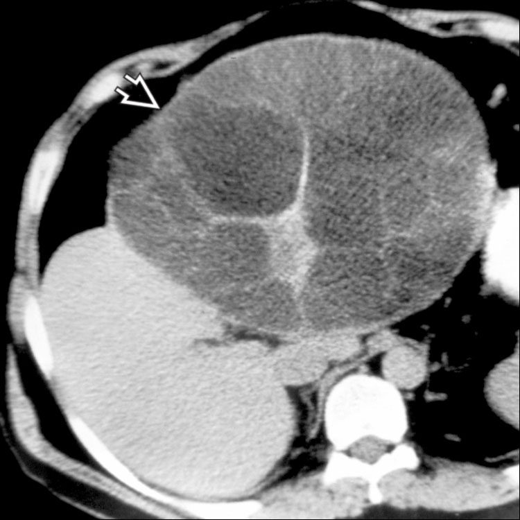
Radiographic Image This CT scan shows a very large hydatid cyst within the liver with internal septations .
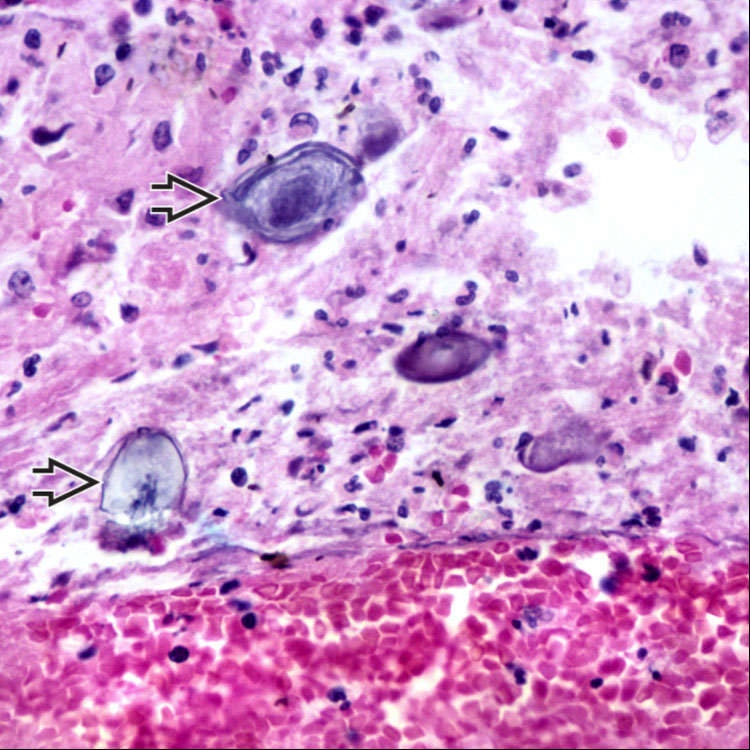
Degenerative Changes Many echinococcal cysts are partially or completely degenerated upon resection and may contain only abundant debris with fragments of degenerated protoscolices .
TERMINOLOGY
Synonyms
• Hydatid disease
Only gold members can continue reading. Log In or Register to continue


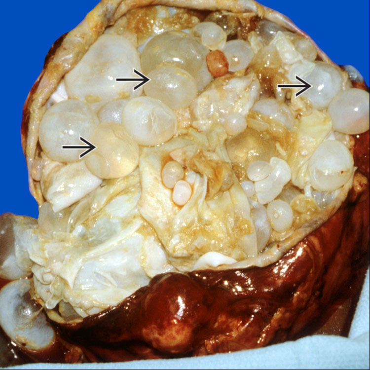
 , with a surrounding fibrous rim.
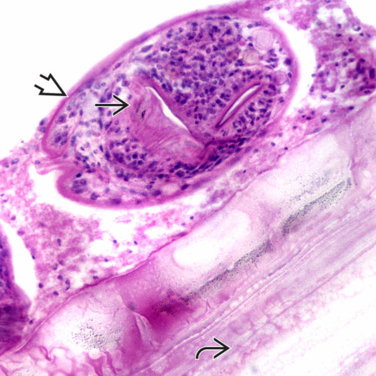
 containing the developing scolices
  . The next layer is composed of acellular, hyalinized material
  .
.
 .
.
 .
.


