Ductal Carcinoma In Situ with Microinvasion
Key Facts
Terminology
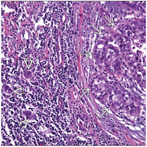
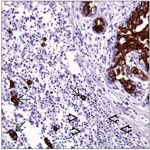
Ductal carcinoma in situ with microinvasion (DCISM)
1 or more separate microscopic foci of tumor cells infiltrating into periductal stroma
No invasive focus measuring > 1 mm in greatest dimension
DCISM is classified as AJCC T1mi
Clinical Issues
DCISM is uncommon, accounting for < 1% of all breast cancers
Up to 50% of patients present with palpable mass
Up to 20% of patients may also experience serosanguineous nipple discharge
Overall prognosis for DCISM is excellent
Outcome may depend on tumor burden and number of foci of microinvasion identified
Image Findings
Radiographic features will be similar to pure DCIS of equivalent size and grade
Microscopic Pathology
Typically, DCISM occurs in extensive area of highgrade DCIS
Periductal stromal changes are frequent finding
Rarely, microinvasion can occur with low-grade DCIS, LCIS, or in absence of carcinoma in situ
Top Differential Diagnoses
DCIS with cancerization of lobules
Invasive cancer with extensive DCIS
Artifactual displacement of tumor cells
TERMINOLOGY
Abbreviations
Ductal carcinoma in situ with microinvasion (DCISM)
Synonyms
Microinvasive ductal carcinoma in situ
Foci of “early stromal invasion”
Definitions
DCIS with extension of cancer cells beyond basement membrane lining ducts and lobules
Largest area of invasion ≤ 1 mm in size
DCISM has been divided into 2 types
Type 1: Single cells
Type 2: Clusters of cells
ETIOLOGY/PATHOGENESIS
Pathogenesis
Transition from DCIS to invasive cancer is poorly understood
No gene expression patterns specific to invasive carcinoma have been identified
Loss of normal luminal cell signaling may lead to dysfunction of myoepithelial and stromal cells maintaining basement membrane
Myoepithelial cells associated with DCIS show abnormal loss of myoepithelial cell markers
Thus invasion may be primarily initiated by loss of normal function of supporting cells rather than gain of function by carcinoma
Several studies have compared biologic properties between DCISM and DCIS
DCISM is characterized by higher proliferative rate and enhanced apoptosis
DCISM shows higher levels of expression for a number of matrix metalloproteases
CLINICAL ISSUES
Epidemiology
Incidence
DCISM is an uncommon entity accounting for < 1% of all breast cancers
Age
Range: 32-78 years (mean: 56.4 years)
Presentation
Up to 50% of cases present as palpable mass
Imaging findings may include abnormal density with or without associated microcalcifications
Up to 20% patients may also experience serosanguineous nipple discharge
Some patients present with Paget disease of nipple
Several studies have shown correlation between extent of DCIS and presence of microinvasion
High-grade DCIS with comedonecrosis is most common architectural pattern reported for DCISM
Presence of extensive high-grade DCIS correlates with presentation as palpable mass more commonly than DCIS without microinvasion
Prognosis
Overall prognosis for DCISM is excellent
5-year survival: 97-100%
Outcome for patients with DCISM may depend on number of foci of microinvasion identified
DCISM with single focus of microinvasion consisting of single cells (type 1) usually has outcome similar to pure DCIS
DCISM with multiple foci of microinvasion consisting of clusters of cells (type 2) may have worse prognosis
Stay updated, free articles. Join our Telegram channel

Full access? Get Clinical Tree


