Drugs Affecting Fertility and Reproduction


Drugs Affecting Fertility and Reproduction


Overview


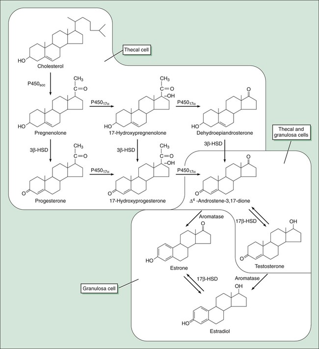
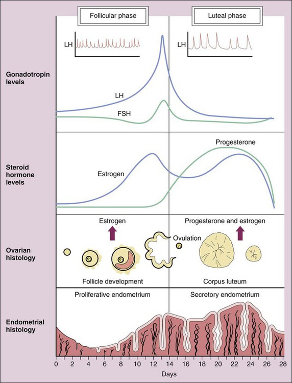
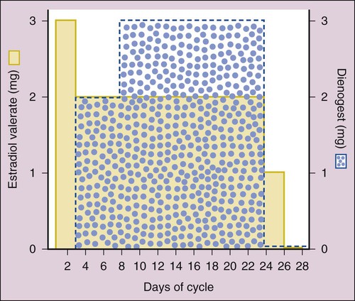
Human reproduction involves a cascade of hormonal secretions, beginning with the secretion of gonadotropin-releasing hormone (GnRH) from the hypothalamus. As described in Chapter 31, GnRH stimulates the pituitary to release two gonadotropins: follicle-stimulating hormone (FSH) and luteinizing hormone (LH). These gonadotropins then stimulate the production of steroids and gametes by the ovary in the female and by the testis in the male.


The three categories of steroids secreted by the gonads are (1) estrogens, which include estradiol, estrone, and estriol; (2) progestins, which include progesterone; and (3) androgens, which include testosterone. Estrogens, progesterone, and testosterone are produced in both males and females, but the relative amounts and patterns of secretion differ markedly between the sexes. Females primarily secrete estrogens and progesterone, whereas males primarily produce testosterone.


Biosynthesis of Gonadal Steroids


As shown in Figure 34-1, pregnenolone is the precursor to progesterone. It is also the precursor to dehydroepiandrosterone and androstenedione (two androgens secreted by the adrenal gland and discussed in Chapter 33) and to testosterone (the major androgen in males). The adrenal and gonadal androgens are converted to estrogens by aromatase, an enzyme that forms the aromatic A-ring necessary for the selective high-affinity binding of estradiol, estrone, and estriol to estrogen receptors.


In females, ovarian thecal cells secrete small quantities of testosterone. In males, about 95% of testosterone is produced by Leydig cells in the testes, and the remainder is derived from the adrenal cortex. Testosterone is synthesized in the testes by the same pathways as in the ovaries. Testosterone is subsequently converted to dihydrotestosterone (DHT) by 5α-reductase in the prostate, hair follicles, and skin. In the plasma, testosterone is primarily bound to sex steroid–binding globulin. In the liver, it is converted to androstenedione and other metabolites, including sulfate and glucuronide conjugates. About 90% of these metabolites are excreted in the urine.


Although both testosterone and DHT activate androgen receptors, DHT has greater receptor affinity and forms a more stable receptor-ligand complex than testosterone. If DHT formation is inhibited, this significantly reduces androgenic stimulation of the prostate gland and hair follicles. The androgen receptor located in target cells interacts with response elements in target genes and thereby stimulates protein synthesis in the same manner as other gonadal steroids.


Physiologic Actions of Estrogens and Progesterone


In females, estrogens and progesterone have multiple actions and interactions that are necessary for reproductive activity. Estrogens are formed in the granulosa cells of the ovary, whereas progesterone is primarily produced by the corpus luteum in response to LH secretion. Estrogens promote the development and growth of the fallopian tubes, uterus, and vagina, as well as secondary sex characteristics such as breast development, skeletal growth, and axillary and pubic hair patterns.


The pattern of hormonal changes occurring during the menstrual cycle is depicted in Figure 34-2. During the follicular phase of the cycle, ovarian follicles are recruited and a dominant estrogen-secreting follicle develops. Estrogen levels gradually increase, whereas progesterone levels remain very low. A surge of LH is released at mid-cycle in response to positive estrogen feedback to the pituitary gland, and this LH surge triggers ovulation. During the luteal phase of the cycle, the follicle becomes the corpus luteum (“yellow body”) that secretes both estrogen and progesterone in response to LH. Together, these hormones prepare the uterus for implantation of a fertilized egg as the endometrium becomes more vascular and secretory. If pregnancy does not occur, the corpus luteum ceases to produce estrogen and progesterone, resulting in menstruation. If pregnancy occurs, the placenta produces human chorionic gonadotropin, which maintains the production of progesterone by the corpus luteum. After about 3 months, the placenta becomes the predominant source of progesterone. This hormone serves to maintain pregnancy and prevents endometrial sloughing and miscarriage.


Estrogens have a number of other actions that are important in reproduction and other bodily functions. They sensitize the myometrium to oxytocin at parturition, and this facilitates labor. They stimulate protein synthesis in the brain and may thereby affect mood and emotions. Estrogens influence the distribution of body fat and thereby contribute to the development of feminine body contours. They enhance blood coagulation by increasing the synthesis of clotting factors, and they prevent osteoporosis by inhibiting bone resorption. In males and females, estrogens are responsible for epiphyseal closure, which halts linear bone growth.


Estrogens and progestins have different effects on serum lipoprotein levels. Estrogens decrease the levels of low-density lipoprotein (LDL) cholesterol and lipoprotein (a) while increasing the levels of high-density lipoprotein (HDL) cholesterol. In contrast, progestins produce a dose-related increase in LDL levels and a decrease in HDL levels.


Progesterone and other progestins increase basal body temperature by 0.5° C to 0.8° C (1.0° F to 1.5° F) at ovulation and throughout the luteal phase. They also affect the emotional state and have mild mineralocorticoid (salt-retaining) properties. Some of the synthetic progestins have other effects that are attributed to their androgenic activity (see later).


Physiologic Actions of Testosterone


LH stimulates testosterone synthesis in Leydig cells, whereas FSH promotes spermatogenesis by Sertoli cells in the seminiferous tubules. These cells provide an environment rich in testosterone, which is necessary for germ cell development. Sertoli cells also produce a protein called inhibin, which acts in concert with DHT as a feedback regulator of FSH secretion by the pituitary.


Testosterone is responsible for the development of secondary sex characteristics in males during puberty. These include growth of the larynx, thickening of the vocal cords, and growth of facial, axillary, and pubic hair. In addition to stimulating growth of the penis, scrotum, seminal vesicles, and prostate gland, testosterone stimulates and maintains sexual function in males. Testosterone and other androgens also do the following: increase lean body mass; stimulate skeletal growth; accelerate epiphyseal closure; increase sebaceous gland activity and sebum production, thereby contributing to the development of acne in both sexes; increase the production of erythropoietin in the kidneys; and decrease the levels of HDL cholesterol.


Estrogens and Progestins


An estrogen or progestin preparation can be used alone for the treatment of various disorders, or the two preparations can be used in combination for hormone replacement therapy (HRT) in postmenopausal women or for contraception in women of childbearing age.


Estrogens


Drug Properties


Chemistry.

The natural estrogens include estradiol and conjugated estrogens. Estradiol is an 18-carbon steroid with an aromatic A-ring (see Fig. 34-1). Conjugated equine estrogens are sulfate esters of estrone and equilin and can be obtained from the urine of pregnant mares. Ethinyl estradiol and mestranol are synthetic derivatives of estradiol.


Preparations.

Micronized estradiol is an orally administered estradiol preparation that has good bioavailability. Also available are vaginal estradiol tablets and a vaginal ring that slowly releases estradiol.


Ethinyl estradiol and mestranol are modified by the addition of an ethinyl group to estradiol, which reduces first-pass metabolism, increases the half-life to about 20 hours, and results in greater oral potency compared with native estradiol. These agents are primarily used in estrogen-progestin contraceptives. Conjugated equine estrogens are hydrolyzed to estrone and equilin before absorption from the gut. They undergo relatively little first-pass metabolism and are converted in the liver to sulfate and glucuronide conjugates that are excreted in the urine. Estrone is also available in a rapidly absorbed formulation for intramuscular administration.


Several long-acting formulations of estradiol are available for transdermal or intramuscular administration. Transdermal estradiol systems slowly release the drug for absorption through the skin. These preparations are formulated for twice-weekly or weekly application. Estradiol cypionate and estradiol valerate are long-acting esters of estradiol that are slowly absorbed after intramuscular administration and provide effective plasma concentrations of estradiol for several weeks. Estradiol valerate is also contained in a variable dose oral contraceptive (NATAZIA), in which the estrogen dose is stepped up and the progestin dose is stepped down during each menstrual cycle (Fig. 34-3).


“After their absorption the natural estrogens are highly bound to sex steroid–binding globulin, are widely distributed, and are concentrated in fat.” They undergo enterohepatic cycling, in which conjugated metabolites are excreted in the bile and converted to free estrogens by intestinal bacteria. The free estrogens are then reabsorbed into the circulation. As with estrone and equilin, estradiol is metabolized in the liver to sulfate and glucuronide conjugates, and these conjugates are primarily excreted in the urine, with small amounts excreted in the feces.


Adverse Effects.

Estrogens occasionally cause breast tenderness, headache, edema, nausea, vomiting, anorexia, and changes in libido. These effects are less likely to occur in women using the lower-dose preparations now recommended for HRT.


The more serious adverse effects of estrogens include hypertension, thromboembolic disorders, and gallbladder disease. The hypertensive effect of estrogens has been partly attributed to increased angiotensinogen synthesis and formation of angiotensin II, whereas thromboembolic complications result from increased hepatic synthesis of clotting factors. Estrogens increase cholesterol excretion in the bile, accounting for their tendency to cause gallstones.


Estrogens are contraindicated during pregnancy and should be avoided in women with uterine fibroids. Estrogens should be used with great caution in women with hepatic diseases, endometriosis, thromboembolic diseases, or hypercalcemia.


Progesterone and Its Derivatives


Progesterone is the primary natural progestin in mammals. Progesterone undergoes extensive first-pass metabolism after oral administration and has a short plasma half-life. To extend the oral bioavailability and half-life, esters of progesterone have been developed. These include megestrol, hydroxyprogesterone caproate, and medroxyprogesterone acetate. Megestrol is administered orally, whereas hydroxyprogesterone caproate is administered as a long-acting intramuscular preparation. Medroxyprogesterone acetate can be given either orally or intramuscularly. Following their absorption, the progesterone esters are bound to albumin in the circulation. The esters are converted to several hydroxylated metabolites and to pregnanediol glucuronide in the liver, and these metabolites are excreted in the urine.


Progesterone esters are used to suppress ovarian function in the treatment of dysmenorrhea, endometriosis, and uterine bleeding. In this setting the progesterone derivatives produce feedback inhibition of gonadotropin secretion by the pituitary gland. In HRT (see later), the progesterone esters are used in combination with estrogens to decrease the incidence of estrogen-induced irregular bleeding and to prevent uterine hyperplasia and endometrial cancer. Hydroxyprogesterone caproate once-weekly injections are approved by the U.S. Food and Drug Administration (FDA) to reduce the risk of preterm delivery in women with a history of at least one spontaneous preterm birth. Studies found that women treated with the drug had significantly fewer preterm deliveries.


Synthetic Progestins


Synthetic progestins are primarily used as oral contraceptives (see later), but they are also used to treat dysmenorrhea, endometriosis, and uterine bleeding in the same manner as the progesterone esters.


Most of the synthetic progestins are derivatives of nortestosterone (testosterone without a methyl group on carbon-19) and have varying degrees of estrogenic, antiestrogenic, and androgenic activity. Norgestrel has more androgenic activity than other progestins, whereas desogestrel and norgestimate have less androgenic and more progestational activity. Drospirenone is a spironolactone derivative with antiandrogenic effects. The synthetic progestins also contain molecular entities that increase their oral bioavailability and duration of action. Their half-lives range from 7 to 24 hours, whereas the half-life of progesterone is only about 5 minutes.


Hormone Replacement Therapy


Menopause refers to the cessation of menstruation that occurs in most women between the ages of 45 and 55. Before menopause, the supply of eggs in a woman’s ovaries declines and ovulation becomes irregular. The ovarian follicles fail to develop and secrete normal amounts of estrogen. When estrogen levels are no longer sufficient to suppress FSH secretion by the pituitary gland, FSH levels rise. When FSH levels are above 40 IU/L, a woman is said to be in menopause.


Although estrogens can be used alone for HRT in women who have had a hysterectomy, estrogen should be used in combination with a progestin for women with a uterus. This is because giving estrogen alone increases the risk of endometrial cancer in these women.


Therapeutic Effects


Studies consistently show that estrogens relieve symptoms of menopause in up to 90% of women. These symptoms include hot flashes or flushes that consist of alternating chills and sweating accompanied by nausea, dizziness, headache, tachycardia, and palpitations (Box 34-1). Episodes of these symptoms often occur several times a day, but night sweats are particularly common. These symptoms occur in association with surges in GnRH and gonadotropins that result from the lack of estrogen feedback inhibition. The gonadotropin surges alter hypothalamic thermoregulatory centers, leading to the symptoms described earlier in this paragraph.


Box 34-1   A Case of Hot Flashes and Loss of Sleep


Case Presentation


A 50-year-old woman tells her health care provider about episodes of hot flashes that occur mostly at night accompanied by sweating and interrupted sleep. She has also noticed vaginal dryness and feeling slightly depressed. Her periods have been increasingly infrequent over the past year. She suspects that she is entering menopause and asks about hormone replacement therapy (HRT). The woman has been healthy throughout her adult life and adheres to a good diet and a regular exercise program. Her mother developed osteoporosis after menopause, and the patient is concerned about maintaining healthy bones. She does not have a family history of premature cardiovascular disease or of reproductive tract cancer. Her physical examination findings and laboratory test results are normal, and she is scheduled for a bone density determination. She is started on a low dose of oral estrogen and a vaginal estrogen cream along with cyclic medroxyprogesterone. Her bone density will be monitored and appropriate therapy provided. She is encouraged to maintain a high level of calcium intake and to increase her vitamin D supplementation. This case is continued in Chapter 36.


Case Discussion


Menopause is defined as the absence of menstruation for 12 consecutive months and is caused by cessation of estrogen production by the ovaries. Common symptoms of menopause include hot flashes, mood swings, sleeplessness, vaginal dryness, and urinary incontinence. Many women experience irregular periods and other symptoms of menopause for several months preceding menopause. Tachycardia, depression, and other symptoms of estrogen withdrawal may also occur. Low doses of estrogens control most menopausal symptoms, and vaginal estrogen preparations effectively relieve vaginal atrophy. Clinical trials have produced conflicting data with respect to HRT and the risk of cardiovascular disease. In some cases, the findings of trials involving older women with heart disease have been erroneously extrapolated to healthy younger women. Recent analysis of these trials suggests that hormonal replacement in younger menopausal women 50 to 59 years of age may protect against cardiovascular disease as well as osteoporosis, and reduces the risk of colorectal cancer. For most healthy menopausal women, short-term HRT is a relatively safe and effective method of controlling menopausal symptoms.


Estrogens also relieve other menopausal symptoms, including urogenital, vulvar, and vaginal atrophy, and they protect against osteoporosis. Estrogens may improve mood and reduce cognitive difficulties, possibly secondary to improved sleep.


Although some epidemiologic studies suggest that HRT reduces the risk of cardiovascular disease, some recent clinical trials of estrogen replacement in postmenopausal women have reached the opposite conclusion. The Nurses’ Health Study was an observational study of a cohort of women that obtained information about the relationship between cardiovascular disease and lifestyle, lipid levels, and HRT. This study concluded that HRT decreased the risk of coronary artery disease and that women with the highest risk of cardiovascular disease obtained the greatest benefit from HRT. This study has the same limitations as all cohort observational studies, and it has been postulated that the women who sought HRT were also more likely to adopt a healthy lifestyle that reduced their risk of heart disease.


The Postmenopausal Estrogen/Progestin Interventions study was a randomized, prospective trial of HRT in 875 women aged 45 to 64. It demonstrated beneficial effects of HRT on risk factors for heart disease by showing that HRT reduced cholesterol and fibrinogen levels. The relatively short duration of the trial (3 years) was not sufficient to determine long-term effects of HRT on morbidity and mortality.


The Heart and Estrogen-Progestin Replacement Study was the first trial to investigate the effects of HRT in older postmenopausal women with existing heart disease. This study found a lack of benefit of HRT on fatal or nonfatal myocardial infarction. In fact, the study found that women were at an increased risk of myocardial infarction during the first year of HRT, although the risk of myocardial infarction decreased in subsequent years. After an additional 3 years of study, no significant differences were noted in cardiovascular outcomes between women on HRT and those not receiving HRT. This trial has been criticized because women in the study were already receiving cardioprotective medications and were allowed to begin or change statin therapy during the study. Also, the average age of women in the study was 67, which is well above the age when women enter menopause.


The Women’s Health Initiative included a randomized trial of more than 16,000 women who received conjugated equine estrogen plus medroxyprogesterone acetate or a placebo. After 5 years the study was halted because the data showed that women receiving HRT had increased risk of coronary artery disease, stroke, and pulmonary embolism. HRT, however, reduced the risk of colorectal cancer and hip fractures. Another arm of this study looked at the effect of estrogen by itself in women without a uterus. This study was stopped when it was reported that estrogen increased the risk of stroke but did not affect the incidence of coronary artery disease in these women. The Women’s Health Initiative study has been criticized for the older age of the participants.


More recently, a trial conducted by the Women’s Health Initiative in younger and generally healthier 50- to 59-year-old women found that that both estrogen alone and estrogen and progestin in combination provided cardioprotective effects with a reduction in coronary events and total mortality. This study added to the growing evidence that HRT is safe and effective in younger menopausal women, particularly when used with the lowest doses and for the shortest duration required to relieve menopausal symptoms.


In summary, low-dose estrogen preparations are still a very useful treatment for relieving menopausal symptoms in younger menopausal women. Estrogens also protect against osteoporosis and certain cancers and may reduce the risk of cardiovascular disease in younger women. The role of estrogens in older women remains uncertain. Older women with existing heart disease should not be placed on estrogen therapy. Other medications are available to prevent osteoporosis in menopausal women and should be used when HRT is discontinued.

< div class='tao-gold-member'>

Stay updated, free articles. Join our Telegram channel

Jul 23, 2016 | Posted by in PHARMACY | Comments Off on Drugs Affecting Fertility and Reproduction

Full access? Get Clinical Tree

Get Clinical Tree app for offline access