(1)
Department of Pathology, Rutgers-New Jersey Medical School, Newark, NJ, USA
Keywords
Congenital anomalies, Fallopian tubeAcute salpingitisChronic salpingitisTuberculous salpingitisSalpingitis isthmica nodosaParatubal cystEctopic pregnancyFallopian tube prolapseEndometriosis, Fallopian tubeAdrenal rest, Fallopian tubeAdenomatoid tumor, Fallopian tubeSerous tubal intraepithelial carcinoma (STIC)Carcinoma of Fallopian tube8.1 Diseases of the Fallopian Tubes
The Fallopian tube can pose some unique challenges for evaluation by pathologists, and there are newer developments in our understanding of the origin of some of the epithelial malignancies of the ovaries now felt to derive from Fallopian tube fimbria (Table 8.1). This chapter addresses these and other diseases of the Fallopian tubes.
Table 8.1
Key points about Fallopian tube pathology
Acid fast bacilli are not reliably identified on acid fast histochemical stains, due to the rarity of organisms. Patients with granulomatous salpingitis may benefit from other diagnostic modalities to rule out tuberculosis |
Implantation site should be sought in cases of tubal abortion with only hematosalpinx seen, to confirm ectopic pregnancy |
For atypical epithelial proliferations in the fallopian tube, a positive p53 indicates a p53 signature and requires increased Ki-67 proliferation index in addition, to diagnose a serous tubal intraepithelial carcinoma (STIC), the putative precursor lesion of high grade ovarian and peritoneal serous carcinoma |
8.2 Congenital Anomalies of the Fallopian Tubes
The Fallopian tubes derive from the upper non-fused portions of the Müllerian ducts. Isolated anomalies are rare, but partial or total atresia has been reported [1]. Atresias are more likely to occur in association with uterine anomalies, such as a unicornuate uterus, than in isolation.
8.3 Infectious and Inflammatory Lesions of Fallopian Tubes
8.3.1 Acute Salpingitis
Acute salpingitis may be secondary to a variety of organisms, including gonorrhea, chlamydia, and occasionally actinomyces (Fig. 8.1), the latter usually in association with IUD usage. Grossly, if the fimbria become adherent, the tube becomes dilated with purulent material, forming a pyosalpinx. Histologically, purulent material, composed of neutrophils and necrotic debris, is seen within the mucosa and the tubal lumen (Fig. 8.2).
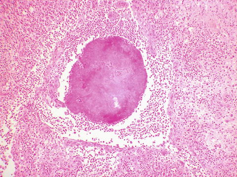
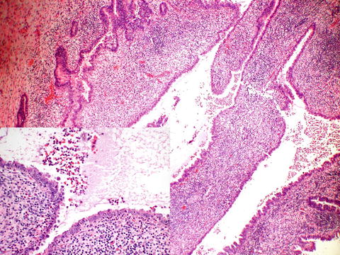

Fig. 8.1
Actinomyces. A typical radiating colony of organisms is seen. Grossly, these colonies are sometimes yellow in appearance, leading to the name “sulphur granules”

Fig. 8.2
Pyosalpinx. The tube is dilated by purulent exudate (pus). Histologically, the purulent material involves both the tubal epithelial folds and the lumen. The inset shows the purulent material, composed of neutrophils and necrotic debris
8.3.2 Chronic Salpingitis
Patterns of chronic salpingitis depend on how the Fallopian tube heals after an episode of acute salpingitis. The fimbria may remain open or may be sealed shut by adhesions. If the tube remains patent, there may be no changes at all, or there may be chronic salpingitis, characterized by a chronic inflammatory infiltrate composed of lymphocytes, plasma cells, and/or histiocytes. The folds of the tube may agglutinate, and although no inflammation is seen, there are numerous blind pouches created, termed follicular salpingitis (Fig. 8.3). This is thought to increase the risk of tubal ectopic pregnancies, as the fertilized ovum can get trapped in one of these areas. If the fimbria are sealed, and pyosalpinx resolves, what is left is flattened epithelium in a tube filled with nonpurulent fluid, a hydrosalpinx (Fig. 8.4).
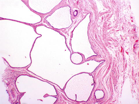
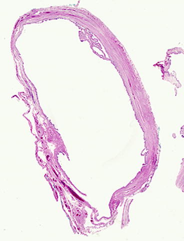

Fig. 8.3
Follicular salpingitis. Numerous blind spaces are seen due to agglutination of the folds of tubal epithelium

Fig. 8.4
Hydrosalpinx. The epithelium is flattened and the folds are lost, leaving behind a dilated sac of fluid
8.3.3 TB Salpingitis
Although rare in developed countries, tuberculous salpingitis is a common cause of infertility in endemic areas, where it may spread to the genital tract through hematogenous or lymphatic spread, or rarely through sexual contact [2]. It has also been reported in association with immunosuppression [3]. Clinically, the tube has been described as sometimes having a beaded appearance [2]. Histopathologically, the hallmark of tuberculosis is granulomatous inflammation (Fig. 8.5), usually with central necrosis called caseation, due to the gross cheesy appearance. However, even in the absence of central necrosis, stains for fungi and acid fast bacilli should be performed on cases of granulomatous salpingitis. Clinicians need to be aware that the stain for acid fast bacilli is very insensitive for confirmation of tuberculosis, due to the rarity of identifiable microorganisms, differing from some other strains of mycobacterium. If suspected clinically, other modalities, such as culture and PCR, should be considered.
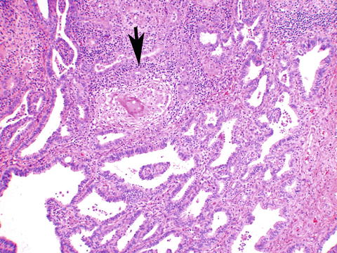

Fig. 8.5


Granulomatous salpingitis. This tube shows chronic salpingitis with fused epithelial folds. A granuloma (arrow) composed of epithelioid histiocytes surrounding a multinucleated giant cell is seen. No necrosis was present in this case
Stay updated, free articles. Join our Telegram channel

Full access? Get Clinical Tree


