35
Diaphragm and Posterior Abdominal Wall
The diaphragm and posterior abdominal wall are two anatomically linked structures that support and protect the abdominal contents. The diaphragm is the primary muscle of breathing and separates the thorax from the abdomen, whereas the muscles of the posterior abdominal wall aid movements of the vertebral column and lower limb. The posterior abdominal wall extends from the posterior attachment of the diaphragm down to the iliac crests. Bony support is provided by the lower ribs, lower thoracic vertebrae, lumbar vertebrae, and pelvis.
Muscles
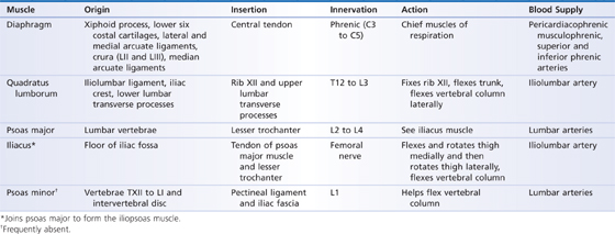
The four muscles (Fig. 35.1A) of the posterior abdominal wall are the diaphragm, quadratus lumborum, iliacus, and psoas (Table 35.1).
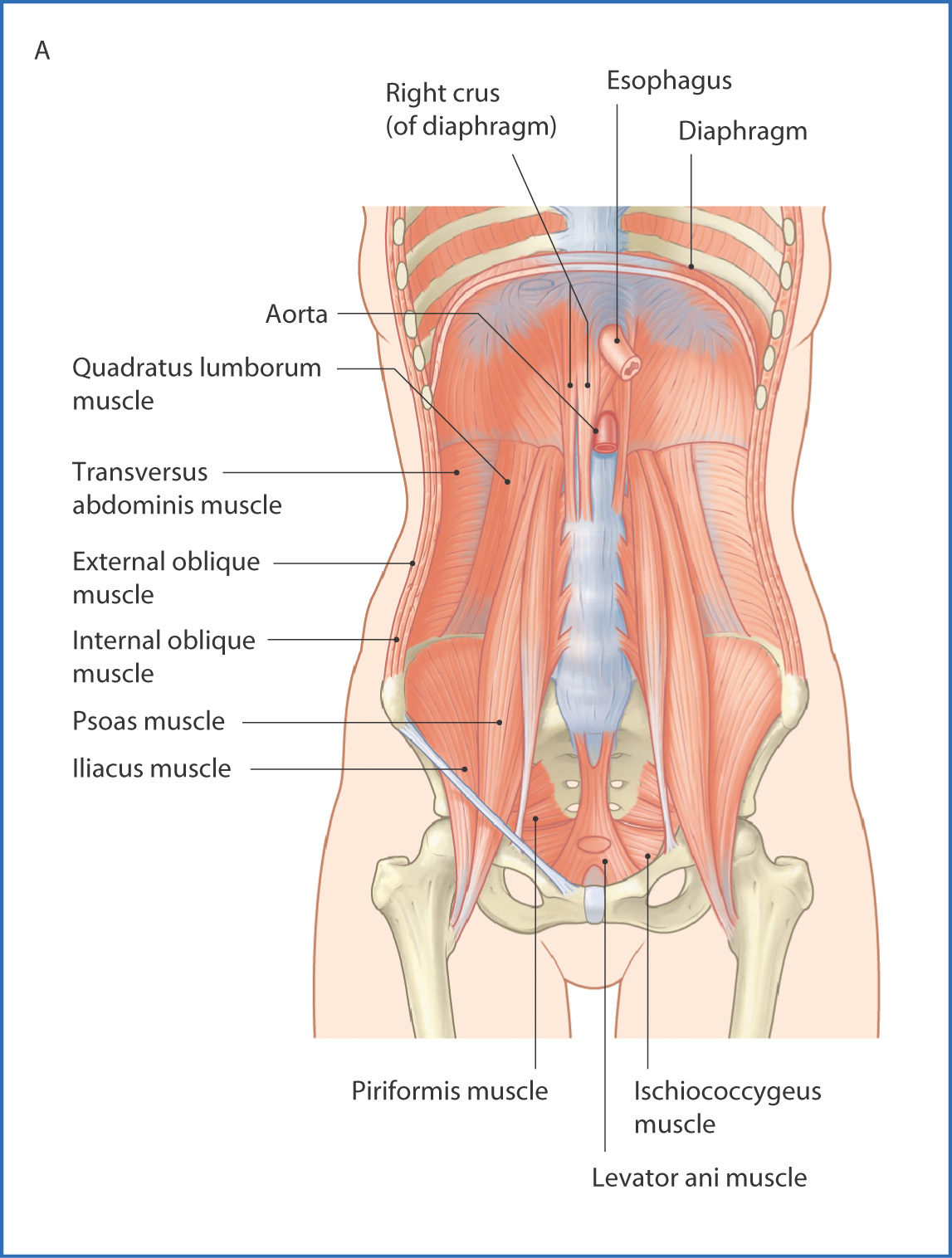
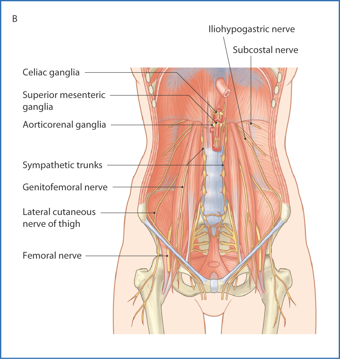
FIGURE 35.1 Muscles (A) and nerves (B) of the posterior abdominal wall.
TABLE 35.1 Muscles of the Posterior Abdominal Wall
DIAPHRAGM
The diaphragm is a domed muscle with a central tendinous region. When viewed from the front (anteriorly), it has two upward-curving domes. The right dome passes over the liver, which causes it to be higher than the left dome. In the central tendinous part of the diaphragm are three major openings for the passage of important structures. From anterior to posterior, these openings are at vertebral levels TVIII, TX, and TXII/LI (Table 35.2).
TABLE 35.2 Openings in the Diaphragm and the Structures Passing through Them
Openings | Structures | Vertebral Levels |
Inferior vena cava (central tendon) | Inferior vena cava, right phrenic nerve | TVIII |
Esophageal hiatus | Esophagus, vagal trunks, esophageal branches of left gastric artery | TX |
Aortic hiatus | Aorta, thoracic duct, azygos and hemi-azygos veins | TXII (behind the diaphragm, between the crura) |
Other structures that pass through the diaphragm | ||
| Left phrenic nerve | Left dome of diaphragm |
| Splanchnic nerves | Crura of diaphragm |
| Sympathetic trunks | Posterior to medial arcuate ligament |
| Subcostal nerve and vessels | Posterior to lateral arcuate ligament |
| Superior epigastric vessels | Anterior between costal and sternal attachments |
| Musculophrenic vessels | Between seventh and eighth costal cartilages |
Motor innervation of the diaphragm is provided by the right and left phrenic nerves (C3 to C5). The phrenic nerves also provide sensory innervation to the central part of the diaphragm. Sensory innervation of the peripheral parts of the diaphragm is provided by the intercostal nerves (T5 to T11) and the subcostal nerves (T12).
Blood is supplied to the diaphragm by the superior phrenic arteries (branches of the thoracic aorta), the musculophrenic and pericardiacophrenic arteries (branches of the internal thoracic arteries), and the inferior phrenic arteries (branches of the abdominal aorta).
Venous drainage is provided by the musculophrenic and pericardiacophrenic veins, which drain to the internal thoracic veins. The right inferior phrenic vein drains to the inferior vena cava; the left inferior phrenic vein drains to the left suprarenal vein, which then empties into the left renal vein.
Lymphatic drainage of the diaphragm is to lymphatic plexuses on the superior and inferior diaphragmatic surfaces. These lymphatic vessels in turn drain to the posterior mediastinal and superior lumbar lymph node groups.
QUADRATUS LUMBORUM MUSCLE
The quadratus lumborum muscle (see Fig. 35.1A) is a rectangular muscle that fills the space between the most inferior rib (rib XII) and the superior part of the pelvis (the iliac crest). It aids breathing by supporting the diaphragm. It is also a lateral flexor of the trunk (see Table 35.1).
PSOAS MAJOR AND ILIACUS MUSCLES
These muscles originate from the internal surfaces of the lower lumbar vertebrae and pelvis and join and insert as the iliopsoas muscle onto the lesser trochanter of the femur. The primary action of the iliopsoas muscle is to flex the thigh at the hip joint (see Table 35.1).
Nerves of the Posterior Abdominal Wall
The subcostal nerve is the anterior ramus of thoracic nerve T12. It enters the posterior wall of the abdomen next to the arcuate ligament of the diaphragm and crosses the quadratus lumborum muscle just posterior to the kidney. The subcostal nerve supplies muscles of the posterior abdominal wall, peritoneum, and skin of the suprapubic region.
Stay updated, free articles. Join our Telegram channel

Full access? Get Clinical Tree


