Fig. 33.1
Severe tinea corporis of the leg due to trichophytun rubrum. It started out much less prominent, but topical and systemic corticosteroid use caused the leg to swell and the erythema and some induratio to spread up the leg from the foot
In infants, granuloma gluteale infantum is a rare condition in the diaper area that often develops following the use of corticosteroid treatment of diaper rash. The literature, however, is unclear as to the true etiology. Fungal cultures have not demonstrated a superinfection with Candida sp. In fact, preexisting candidal dermatitis has been implicated as a possible contributing factor.
Steroid Rosacea and Acne
One common complication of topical steroid use is a worsening of rosacea. Oftentimes this is found in patients of fair skin with preexisting disease. This typically occurs on the face in chronic users of topical cortciosteroids. A patient would often apply a low-dose topical corticosteroid to treat the initial malady. With judicious use of steroid, tachyphylaxis can develop, which subsequently can lead to increased frequency of application. With tachyphylaxis comes the risk of rebound recurrence. Patients often demonstrate a diffuse pruritic erythematous rash with papules and pustules. Possible mechanisms include dilation of blood vessels, rebound release of proinflammatory cytokines, and an accumulation of nitric oxide.
Treatment of steroid-induced rosacea is challenging. Ideally, cessation of steroid use is the treatment of choice, however, patients often have difficulty with the rebound tachyphylaxis. Many adjunctive treatments have been used, including the addition of topical immunomodulators such as tacrolimus, as well as topical and oral antibiotic treatment.
Interestingly, studies have shown that the presumed effect of tachyphylaxis is, in fact, a misnomer. In fact, physician and patient both often presume the presence of tachyphylaxis when in fact there was improvement or no change in the clinical condition. This may be related to the therapeutic efficacy of the topical corticosteroid, where perhaps physicians and patients expected a greater response from a weaker strength of medication.
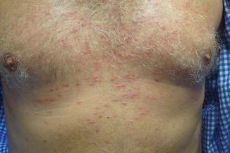
Steroid-induced acne tends to have monotonous symmetric papules without comedones or cysts. Steroid-induced folliculitis (Fig. 33.2) is indistinguishable clinically, except often in an age group not prone to acne, and often more symptomatic with pruritis and a flare with heat.


Fig. 33.2
Erythematous, monotonous papules in a symmetrical distribution over the chest in a man treated for months with triamcinilone cream. As with steroid acne, there are no comedones or cysts
Perioral Dermatitis
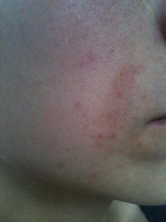
In a similar vein to steroid-induced acne and rosacea is that of perioral dermatitis. This is most commonly found in women, although men have been shown to demonstrate it as well. The typical picture is once again a chronic topical corticosteroid user who develops perioral pruritis, erythema, and papules. The papules may be pinhead-sized and can coalesce into plaques. There is often a perioral halo of clear skin, and the disease will only reach the lips in the corners (Fig. 33.3). Treatment once again is tapering the steroids, with possible adjunctive immunomodulators. Some authors refer to it as periorificial since it can occur around the nose and genital area when topical steroids are used there.


Fig. 33.3
Peroral dermatits on the left cheek, with tiny papules becoming plaque-like in places. Upper border is the corner of lip where the dermatits barely reaches. Hydrocortisone cream had been used for weeks for an unknown skin ailment when this acneiform problem appeared
Alterations to Pigment and Hair
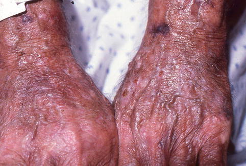
As mentioned previously, corticosteroids affect a plethora of cell types. An additional postulated cell type is the melanocyte. Steroids have been postulated to potentially decrease the synthesis of melanin from melanocytes. As a result, patients with intralesional injection of glucocorticoids have been shown to develop hypopigmentation along the distribution of the steroid. Additionally, specifically with patients of darker complexions, long-term use of topical glucocorticoids have been shown to produce patchy hypopigmentation (Fig. 33.4). The hypopigmentation typically resolves with discontinued use of the steroid.


Fig. 33.4
Increased skin transparency over the backs of hands with patchy areas of hypopigmentation, purport, and small erosions. Theses changes were the result of prolonged use of desoximetasone cream for an unknown skin disease
Glucocorticoids have also been shown to promote the development of hirsutism and hypertrichosis. Studies have shown that children with long-term overuse of glucocorticoids have developed diffuse hypertrichosis. These same children have also been found to exhibit other adverse effects of prolonged corticosteroid use including growth retardation and adrenal suppression.
Hypertrichosis is a relatively rare phenomenon and is typically demonstrated in patients with prolonged use of glucocorticoids. The presence of hypertrichosis is an ominous sign and should be a clue to the physician to look for other sequelae of prolonged steroid use.
Skin Atrophy
Perhaps one of the more widely studied complications of prolonged topical steroid use is dermal atrophy. In fact, some studies have demonstrated that all topical steroids cause skin atrophy at varying degrees. This is due to a multitude of contributing factors. First, glucocorticoid activity has been shown to suppress cell division in the basal skin layer. Additionally, it has been shown to prevent fibroblast mitoses as well as decreases in collagen synthesis, as well as the creation of mucopolysaccharides. Pathological studies have also shown a significant decrease in mast cells.
Clinically, patients present with a thinning of the skin, increasing its transparency, as shown in Fig. 33.4. Age, location, and potency of steroid all contribute to the atrophic affect. Areas of the thinnest skin, such as intertriginous areas, are more susceptible.
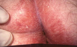
Often concurrently seen with skin atrophy are the presence of telangiectasias (Fig. 33.5). These typically develop due to the stimulation of dermal vascular endothelial cells by glucocorticoids.


Fig. 33.5
Dramatic telangiectasias, milia, and atrophy in a patient using a fluorinated corticosteroid ointment for months for itching in the rectal area
Currently, the best treatment for steroid-induced skin atrophy is prevention. All topical steroids should be immediately discontinued. Ideally, potent steroids should be avoided in areas of thin skin and given for the shortest period whenever possible. Topical tretinoin has shown promise, however, studies have had conflicting results. Its potential efficacy is related to in vitro studies, which have demonstrated the opposite effect of tretinoin on the skin, including an increase in mast cell count and collagen synthesis.
Stay updated, free articles. Join our Telegram channel

Full access? Get Clinical Tree


