20
Cubital Fossa and Elbow Joint
Cubital Fossa
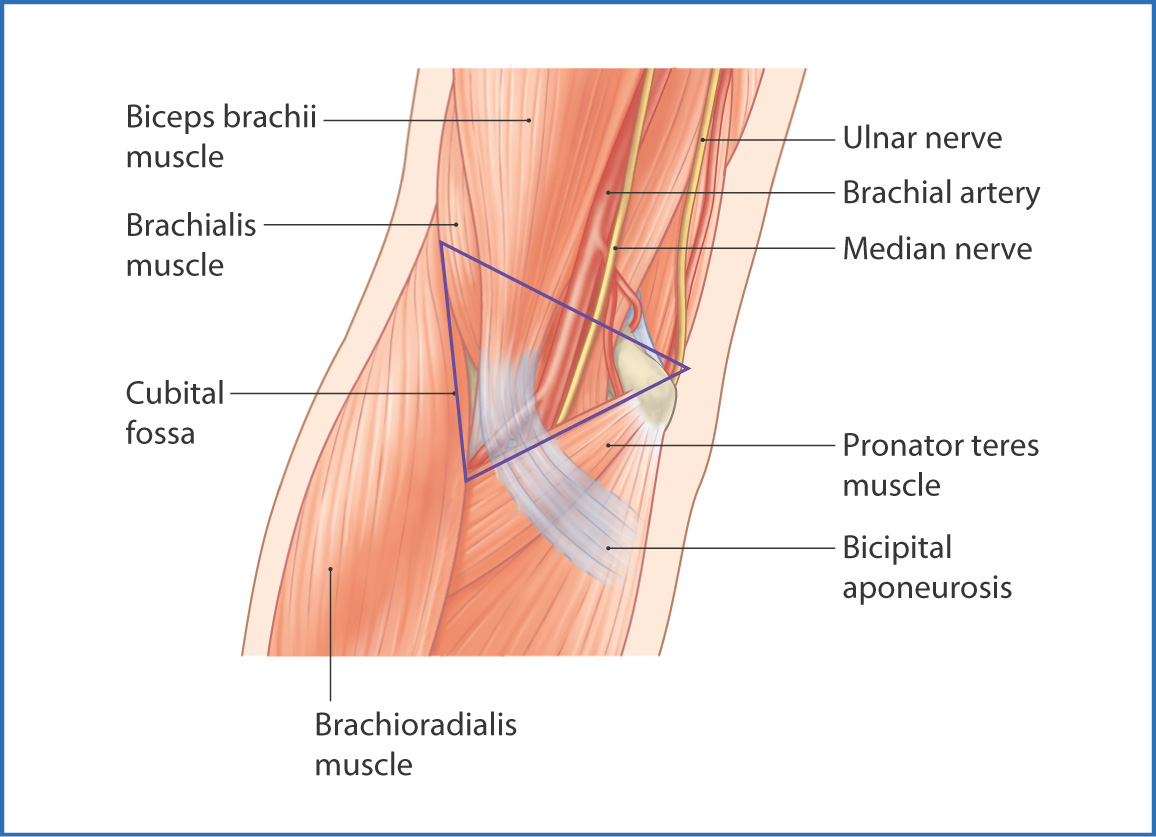
The cubital fossa is a triangular area on the anterior surface of the elbow joint formed by the borders of the arm and forearm muscles:
- The apex points inferiorly toward the hand.
- The medial side is formed by the pronator teres muscle.
- The lateral border is the brachioradialis muscle.
- The superior border is an imaginary line joining the medial and lateral epicondyles of the humerus.
- The floor is formed by the supinator and brachialis muscles.
- The roof is the deep fascia of the forearm reinforced by the bicipital aponeurosis.
- The medial side is formed by the pronator teres muscle.
The superficial fascia of the cubital fossa contains a variable arrangement of superficial veins, as well as the medial and lateral cutaneous nerves of the forearm (for origin see Chapter 16). The cephalic vein is lateral, the basilic vein is medial, and the median cubital vein joins the two. The median vein of the forearm ends in the median cubital vein. The contents of the cubital fossa from lateral to medial are the tendon of the biceps brachii, the brachial artery, and the median nerve (Fig. 20.1).

FIGURE 20.1 Cubital fossa (outlined) and neighboring structures.
The Elbow Joint
The elbow is a synovial joint at the junction of the arm and forearm. It joins the distal end of the humerus to the proximal ends of the radius and ulna and is primarily mobile in only one axis, which allows active flexion up to about 135° to 145°. The elbow joint comprises three distinct articulations, all contained within a single joint capsule:
- the humero–ulnar joint between the trochlea of the humerus and the trochlear notch of the ulna (a hinge joint)
- the humeroradial joint between the capitulum of the humerus and the head of the radius (a ball-and-socket joint)
- the proximal radio–ulnar joint between the head of the radius and the radial notch of the ulna
- the humeroradial joint between the capitulum of the humerus and the head of the radius (a ball-and-socket joint)
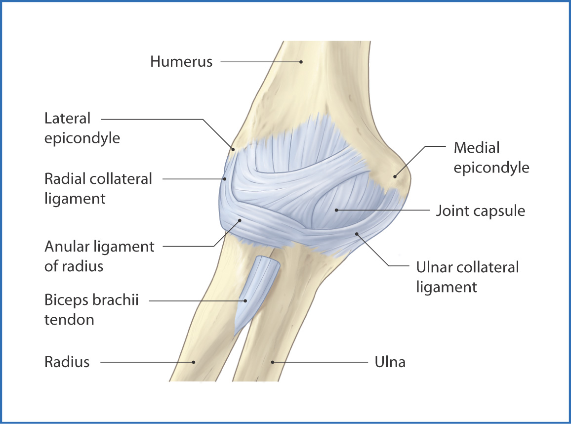
The humero-ulnar and humeroradial joints act as a single hinge joint during extension and flexion of the forearm; the proximal radio-ulnar joint allows rotation of the head of the radius during pronation and supination of the forearm. These three articulations are enclosed within the joint capsule of the elbow. This is thickened laterally to form the radial collateral and ulnar collateral ligaments, which stabilize the elbow joint and prevent lateral flexion (Fig. 20.2):
- The radial collateral ligament is fan-like and joins the lateral epicondyle of the humerus to the anular ligament of the radius.
- The ulnar collateral ligament is triangular and extends from the medial epicondyle of the humerus to the coronoid process and olecranon of the ulna.

FIGURE 20.2 Ligaments of the elbow.
Distally, the joint capsule forms the anular ligament of the radius, which is a strong band of connective tissue lined by hyaline cartilage that forms a ring-like shape around the head of the radius. This unique shape allows the head of the radius to rotate while the ulna remains stationary, an action needed during pronation and supination of the forearm. The distal radio-ulnar joint is not part of the elbow joint complex but moves in conjunction with the proximal radio-ulnar joint and facilitates pronation and supination of the forearm and hand (Table 20.1).
TABLE 20.1 Joints of the Elbow and Forearm

Stay updated, free articles. Join our Telegram channel

Full access? Get Clinical Tree


