• Risks, particularly for large congenital melanocytic nevus
~ 5% risk of developing cutaneous melanoma
Neurocutaneous melanosis
Microscopic
• Melanocytes usually extend into lower reticular dermis and sometimes subcutaneous tissue
• Infiltration of
Arrector pili muscle
Adnexal structures
Nerves
• Clustering around blood vessels
• Cells become smaller and more dispersed with depth
• Nodular proliferations (more common in large nevi)
Based in dermis, no epidermal involvement or necrosis
Top Differential Diagnoses
• Acquired melanocytic nevi: Not present at birth
Usually limited to upper 1/2 of reticular dermis)
• Atypical (dysplastic/Clark) melanocytic nevi: Not present at birth
Junctional component shows cytologic atypia, bridging, and lateral extension beyond intradermal cells, which are often limited to upper 1/2 of reticular dermis
• Malignant melanoma arising in congenital nevus
In differential diagnosis of dermal nodular proliferation, very rare if age is < 1 yr
Well-demarcated, highly cellular nodules of atypical epithelioid to spindled cells with cytologic atypia; nuclear pleomorphism and necrosis often present
Congenital Melanocytic Nevus This light tan, medium-sized congenital melanocytic nevus, which measured 10 x 8 cm, has coarse terminal hairs within it. The patient also has keratosis pilaris and an acquired melanocytic nevus .
Compound, Predominantly Intradermal Congenital Nevus This congenital compound nevus has nests of larger, more heavily pigmented superficial cells progressing to smaller, less pigmented cells deeper in the dermis , consistent with maturation with dermal descent.
Congenital Intradermal Nevus This congenital intradermal melanocytic nevus extends deeply throughout the dermis, with clustering of melanocytes around vessels and adnexa.
Congenital Junctional Nevus Some congenital nevi, especially if small, may be purely junctional, with scattered nests and single melanocytes at the tips and sides of rete, mimicking an acquired junctional melanocytic nevus.


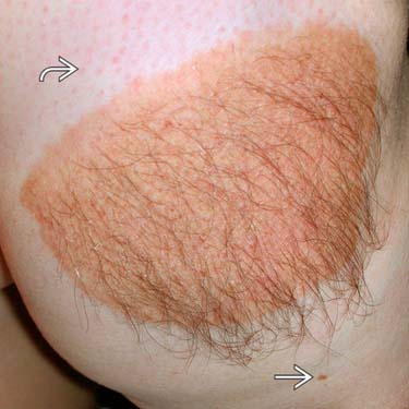
 and an acquired melanocytic nevus
  .
.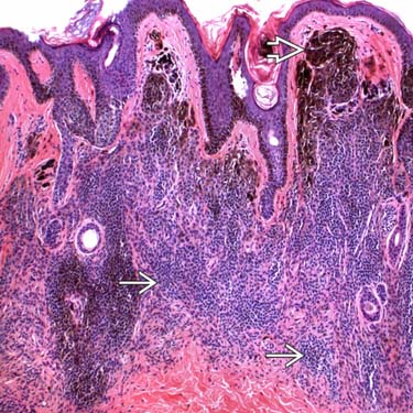
 progressing to smaller, less pigmented cells deeper in the dermis
  , consistent with maturation with dermal descent.
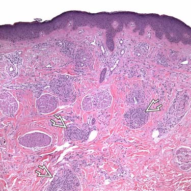
 around vessels and adnexa.
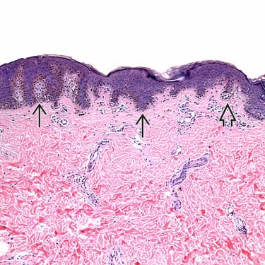
 and single melanocytes
  at the tips and sides of rete, mimicking an acquired junctional melanocytic nevus.


