Congenital Anomalies and Malformation Syndromes


Congenital Anomalies and Malformation Syndromes


Joseph R. Siebert, Ph.D.


The study of congenital anomalies continues to be hampered by misunderstandings at a number of levels. In many circles, for example, the statement that “the baby was born with a genetic deformity” is often heard. In fact, this is often not the case, for many congenital anomalies are neither genetic in origin nor do they constitute a physical deformation per se. Another common, but erroneous, opinion is that hundreds or even thousands of substances in the environment cause birth defects. In fact, only 30 to 40 exogenous substances (i.e., teratogens) have been proven to have this potential. But if these issues continue to hamper our dealings professionally, they also tug at the souls of grieving parents who ask “What caused my baby’s problem?” or “How did this happen?” or “Will it happen again?” These questions take on added complexity when multiple anomalies are encountered in a single patient. It can fall to the pathologist, as well as clinical specialists, to help explain these findings. The purpose of this chapter, then, is to provide a broad context for understanding the basic patterns of anomalies. As such, descriptions will emphasize gross features over microscopic appearances or discussions of intricate pathologic processes. For help with these latter matters, the reader is referred to chapters that cover specific organ systems.


ETIOLOGY AND PATHOGENESIS

The question of causation—etiology—is not at all simple, for etiology may be heterogeneous, that is, multiple factors may bring about a given defect. Holoprosencephaly is a powerful example, for it arises sporadically or is associated with several gene mutations, aneuploidies (e.g., trisomy 13), diseases such as maternal diabetes mellitus, and teratogens (e.g., ethyl alcohol). Robin sequence (micrognathia, cleft palate, glossoptosis) is another example, in which causes may be chromosomal, teratogenic, monogenic, disruptive (i.e., amniotic bands), or unknown.

The mechanism—or pathogenesis—responsible for a defect may be varied as well. In Robin sequence, for example, deformations may occur secondary to intrauterine constraint produced by oligohydramnios. However, reduced amniotic fluid volume may occur from premature rupture of membranes (particularly chronic leakage), renal anomalies, or placental or maternal factors.


CONCEPTS AND TERMS OF MORPHOGENESIS

In 1982, a set of standardized terms for describing human developmental abnormalities was established (1). The definitions are essential for pediatric pathologists, pediatricians, medical geneticists, and others dealing with congenital anomalies. Several additional discussions of the terminologic, historic, diagnostic, nosologic, and morphologic aspects of congenital anomalies in humans are available (2,3).

Hypoplasia refers to underdevelopment and hyperplasia to overdevelopment of an organism, organ, or tissue and result from a change in cell number. Hypotrophy and hypertrophy refer to a decrease and increase, respectively, in the size of an organ, tissue, or cells. Agenesis is the absence of a part of the body caused by a presumed absence of the anlage, or primordium. Aplasia refers to absence of a rudimentary structure caused by failure of the anlage to develop completely. Aplasia can be regarded as an extreme degree of hypoplasia. Atrophy describes the shrinkage of a previously normally developed tissue mass or organ because of a decrease in cell size or cell number.

A developmental field is the portion of the embryo that reacts as a coordinated unit to inductive effects with differentiation and growth (4). Developmental fields represent, then, the major branches on the morphogenetic tree. It has been suggested that the embryo itself constitutes the primary developmental field (5) and that other more constricted ones are operational at later stages of development. A monotopic field defect represents a defect in organogenesis and includes contiguous anomalies (e.g., cyclopia and holoprosencephaly; cleft lip and cleft palate). Such alterations are more likely to
arise late in gestation and produce more confined defects (6). By contrast, a polytopic field defect is thought to result from an earlier defect during blastogenesis—the first 4 weeks of development—and occurs if abnormal inductive processes produce more distantly located and diverse defects (6,7).

The midline also acts as a developmental field (8). It represents the normal plane of cleavage in monozygotic twinning and the plane around which symmetry of visceral position is determined. It is an especially vulnerable site in terms of developmental anomalies. Morphogenetic events involving the midline include fusions, segmentation, programmed cell death with morphogenetic “necroses” or resorptions, rotations, and other developmental movements. In some anomalies involving the midline, the incidence of monozygotic twinning may be increased (e.g., sirenomelia, cloacal anomalies). Other examples of midline anomalies include the holoprosencephaly complex, agenesis of the corpus callosum, cleft lip, cleft palate, midface cleft complex, spina bifida, omphalocele, congenital heart defects, hypospadias, and imperforate anus.

A malformation is “a morphologic defect of an organ, part of an organ, or larger region of the body resulting from an intrinsically abnormal developmental process” (1).

A disruption, or secondary malformation, is “a morphologic defect of an organ, part of an organ, or a larger region of the body, resulting from the extrinsic breakdown of, or interference with, an originally normal developmental process” (1). Disruptions are causally heterogeneous and may bear close resemblance to malformations anatomically. In a given case, the distinction between a disruption and a malformation may be made on the basis of the associated malformations or the history of gestational exposure to a teratogenic agent or event. The general prevalence of birth defects is given as 3% to 5%.

A deformation is “an abnormal form, shape, or position of a part of the body caused by mechanical forces.” It may be extrinsic, due to intrauterine constraint (e.g., lack of amniotic fluid), or intrinsic, due to a defect of the nervous system that causes hypomobility (1). Examples of deformities are talipes equinovarus and arthrogryposis. About 1% to 2% of newborn infants have deformations of some sort.

Dysplasia represents “an abnormal organization of cells into tissue(s) and its morphologic result(s)” (1). Dysplasia is therefore a process and the consequence of dyshistogenesis, an abnormal differentiation of tissue structure. This is in contrast to a malformation, which is a defect in morphogenesis of the organ structure. Dysplasias may or may not be metabolically induced, may involve one or several germ layers, and may be generalized or localized; they often demonstrate a sporadic pattern of occurrence (1).

Mild dysplasias, common in the normal population, include freckling, capillary hemangioma over the glabella and metopic suture area of the forehead, café au lait spots, moles, and nevi. If they are mendelian traits, they usually represent autosomal dominant mutations. Dysplasias are components of every aneuploidy syndrome and probably are one reason for the increased incidence of associated cancers. Dysplasias can be induced environmentally by radiation, viruses, and carcinogens.

Anomalies sometimes occur as groups of defects, which require additional classification. The terms described below help in categorizing anomalies but are only aids. Placing a name on a cluster of anomalies helps in organizing thoughts about a given condition but does not identify cause or mechanism or suggest recurrence risk (9).

That being said, a syndrome is “a pattern of multiple anomalies thought to be pathogenetically related and not known to represent a single sequence or a polytopic field defect” (1). No structural anomaly of any malformation syndrome is obligatory, and no one component is pathognomonic of any syndrome.

A sequence is a “pattern of multiple anomalies derived from a single known or presumed prior anomaly or mechanical factor” (1). In the Potter sequence, the pathogenetic event is oligohydramnios arising from a genetic or nongenetic cause; the causal event represents a malformation (e.g., renal agenesis or dysplasia, as in polycystic kidney) or a mechanical factor (e.g., amniotic fluid leakage). Lack of amniotic fluid restricts fetal movement and causes fetal compression, producing the typical changes of Potter sequence (Figure 4-1).

A malformation complex consists of “those groups of heterogeneous disorders with overlapping characteristics that are difficult to separate into specific conditions,” for example, facio-auriculo-vertebral spectrum and hypoglossia-hypodactylia.

An association consists of “a nonrandom occurrence in two or more individuals of multiple anomalies not known to be a polytopic field defect, sequence, or syndrome” (1). Associations have also been defined as the results of “disruptive events acting on developmental fields” (10). However, the diversity of findings in associations highlights how much remains unknown about possible developmental fields. The term “association” is a temporary category that should change as conditions become better understood. An example is CHARGE syndrome, which for many years was classified as an association, until mutations in the CHD7 gene were identified in numerous patients (11).

Jones has offered a valuable policy for naming patterns of malformations (12):


  • When the etiology is known and easily remembered, the appropriate term should be used to designate the disorder.


  • Time-honored designations should be continued unless there is good reason to change.


  • In the absence of a reasonably descriptive designation, eponyms, some of them multiple, may be used until the basic defect for the disorder is recognized. However, use of an eponym should thereafter be limited to one proper name.


  • The use of the possessive form of an eponym should be discontinued, because the author neither had nor owned the disorder.


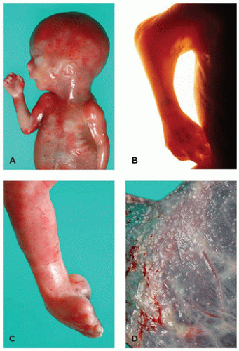
    FIGURE 4-1 • Potter sequence. A: A 22-week fetus with a history of severe oligohydramnios (renal system normal; no history of premature rupture of membranes). Note the blunt nose, small mandible, and flattened ear. B: Marked skin webbing (pterygium) of right elbow developed secondary to prolonged immobilization of joint. C: Medial rotation of foot at ankle joint (talipes equinovarus) resulted from intrauterine constraint. D: Fetal surface of placenta shows amnion nodosum, a finding common in cases of oligohydramnios.


  • Designation of a disorder by one or more of its manifestations does not necessarily imply that they are either specific or consistent components of that disorder.


  • Names that may have an unpleasant connotation for the affected individual or family should be avoided.


  • The syndrome should not be designated by the initials of the originally described patients.


  • Names that are too general for a specific syndrome should be avoided.


  • Unless acronyms are extremely pertinent or appropriate, they should be avoided.


DEFORMATIONS


Amniotic Fluid Volume

Oligohydramnios, or anhydramnios, is an ominous sign, as it effectively reduces the space available to the fetus and is associated with a wide variety of fetal deformations involving the limbs and craniofacial complex. With reduced inhalation of fluid comes pulmonary hypoplasia, which is lethal when severe. Reduced fluid volume comes about primarily from leakage (e.g., premature rupture of membranes) or renal anomalies with reduced production of fetal urine. Placental abnormalities, with decreased fetal blood flow, can cause “prerenal” oligohydramnios.


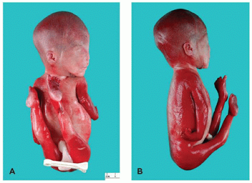
FIGURE 4-2 • Severe arthrogryposis in a 23-week fetus. Extraordinary flexion and contracture deformities and marked flattening of the face are apparent. Autopsy revealed no other fetal anomalies (karyotype 46,XX). Etiology is heterogeneous in this condition. A: Frontal view. B: Lateral view.


Abnormalities of Uterus and Placental Implantation

A bicornuate uterus may cause fetal compression and constraint, resulting in a deformed fetus. Uterine malformations may also predispose the fetus to malformations arising from abnormalities in implantation, placentation, body stalk formation, and late fetal cord compression or torsion. With these occurrences comes an increased risk of stillbirth (see Chapter 18).


Neurogenic, Skeletal, and Other Causes of Deformations

Central nervous system (CNS) and skeletal muscle defects (e.g., amyoplasia) may result in deformations. The most common congenital limb deformities are tibial bowing, mild metatarsus varus, talipes equinovalgus and varus, and the flexural contractures of arthrogryposis (Figure 4-2). Skeletal dysplasias may be associated with deformities of prenatal or postnatal onset. Twins and multiple fetuses may interfere with each other’s physical development and manifest deformities.


DISRUPTIONS


Ionizing Radiation

Studies of radiation exposure to pregnant women during medical treatments or warfare have provided valuable information regarding radiation-induced anomalies. It is commonly held
that pregnant women should avoid all unnecessary radiation exposure. However, data regarding exact doses of radiation are often unavailable, and so actions based upon those fears (i.e., elective abortion after an exposure or suspected exposure) may be unwarranted. Counselors must use extreme caution when dealing with questions regarding radiation exposure.

Guidelines are available for this purpose (13). During pregnancy, the acceptable cumulative dose of ionizing radiation during pregnancy is 5 rads. With few exceptions, diagnostic studies produce dosages less than this level. A two-view radiograph of mother’s chest, for example, exposes the fetus to just 0.00007 rads. Therefore, a mother would need the equivalent of 500 chest examinations before the fetus would be exposed to a harmful level of radiation. Because 8 to 25 weeks, and especially 10 to 17 weeks, of gestation is a highly sensitive period for CNS teratogenesis, unnecessary exposures directly to the fetus should be avoided during this time. Prenatal radiation exposure may produce a slight increase in the risk of childhood leukemia or small change in the frequency of gene mutations, but these are quite rare and not an indication for pregnancy termination.

When maternal radiation occurs at higher than diagnostic levels, the exposed fetus may exhibit generalized growth retardation, microcephaly, skull defects, eye anomalies, spina bifida, cleft palate, micromelia, clubfoot, and genital and other anomalies (14). Altered cognitive status, ranging from reduced intelligence quotient (IQ) to frank mental retardation and seizures, is recognized; MRI examinations in these patients are suggestive of neuronal migration defects (15).


Teratogenic Disruptions

A list of teratogenic agents in humans is shown in Table 4-1. Additional resources on this topic are available (16,17). While some viruses are responsible for malformations, a number of others cause congenital/neonatal or maternal disease, but not malformations per se. These include adenovirus, echovirus, hepatitis B and C virus, influenza, and West Nile virus, and are excluded from Table 4-1. Teratogens exert their influence during especially vulnerable times (“critical periods”), when specific tissues are undergoing development (Table 4-2).


Thalidomide Embryopathy

Thalidomide, a drug once used in the treatment of morning sickness, was first recognized as a teratogen by Lenz and McBride in separate reports in 1961. Exposure to thalidomide during the critical period (days 23 to 28 of gestation) results in a number of defects, the most notable of which are limb defects ranging from triphalangeal thumb to tetra-amelia or phocomelia of the upper and lower limbs, at times with preaxial polydactyly of six or seven toes per foot. Congenital heart defects, urinary tract anomalies, genital defects, gastrointestinal anomalies, eye defects, ear malformations, and dental anomalies may also occur. The drug is also used in the treatment of leprosy (i.e., erythema nodosum leprosum), certain autoimmune disorders, and cancer. For this reason, cases of thalidomide embryopathy still occur, especially in countries with a high prevalence
of leprosy and access to the drug. The mechanism of action continues to be studied. The drug has anti-inflammatory properties, and some have suggested that defective angiogenesis in developing limb buds may also be operational (19).


TABLE 4-1 TERATOGENIC AGENTS IN HUMANS


Radiation


Atomic weapons


Radioiodine


Greater than therapeutic levels


Infectious Agents


Cytomegalovirus


Herpes simplex virus 1 and 2


Lymphocytic choriomeningitis virus (LCMV)


Parvovirus B-19 (erythema infectiosum)


Rubella virus


Syphilis


Toxoplasmosis


Varicella virus


Venezuelan equine encephalitis virus


Maternal and Metabolic Imbalance


Alcohol abuse


Amniocentesis, early


Chorionic villus sampling (before day 60)a


Cretinism, endemic


Diabetes mellitus


Folic acid deficiency


Hyperthermia


Myasthenia gravis


Phenylketonuria


Rheumatic disease and congenital heart block


Sjögren syndrome


Virilizing tumors


Drugs and Environmental Chemicals


Aminopterin, methylaminopterin


Androgenic hormones


Captopril (renal failure)


Carbamazepinea


Chlorobiphenyls


Cocaine


Corticosteroidsa


Coumarin and other anticoagulants


Cyclophosphamide


Diethylstilbestrol


Diphenylhydantoin


Enalapril (renal failure)


Etretinate and other retinoic acid compounds


Fluconazole (high dose)


Iodides


Lithiuma


Mercury (organic forms)


Methimazole (scalp defects, choanal atresia)a


Methylene blue (by intra-amniotic injection)


Misoprostola


Nicotine (smoking, passive exposure, nicotine patch)


Penicillamine


Phenobarbitola


Sartans


Tetracycline


Thalidomide


Toluene abuse


Trimethadione


Valproic acid


a Agents produce <10 defects per 1000 exposures.


Modified from Shepard TH, Lemire RJ. Catalog of Teratogenic Agents, 13th ed. Baltimore, MD: The Johns Hopkins University Press; 2007.


TABLE 4-2 CRITICAL PERIODS IN HUMAN TERATOGENICITY


Teratogen


Gestational Age (Days)


Malformation


Rubella virus


0-60


Cataract; heart defect


0-120+


Hearing deficit


Thalidomide


21-40


Limb reduction defects


Hyperthermia


18-30


Anencephaly


Male hormones in females (androgens, exogenous drugs, tumors)


Prior to 90


Clitoral hypertrophy; labial fusion


After 90


Clitoral hypertrophy


Anticoagulants


Prior to 100


Hypoplasia of nose; stippling of epiphyses


After 100


Possible mental retardation


Diethylstilbestrol


After 14


Vaginal adenosis (50%)


After 98


Vaginal adenosis (30%)


After 126


Vaginal adenosis (10%)


Radioiodine therapy


After 65-70


Atrophy (“thyroidectomy”) of fetal thyroid gland


Goitrogens, iodides


After 180


Fetal goiter


Tetracycline


After 120


Staining of enamel of primary teeth


After 250


Staining of crowns of permanent teeth


Modified from Shepard TH. Proven and suspected human teratogens-How can we sort them out? In: Crichton JU, ed. Safe Drugs for Canadian Children—Report of the Third Canadian Ross Conference on Pediatric Research. Montreal, Canada: Ross Laboratories; 1978:9-25 (Ref. 18).


Folic Acid Deficiency

Deficiency of folic acid results in up to 70% of neural tube deficits (NTDs), particularly anencephaly. Preconceptional intake of 0.4-mg folic acid daily reduces the incidence of NTDs by up to 90% and congenital heart defects by approximately 40% (20). The fortification of wheat flour with folic acid in the United States has resulted in a decrease in the incidence of NTDs; a similar success might also be expected with similar fortification and surveillance worldwide (21,22). In addition to aiding in the prevention of NTDs, prenatal supplementation with folic acid prevents pregnancy-induced megaloblastic anemia (23).


Folic Acid Antagonists and Derivatives

Aminopterin and methotrexate, its methyl derivative, are folic acid antagonists that have several applications and may cause a variety of anomalies. Aminopterin is used as a pesticide; both drugs are used to treat certain cancers or to end an unwanted or ectopic pregnancy, making exposure during early pregnancy possible. Craniofacial anomalies include severe hypoplasia of frontal, parietal, temporal, or occipital bones; wide fontanelles; upsweep of frontal scalp hair; broad nasal bridge; shallow supraorbital ridges; prominent eyes; cleft palate; apparently low-set ears; micrognathia; maxillary hypoplasia; and epicanthal folds. The limbs are relatively short, and dislocation of hips, short thumbs, partial syndactyly of third and fourth fingers, dextroposition of the heart, and hypotonia may occur (24).


Fetal Iodine Deficiency

The pregnant woman and her developing fetus both have an increased need for iodine. The woman deficient in dietary intake of iodine therefore puts both herself and her fetus at risk (25). The use of iodized salt has done much to reduce the risk of associated mental retardation worldwide, but is of little help to women who do not have access to this food or those who must reduce their salt intake during pregnancy.

Iodine deficiency has been called the single greatest preventable cause of intellectual disability (26). Deficiency in the fetus results in mental retardation, spastic diplegia, deafness, and strabismus (27,28) and develops from severe maternal iodine deficiency (<20 µg/day) during the first half of gestation. This is especially prevalent in regions with reduced iodine content in the soil, particularly certain European countries and mountainous areas, such as New Guinea, the Himalayas, and the Andes (29). The World Health Organization recommends a daily iodine intake of between 150 and 300 µg (30).


Trimethadione Syndrome

Trimethadione is a drug used in the treatment of seizures. A large percentage of cases are associated with pregnancy loss or abnormalities in offspring, which include delayed growth and mental development, and craniofacial (e.g., malformed ears, cleft palate), skeletal, cardiac, gastrointestinal, or genitourinary anomalies (31,32).


Valproic Acid Embryopathy

The teratogenicity of valproic acid has been well documented (16). Although valuable as an antiepileptic and mood-altering drug, valproic acid administration during pregnancy
is associated with a host of anomalies, including microcephaly, trigonocephaly, porencephaly, spina bifida, other CNS defects, facial anomalies, cardiac defects, limb reduction anomalies, and genitourinary defects (33). Dosages associated with malformations have generally been 750 to 1,000 mg/day (although low-dose effects are also suspected) and exposures verified during the first trimester.


Warfarin Embryopathy

Although the contraindications of warfarin usage during pregnancy are well recognized, women, for example those with prosthetic heart valves, may take the drug during the first trimester, before pregnancy is recognized. Such exposure can result in intrauterine death or an embryopathy characterized by restricted growth, hypoplastic nose, limb defects (shortening, brachydactyly, nail hypoplasia), gastroschisis, cardiac defects, and stippled epiphyses or chondrodysplasia punctata (34,35). If exposure occurs later in gestation, CNS hemorrhage, with subsequent brain damage and mental retardation, may occur.


Synthetic Progestin Embryopathy

Exposure to synthetic progestins (e.g., 17-α-ethinyl-19-nortestosterone) early in gestation can induce enlargement of the clitoris or labioscrotal fusion in female fetuses and hypospadias in males (36). The incidence of ectopic pregnancy is increased in women who experience contraception failure from either oral progestins or implants (37). Diethylstilbestrol may cause vaginal adenosis, clear cell adenocarcinoma of the vagina or cervix, or breast cancer in prenatally exposed females and reproductive anomalies in exposed males (38,39). The use of a variety of exogenous sex hormones is not associated with increased risk of major malformations, with the exception of esophageal atresia, which carries a risk ratio of 2.87, or approximately 6 per 10,000 live births (40).


Mercury Embryopathy

Exposure of the developing human to mercury compounds has serious effects, most notably an increased incidence of growth restriction, microcephaly, and CNS damage, with consequent deficits that include blindness, hypotonia or spasticity, deafness, dysarthria, chorea, athetosis, and strabismus. In one study of maternal exposure to inorganic mercury, the prevalence of miscarriage and stillbirth was not increased (41). Both maternal ingestion and occupational exposure are recognized routes of exposure. The classic condition is Minamata disease, an epidemic that affected women living on the island of Minamata, Japan, who ingested shellfish contaminated with methylmercury (42). Pregnant women continue to be exposed, especially those living in areas of heavy industrial pollution or downstream of gold mines, where contamination of soil and water occurs (43), or those ingesting contaminated marine food (44).


Isotretinoin Embryopathy

Isotretinoin (i.e., Accutane) is a synthetic vitamin A analog, 13-cis-retinoic acid. Because it inhibits sebaceous gland function, the drug is valuable in the treatment of cystic acne. Administration to pregnant women is associated with a variety of serious anomalies. Miscarriage, perinatal mortality, and premature birth are reported, and survivors may have a variety of malformations or decreased mental status. Ear anomalies are common, including dysplastic, hypoplastic, or absent ears; agenesis of the external ear canal is variable. CNS abnormalities (microcephaly, hydrocephalus, porencephaly, Dandy-Walker malformation, neuronal migration defects) and conotruncal congenital heart defects are recognized (45).


Alcohol Embryopathy

Alcohol is a common and important teratogen in humans, but its influence was not fully appreciated until 1968 (46). In 1973, Jones and Smith named the condition “fetal alcohol syndrome” (FAS) (47). Effects include structural, behavioral, and neurocognitive deficits, and so a number of other designations have been used, including the earlier “fetal alcohol effect” and current “fetal alcohol spectrum disorders” (48). In a sense, the term “fetal alcohol syndrome” is unfortunate, for, although popular, it implies that alcohol exerts its primary influence on the fetus; in fact, teratogenic damage to the embryo is far more significant, hence the more accurate term “alcohol embryopathy.”

A maternal history of alcohol consumption is often difficult to ascertain, but nevertheless, clinical criteria for making the diagnosis are available. Major characteristics of affected infants and children include distinctive facies (epicanthal folds, short palpebral fissures, midface hypoplasia, thin vermilion border of the upper lip, absent to indistinct philtrum, and short, upturned nose), growth restriction, malformations, and psychomotor abnormalities (49). Patients generally present with prenatal and postnatal growth retardation and CNS dysfunction, including mental retardation, hyperactivity, sleep disorders, spastic tetraplegia, seizures, and behavioral difficulties (Table 4-3). Additional disabilities may include academic or legal difficulties, inappropriate sexual behavior, and other problems related to alcohol or other drug use. Joint, limb, and conotruncal cardiac anomalies are often present; limb defects include shortness of the metatarsals and metacarpals or severe ectrodactyly (52). The unusual hirsutism that is present at birth may disappear with age. Structural brain malformations, chiefly hypoplasia or agenesis of the corpus callosum, lissencephaly, and holoprosencephaly, as well as ocular abnormalities, have been described (50,53). Recently, an abnormally angled corpus callosum, diagnosed by transfontanellar ultrasound, has been noted in a large percentage of affected infants (54). Cystic hygromas are found in patients with FAS, but also with a number of other conditions (Table 4-4). FAS has been reported in both monozygotic and dizygotic twins; the higher incidence in the
former has suggested a genetic influence (56). Despite small head circumference and initially slow psychomotor maturation, some infants with FAS may progress and develop intelligence within the normal range. Endocrine investigations usually show normal or near-normal levels of growth hormone, cortisol, and gonadotropins (see Chapter 21) (57). FAS is also a carcinogenic syndrome and is associated with tumors virtually identical to those seen in the fetal diphenylhydantoin (Dilantin) syndrome (see below).


TABLE 4-3 CHARACTERISTICS OF FAS


Somatic and Cutaneous Findings


Prenatal and postnatal growth retardation, with diminished adipose tissue content


Hirsutism


Cutaneous hemangiomas


Central Nervous System


Micrencephaly


Neuronal migration defects (heterotopia)


Absent or hypoplastic corpus callosum


Ventriculomegaly


Holoprosencephaly


Hypoplastic cerebellum


Dysplastic brainstem


Lissencephaly


Craniofacial


Microcephaly


Ocular hypertelorism


Short palpebral fissures, sometimes downslanting


or with epicanthal folds


Microphthalmia, other eye anomalies


Posteriorly rotated ears, with hypoplastic concha


Low nasal bridge


Hypoplastic midface, with hypoplastic maxillae


Retro- or micrognathia


Cleft lip and/or palate


Smooth vermillion border


Long, indistinct philtrum


Small teeth


Cardiovascular


Congenital heart disease, often conotruncal (e.g., tetralogy of Fallot)


Atrial and/or ventricular septal defects


Gastrointestinal Tract


Esophageal, duodenal, or anal atresia


Tracheoesophageal fistula


Pyloric stenosis


Urogenital System


Hypospadias


Hypoplastic labia


Small rotated kidneys


Hydronephrosis


Musculoskeletal Systems


Abnormal palmar creases


Hypoplastic nails


Reduction defects of limbs and digits


Pectus excavatum or carinatum


Scoliosis


Klippel-Feil anomaly


Diaphragmatic hernia


Umbilical hernia


Behavioral


Developmental delay, mental retardation


Irritability (in infancy)


Hyperactivity (in childhood)


Hypotonia, reduced coordination


Modified from Clarren SK, Smith DW. The fetal alcohol syndrome. N Engl J Med 1978;298:1063-1067; Clarren SK, Alvord EC, Sumi SM, et al. Brain malformations related to prenatal exposure to ethanol. J Pediatr 1978;92:64-67; Potter BJ, Hetzel BS. Fetal alcohol syndrome. In: Hetzel BS, Smith RM, eds. Fetal Brain Disorders: Recent Approaches to the Problem of Mental Deficiency. New York, NY: Elsevier North Holland; 1981 (Refs. 49-51).


TABLE 4-4 DISORDERS ASSOCIATED WITH NUCHAL CYSTIC HYGROMA


Single Gene Disorders


Familial webbing of neck


Lymphedema distichiasis syndrome


Roberts syndrome


Bieber syndrome


Lethal multiple pterygium syndrome


Noonan syndrome


Chromosome Disorders


45 X (Ulrich-Turner syndrome or monosomy X)


X-chromosome polysomy


13q-


18p-


Trisomy 18


Trisomy 21


Trisomy 22 mosaicism


Teratogenic Disorders


Alcohol embryopathy


Fetal amethopterin syndrome


Fetal trimethadione syndrome


Modified from Gilbert-Barness EF, Opitz JM. Congenital anomalies and malformation syndromes. In: Wigglesworth JS, Singer DB, eds. Textbook of Fetal and Perinatal Pathology. Oxford, UK: Blackwell Scientific Publications; 1991 (Ref. 55).


Nicotine

Prenatal nicotine exposure, originating most often from maternal smoking, passive exposure to smoke, or use of nicotine-containing agents, is associated with abnormal placentation, abruption, reduced fetal growth and length of gestation, and stillbirth or neonatal mortality (58). These effects are substantial. If women did not smoke during pregnancy, it has been estimated that the prevalence of small-for-gestation babies could be reduced by 30%, abnormal placentas by 10%, premature deliveries (<37 weeks) by 30%, and premature or perinatal deaths by 15% (59). Prenatal exposure to cigarette smoke is associated with both congenital and long-term effects, including cryptorchidism, limb deficiencies, neurocognitive deficits, and decreased reproductive success in males; exposure is a recognized risk factor for increased respiratory disorders and infection, obesity, anxiety disorders, and sudden infant death syndrome.


Diphenylhydantoin Embryopathy

The antiseizure drug diphenylhydantoin and derivatives (phenytoin, Dilantin) causes a syndrome of intrauterine growth restriction, microcephaly and mental retardation, cleft palate, congenital heart defect, digital hypoplasia, and characteristic facial appearance consisting of wide-set eyes, epicanthal folds, broad sunken nasal bridge, upturned nose, and widened lips (60,61). Intellectual development may be delayed as well. Human exposure during the 5th to 6th week results in cleft lip and maxillary hypoplasia (62). The teratogenic mechanism(s) remains unclear despite abundant research.

Fetal exposure to diphenylhydantoin is also known to be carcinogenic. Neuroblastoma, ganglioneuroblastoma, extrarenal Wilms tumor, and malignant mesenchymoma have been observed in individuals exposed to diphenylhydantoin in utero (63).


Metabolic Disruptions


Phenylketonuria

Unmanaged maternal phenylketonuria (PKU) leads to intrauterine and postnatal growth restriction, microcephaly and mental retardation, cardiovascular defects, dislocated hips, and other more minor anomalies. The incidence of fetal defects is greatly decreased in mothers whose PKU is well controlled during pregnancy. It has been suggested that impaired accretion of two fatty acids, arachidonic and docosahexaenoic acids (structural components of the CNS), contributes to the small head, reduced vision, and mental retardation (64). In severe forms, the accumulation of phenylalanine results in the formation of CNS fibrils that resemble amyloid, suggesting that the disease may be one of abnormal protein folding (65,66). Infants of phenylketonuric mothers are heterozygous, and because phenylketonuric heterozygotes are generally normal, the defect in the fetus must be attributed to the maternal metabolic disturbance.


Diabetes Mellitus

A large number of pregnancy complications are recognized in women suffering from diabetes mellitus. Stillbirth and perinatal mortality in insulin-dependent women occur at five times the background rate; neonatal mortality is increased 15 times and infant mortality three times over the general population (67). Macrosomia complicates vaginal delivery. Type I maternal diabetes is also associated with an increased incidence of preeclampsia and pregnancy-induced hypertension (68). The effects of gestational diabetes are the subject of continuing study, but in general tend to be fewer and less severe than those arising from pregestational diabetes.

Maternal diabetes mellitus is also associated with a number of fetal anomalies, with an incidence variably estimated at two to eleven times that of the normal population (69). Defects may involve virtually every organ system, prominent examples being anencephaly, holoprosencephaly, arhinencephaly, or myelomeningocele, congenital heart defects, caudal dysgenesis (formerly “caudal regression”)/sirenomelia, imperforate anus, radial aplasia, and renal abnormalities, including renal agenesis and dysplasia (Figure 4-3). Malformations (Table 4-5) are the most important cause of mortality in infants of diabetic mothers.


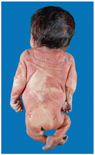
FIGURE 4-3 • Infant of diabetic mother. Pelvic girdle is reduced noticeably in this 31-week-old male with absent lumbosacral spine and malformed pelvis, indicative of caudal dysgenesis (formerly caudal regression syndrome).

The exact role of glucose metabolism in diabetic embryopathy is highly complex and beyond the scope of this review. Current studies are centered on metabolic derangements, including oxidative stress, that inhibit regulatory genes, resulting in apoptosis of progenitor cells and malformations (70,71).


Infectious Disruptions

Infections, particularly toxoplasmosis, rubella, cytomegalovirus (CMV), herpes simplex, varicella, syphilis, and others (TORCHS), may cause fetal disruptions (see Chapter 6). The earlier in pregnancy the infection occurs, the greater is the likelihood of embryonic death or development of anomalies. The most frequent fetal abnormalities are intrauterine growth restriction, microcephaly and mental retardation, deafness, cataracts, retinopathy, microphthalmia, glaucoma, myopia, and congenital heart defects.

Periventricular calcifications and chorioretinitis are frequent in toxoplasmosis. Other organisms that may be implicated in human congenital anomalies are herpes hominis type 2, which is associated with a severe congenital brain
defect, varicella, Venezuelan equine encephalitis, Coxsackie virus, and syphilis. Acquired immune deficiency syndrome (AIDS) is acquired by transplacental means or during labor, delivery, or breast feeding and constitutes an enormous problem worldwide (72). In 2005 in the United States, 92% of cases of children with AIDS were attributed to maternal transmission of the human immunodeficiency virus (HIV); the incidence of neonatal HIV infection has fallen substantially in the United States with the implementation of prenatal testing, antiretroviral therapy, C-section, and avoidance of breast feeding (73).


TABLE 4-5 CONGENITAL ANOMALIES ASSOCIATED WITH MATERNAL DIABETES MELLITUS


Central Nervous System


Anencephaly


Holoprosencephaly


Arhinencephaly


Occipital encephalocele


Myelomeningocele


Cardiovascular System


Atrial, ventricular septal defect


Transposition of the great vessels


Single ventricle, hypoplastic left heart


Ebstein anomaly of tricuspid valve


Pulmonic stenosis, mitral atresia


Hypertrophic cardiomyopathy


Musculoskeletal System


Amelia of upper limbs


Caudal dysgenesis (regression)/sirenomelia


Costovertebral segmentation defects


Urogenital System


Renal adysplasia


Unilateral renal agenesis


Craniofacial Complex


Bilateral auricular atresia


Cleft lip/palate


Bifid tongue


Microtia/anotia


Hemifacial microstomia


Other Abnormalities


Hypoplastic lungs


Thymic aplasia


Omphalocele


Anorectal stenosis/atresia


Modified from Castori M. Diabetic embryopathy: a developmental perspective from fertilization to adulthood. Mol Syndromol 2013;4:74-86.


Amnion Rupture Disruption Sequence

Early amnion rupture (or ADAM complex) may result in severe defects of the fetus, including asymmetric clefts, body wall defects, often with extrusion of viscera, and highly variable amputations (Figure 4-4). When amnion adheres to the head, marked distortions of craniofacial structures are found, with widely separated eyes, displacement of the nose onto the forehead, and exencephaloceles; swallowing of amniotic bands may produce bizarre orofacial clefts that often follow a linear pattern. Marked deformations, growth deficiency, and a short umbilical cord are also observed in this condition (74). The fetus may also be adherent to the placenta, making diagnosis straightforward. However, when strands of amnion are not identified, diagnosis is hampered, although the pattern of defects may still imply this mode of pathogenesis. In the macerated fetus, strands of tissue resembling sloughed epidermis may be identified as amnion by microscopy.

Amnion may also be absent from the fetal surface of the placenta, or free membranes. The least severe form of amniotic band disruption is a constriction groove (“Streeter band”) on a limb. The temporal relationship of abnormalities in early amnion rupture sequence is shown in Table 4-6. Amnion rupture is thought to be rather common, affecting perhaps 1 of every 1,200 liveborn and stillborn fetuses. If this is the case, many occurrences apparently have few or no sequelae. Rare families with amniotic bands in relatives have been reported, but the recurrence risk appears to be negligible (76). Causes for premature rupture are not understood. The forces of uterine contraction have been implicated, but recent studies have suggested that a process of programmed weakening of membranes may operate prior to delivery (77). This observation could help explain familial recurrences (see Chapter 9 for additional details).


Chorion and Yolk Sac Rupture Sequence

While rupture of the amnion is well recognized, some have hypothesized that similar defects might arise from rupture of the chorion or yolk sac. Rupture during the 3rd week of gestation and subsequent mechanical compression of the fetus could interfere with normal cardiac descent, resulting in cleft sternum, ectopia cordis, and thoracic and pulmonary hypoplasia (78). Such cases reflect the complex nature of embryogenesis in the region. Another published example involved an infant with rudimentary occipital meningocele and transverse defects of the hands, who, by microscopy, had intestinal mucosa adherent to the scalp (79). Possible explanations included a genetic defect similar to disorganization in the rodent; homeotic transformation; abnormal juxtaposition of epidermis and yolk sac remnant (or omphaloenteric duct); or adhesion of endoderm and ectoderm to the embryo.


Ischemic and Vascular Disruptions

Interference with blood supply may result in ischemic disruptions. Cutis marmorata telangiectatica congenita is a vascular disruption characterized by atypical capillaries, venules, and veins in different cutaneous layers. Clinically, the lesions manifest as telangiectasia, capillary hemangiomata, cutis marmorata, venous hemangiomata, and varicose veins, depending on the type of vessels involved and

the layer of skin affected. Secondary thrombosis with subsequent localized atrophy and ulceration may occur. Cutis marmorata telangiectatica congenita occurs sporadically, with female preponderance and occasional minor manifestations in close relatives.


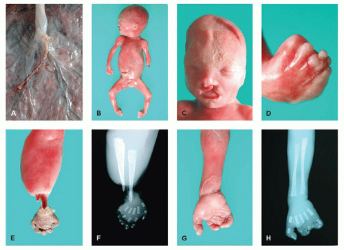
FIGURE 4-4 • Amnion rupture sequence. A: Close-up view of fetal surface of placenta shows tiny remnant of amnion. B: A 22-week male fetus with multiple amputation defects. C: Face with unilateral cleft lip. D: Right foot with syndactyly and multiple amputations of the digits. E: Exposed radius and ulna and necrosis of hand reflect the evolution of a band-induced amputation. F: Radiograph corresponding to (E). G: Right hand with multiple amputation defects. H: Radiograph corresponding to (G).


TABLE 4-6 TIMING OF ANOMALIES ASSOCIATED WITH EARLY AMNION RUPTURE


Age at Occurrence


Craniofacial Defect


Limb Defect


Other Abnormality


3-4 weeks


Anencephaly


Encephalocele


Meningocele


Facial deformation


Unusual, often linear clefting


Proboscis


Orbital/eye defect


Complete absence of limb


Placenta adherent to head or abdomen; short umbilical cord


5-6 weeks


Cleft lip


Choanal atresia


Limb deficiency


Polydactyly


Syndactyly


Abdominal wall defect


Thoracic wall defect


Scoliosis


7 or more weeks


Cleft palate


Micrognathia


Ear deformity


Craniosynostosis


Amniotic bands


Amputation


Hypoplasia


Pseudosyndactyly


Foot deformity


Dislocation of hip


Distal lymphedema


Omphalocele


Short umbilical cord


Ambiguous genitalia


Second, third trimester


Oligohydramnios deformation sequence, with flattened ears, blunt nose, small mandible


Pena-Shokeir phenotype


Constriction bands, with lymphedema distal to site of constriction and possible autoamputation


Pulmonary hypoplasia


Constriction of umbilical cord by bands may cause death when severe


Altered dermal or hair pattern


Modified from Gilbert-Barness EF, Opitz JM. Congenital anomalies and malformation syndromes. In: Wigglesworth JS, Singer DB, eds. Textbook of Fetal and Perinatal Pathology. Oxford, UK: Blackwell Scientific Publications; 1991; Higginbottom MC, Jones KL, Hall BD, et al. The amniotic band disruption complex: timing of amniotic rupture and variable spectra of consequent defects. J Pediatr 1979;95:544-549 (Refs. 55,75).


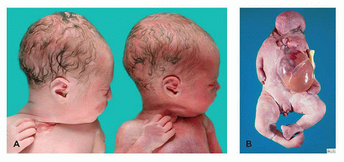
FIGURE 4-5 • Complications of monochorionic twinning. A: Pale, donor twin (left) and congested, recipient cotwin (right) in twin-twin transfusion syndrome. B: Acardiac cotwin in TRAP. Note the absence or malformation of structures of the upper body, omphalocele, and more normal lower extremities (but with anomalies of numerous digits).

In the Klippel-Trenaunay-Weber syndrome (see below), which usually occurs sporadically, dysplasia and capillary or cavernous hemangiomatosis and phlebectasia and varicosities with oligodactyly, syndactyly, and gigantism of digits have been observed. Congenital or postnatal hypertrophy of one or more limbs is frequent. Visceral hemangiomata may occur.

In addition to the well-recognized difficulties that arise in singletons, twins or other multiple gestations are especially at risk. Cord entanglement occurs in twins and may disrupt blood flow. Because of the variability in distance between cord insertion sites, more complications are observed in monochorionic monoamniotic than in monochorionic diamniotic placentas (21). Two additional examples of vascular disruption involve monochorionic twinning, namely twin-twin transfusion syndrome and twin reversed arterial perfusion (TRAP).

Sep 23, 2016 | Posted by in PATHOLOGY & LABORATORY MEDICINE | Comments Off on Congenital Anomalies and Malformation Syndromes

Full access? Get Clinical Tree

Get Clinical Tree app for offline access