Congenital and Acquired Systemic Infectious Diseases


Congenital and Acquired Systemic Infectious Diseases


Haresh Mani, M.D.

David Parham, M.D.

Jennifer Dien Bard, Ph.D., D(ABMM), F.C.C.M.


As it takes two to make a quarrel, so it takes two to make a disease, the microbe and its host.—Charles V. Chapin, 1856-1941

Infections are the leading global cause of death in the pediatric population. The optimism that accompanied the advent of antibiotics in the mid-20th century was premature, and even advances such as immunization and improved sanitation have not stopped microbial reemergence. The importance of infectious diseases is underscored by ever-increasing antimicrobial resistance and resurgence of infections such as tuberculosis (TB), malaria, and syphilis, once thought eradicated from developed countries. International travel has removed boundaries from the spread of infection, as exemplified by the rapid spread of infections such as severe acute respiratory syndrome (SARS) across countries. In this chapter, we aim to provide an overview of systemic infections that the pediatric pathologist is likely to encounter. Infections that are predominantly confined to a single organ system (e.g., poliomyelitis, hepatitis) are not considered in this chapter, even though they may occasionally cause systemic manifestations. The reader is directed to chapters dealing with specific organ systems for information on such infections. Further, since it is impossible to comprehensively detail all facets of various systemic infections in a single chapter, we have generously referenced resources for the reader with specific interests. Detailed information is also available in standard textbooks including Feigin and Cherry’s Textbook of Pediatric Infectious Diseases (1), Connor and Chandler’s Pathology of Infectious Diseases (2), and the American Academy of Pediatrics Red Book (www.aapredbook.com).


THE PATHOGENESIS OF INFECTIOUS DISEASES

An infection is the result of an encounter between an infectious agent and a susceptible host. The microorganism is the sine qua non, but the occurrence of the disease, its pathophysiology, and its outcome are determined by host and environmental factors. A discussion of microbial virulence factors is beyond the scope of this chapter; various reviews cover the topic in considerable depth (3,4).


HOST FACTORS

Host genetic factors, immune status, age, and geographic location determine the likelihood and severity of infection. The heritability of infectious disease susceptibility is a complex factor modified by developmental and maturational changes in host defense, from embryo through adolescence, with resultant differences in response to infection (5,6). Polymorphisms in genes coding for proteins that recognize bacterial pathogens [such as Toll-like receptor 4, CD14, Fc(gamma)RIIa, and mannose-binding lectin] and the response to bacterial pathogens [with elaboration of cytokines such as tumor necrosis factor-α, interleukin (IL)-1α, IL-1β, IL-1 receptor agonist, IL-6, IL-10, heat shock proteins, angiotensin I-converting enzyme, plasminogen activator inhibitor-1] can influence response to bacterial stimuli (7).

Immunologic maturity and immunodeficiencies (quantitative and qualitative) also determine infection susceptibility. Since they have developmentally immature immune systems, neonates and infants are at a relative immunologic disadvantage. The immaturity of the fetal immune system helps prevent “premature rejection” by the host (the mother). Paradoxically, this potential benefit also increases the risk of infections for the fetus and the prematurely born neonate. Term newborns have a higher frequency of microbial infections than older children and adults; extremely premature newborns (<28 weeks of gestation) have a 5- to 10-fold higher frequency than term newborns. Immunologic immaturity also obscures clinical symptoms in neonatal sepsis. Recent advancements in developmental immunology provide a framework for understanding the mechanisms underlying the propensity of infections in the preterm, near-term, and term newborn (8). The immune environment during early life favors innate over acquired immunity. Innate immunity against pathogens represents the critical first-line barrier of host defenses, as newborns have a naïve adaptive immune
system. However, innate immune mechanisms are also relatively impaired in neonates as compared to older children and adults, thereby increasing neonatal susceptibility to infections (9). Further, the neonate is unable to produce antibody to thymus-independent antigens such as bacterial polysaccharides, although transplacentally acquired maternal antibodies confer some protection for the first few months of life. Neonatal B-cells have an immature phenotype, and the neonatal spleen has a different cellular composition; unlike adults, neonatal accessory cells, that is, macrophages and dendritic cells, appear to produce less stimulatory cytokines and an overabundance of inhibitory cytokines (10).

Children with immunodeficiencies (primary or acquired) have an increased risk of infections (Tables 6-1 and 6-2). Impaired splenic function (due to asplenia, disease, or splenectomy) significantly increases the risk of life-threatening bacterial sepsis, especially with capsulated organisms, necessitating pneumococcal and meningococcal immunizations. Secondary factors such as comorbidities (Table 6-3), medications, and nutritional status also dictate clinical course. Most organisms causing disease in a setting of immunodeficiency are “opportunists” and are ubiquitous in the internal or external environment where they ordinarily do no harm. Systemic or repeated local infections may be the first manifestation of unsuspected primary immunodeficiency. A family history of unusual infections, age at presentation, specific type of infection (e.g., Aspergillus in chronic granulomatous disease, Pneumocystis in severe combined immunodeficiency), recurrent otitis media, need for IV antibiotics, and lymphopenia are additional warning signs (11).


TABLE 6-1 INFECTIONS ASSOCIATED WITH PRIMARY IMMUNODEFICIENCIES


Infection


Host Factor


General associations


Recurrent respiratory and pyogenic infections by extracellular bacteria: Streptococcus pneumoniae, Haemophilus influenzae, Staphylococcus aureus


Antibody deficiencies


Chronic or severe infections with intracellular pathogens: viruses, mycobacteria, Pneumocystis carinii, Toxoplasma gondii, and others


Deficiencies of T lymphocytes


Specific associations


Chronic viral encephalitis


X-linked agammaglobulinemia


Echovirus dermatomyositis


Polio vaccine-induced paralysis


Severe parainfluenza infection


Severe combined immunodeficiency


Severe varicella


Cartilage-hair hypoplasia


Severe Epstein-Barr virus infection


X-linked lymphoproliferative syndrome


Recurrent meningococcal sepsis


Complement deficiencies


Disseminated gonococcal infection


Staphylococcal skin infections


Neutrophil abnormalities


Mucosal and periodontal infections


Infections with Aspergillus sp., S. aureus, Pseudomonas cepacia, Chromobacterium violaceum


Chronic granulomatous disease


BCGosis and atypical mycobacteria


NF-γ and IL-12 deficiencies


Persistent mucocutaneous candidiasis


Chronic mucocutaneous candidiasis


Chronic/recurrent giardiasis


IgA deficiency


BCG, Bacille Calmette-Guérin; INF, interferon; IL, interleukin; IgA, immunoglobulin A.


ENVIRONMENTAL FACTORS

Over 50 years ago, Haldane proposed that the prevalence of thalassemia in malaria-endemic areas was due to the heterozygotic advantage it conferred against malaria, despite its otherwise deleterious effects. Table 6-4 outlines examples of the influence of geographic, political, and socioeconomic factors on infectious diseases that account for a major part of the world’s infant morbidity and mortality. Infectious disease risks associated with international travel are diverse and depend on the destination, planned activities, and baseline medical history. Common infections associated with travel to developing countries include malaria, diarrhea, respiratory infections, and cutaneous larva migrans (12). Children have special needs and vulnerabilities that should be addressed when preparing for travel abroad (13). Natural disasters also exacerbate the risk of infections due to changes in the environment, in human conditions, and in the vulnerability to existing pathogens and are associated with many infectious diseases including diarrheal diseases, acute respiratory infections, malaria, leptospirosis, measles, dengue fever, viral hepatitis, typhoid fever, meningitis, tetanus, and cutaneous mucormycosis (14).

On a less global scale, certain local environments must frequently be considered as contributors to disease. Such


nosocomial environments as intensive care units, neonatal nurseries, day care centers, schools, and summer camps play a role either by serving as reservoirs for pathogenic microbes or by facilitating their spread in a susceptible population. Biofilm formation may also be important in bacterial pathogenesis (15). Hospitalized or chronically ill children are exposed additionally to equipment and pharmaceutical agents that may be the source of iatrogenic infections. Lack of personal hygiene and exposure to pets, soil, and natural environments are additional factors to consider in children.


TABLE 6-2 CONGENITAL IMMUNODEFICIENCIES WITH AN INCREASED RISK OF SEPSISa


Immunodeficiency


Characteristic Susceptibilityb


Estimated Sepsis Occurrence, %c


Innate immune defects


Complement deficiency


Neisseria


28


Mannose-binding lectin


Neisseria, S. pneumoniae, H. influenzae


NEMO deficiency


Klebsiella, S. pneumoniae, mycobacteria


86


IRAK4 deficiency


Gram-positive bacteria


75


TLR-4


Gram-negative bacteria


Caspase-12 defect


CGD


S. aureus, Salmonella, Burkholderia, Candida, Aspergillus, Nocardia


21 (X-linked)


10 (autosomal)


Leukocyte adhesion deficiency


Pseudomonas aeruginosa


28


Specific granule deficiency


Severe chronic neutropenia


3d


Type 1 cytokine axis defects


Mycobacteria, Salmonella


Chediak-Higashi syndrome


S. aureus


Adaptive immune defects


SCID


Usual opportunistic infections and severe viral infections (RSV, VZV, HSV, CMV)


5e


Agammaglobulinemia


HiB, S. pneumoniae, enteric infections, Pseudomonas


10f


Hyper-IgM


Severe sinopulmonary infections, Pneumocystis, Pseudomonas, Escherichia, Giardia, Cryptosporidium


13


Hyper-IgE


Recurrent pneumonias (S. aureus, S. pneumoniae, HiB), eczema


CVID


Respiratory infections (HiB, S. pneumoniae), GI infections (C. jejuni, Salmonella, Giardia)


1g


Transient hypogammaglobulinemia of infancy


IgG subclass deficiency


Ataxia-telangiectasia


Gram-positive bacteria, severe sinopulmonary infections


5


IPEX


Enteric bacteria


Wiskott-Aldrich syndrome


36


NK-cell deficiency


Severe viral infections (CMV, HSV, VZV) beyond infancy


aSepsis, caused by bacteremia, fungemia, or viremia.

b Characteristic infectious susceptibility that can be useful in considering a particular diagnosis.

c Occurrence of sepsis within a particular population.

d Bacteremia in additional 15%.

e As high as 30% in reticular dysgenesis and 16% in Omenn syndrome.

f As a presenting manifestation before immunoglobulin replacement therapy.

g Higher in the Good syndrome variant (16%).


NEMO, nuclear factor-B; TLR, toll-like receptor; CGD, chronic granulomatous disease; SCID, severe combined immunodeficiency; Ig, immunoglobulin; CVID, common variable immunodeficiency; IPEX, immunodysregulation, polyendocrinopathy, enteropathy, X-linked.


Sources: Orange JS. Congenital immunodeficiencies and sepsis. Pediatr Crit Care Med 2005;6(3 Suppl):S99-S107.; Randolph AG, McCulloh RJ. Pediatric sepsis: important considerations for diagnosing and managing severe infections in infants, children, and adolescents. Virulence 2014;5(1):179-189.


TABLE 6-3 PRIMARILY NONIMMUNE DISORDERS INFLUENCING INCIDENCE AND SEVERITY OF INFECTION


Site


Predominant Organisms


Proposed Mechanism


Metabolic disorders


Diabetes mellitus


Skin, GU tract


S. aureus, E. coli, yeasts, Zygomycetes


Impaired phagocytosis, neutrophilic chemotaxis, and opsonization


Galactosemia


Bacteremia, meningitis


E. coli, Viridans group streptococci


Impaired phagocytosis due to hypoglycemia


Uremia


Pneumonia, septicemia


Unspecified


Impaired macrophage function


Iron deficiency


Unspecified


Unspecified


Impaired bacterial killing


Nephrotic syndrome


Peritonitis


S. pneumoniae, enteric bacilli


Unknown, protein loss (?)


Intravenous lipid


Pulmonary arteritis, fungemia


Malassezia furfur


Lipophilic organism


Circulatory alterations


Congenital/rheumatic heart disease


Endocarditis, pericarditis


Viridans group streptococci, S. aureus


Endocardial damage due to jet effects, turbulence


Sickle cell disease


Meningitis, systemic osteomyelitis


S. pneumoniae, Salmonella spp.


Ischemia, functional asplenia, defective opsonization


Exudative enteropathy


Pneumonia, gastroenteritis


S. pneumoniae, enteric bacilli, Giardia


Intestinal loss of immunoglobulins and lymphocytes


Cystic fibrosis


Bronchitis, bronchiectasis, pneumonia


S. aureus, Pseudomonas


Defective ciliary movement, mechanical obstruction due to hyperviscosity of mucus


Immobile cilia syndromes


Otitis, sinusitis, bronchitis, bronchiectasis


H. influenzae, Neisseria, staphylococci, streptococci, Pseudomonas


Defective ciliary motility


GU obstruction/malfunction


Pyelonephritis, cystitis


E. coli, Proteus, Enterobacteria


Urinary stasis, instrumentation, trauma


Barrier defects


Eczema, exfoliative dermatitis


Impetigo, sepsis


Staphylococci, β-hemolytic streptococci


Mechanical loss of skin barrier


Burns


Skin, sepsis


Pseudomonas, S. aureus, S. epidermidis, fungi, VZV, HSV


Changes in flora, physiochemical properties of the skin


Skull fractures


Meningitis


S. pneumoniae


Direct access to CSF via respiratory passages and sinuses


Neural tube defects


Meningitis


S. pneumoniae, enteric bacteria, staphylococci


Direct access to CSF from skin


Foreign bodies


Arterial and venous catheters


Phlebitis, omphalitis, endocarditis, arteritis, liver abscess


S. epidermidis, Pseudomonas, yeasts


Barrier bypass, nidus for infection


CSF shunts


Meningitis, peritonitis, septicemia, endocarditis, phlebitis


S. epidermidis, S. aureus, enteric organisms


Barrier bypass, nidus for infection


Prostheses


Endocarditis


S. aureus, S. epidermidis


Nidus for infection


Aspiration


Pneumonia, lung abscess


Anaerobes


Aspiration of infected material, bronchial obstruction by foreign bodies, necrosis of airway epithelium


Splenectomy


Fulminant septicemia


S. pneumoniae, Salmonella


Malnutrition


Pneumonia


Measles, HSV, staphylococci, enteric bacteria, Pneumocystis


Depression of complement, cell-mediated immunity, and phagocytosis


CSF, cerebrospinal fluid; GU, genitourinary.


? = Pathogenesis suspected, but largely unknown at present.


TABLE 6-4 INFLUENCE OF ENVIRONMENTAL FACTORS ON INFECTIOUS DISEASES


Example


Comment


Geographic


Climate


Malaria


Requires summer average over 21°C


Geologic characteristics


Onchocerciasis


Insect vector develops in fast-flowing rivers


Animal reservoirs


Toxoplasmosis


Oocysts produced in cats


Vector availability


Chagas disease


Transmitted by triatomid insects


Political and economic


Population density/overcrowding


Tuberculosis


Complex relationship, including ease of droplet transmission, malnutrition, presence of other chronic diseases


Political structure and stability


Measles, polio


Can be eradicated by effective and sustained public health and immunization programs


War, refugee status


Malaria


Refugee-borne global spread of disease


Socioeconomic status


Tetanus


Most common cause of neonatal death in countries with lowest per capita income


Cultural/behavioral patterns


AIDS


Sexual and parenteral transmission of disease in homosexuals and drug addicts


Nosocomial sources of infection


Pediatric ICU


S. aureus, E. coli, Klebsiella, Enterobacter, Serratia


Pediatric ICU-acquired infections less common than adult ICU-acquired


Neonatal ICU


Staphylococci, E. coli


Susceptibility increased because of absence of normal flora


Day care


Diarrheal illnesses, hepatitis A, H. influenzae, upper respiratory viruses


Close person-to-person contact in a highly susceptible population with behavior patterns facilitating transmission


Contaminated ventilatory equipment


Pseudomonas, Serratia


Aerosols and nebulizers are reservoirs; cystic fibrosis patients especially affected


Contaminated IV fluids


E. coli, Erwinia, Pseudomonas


Contamination of containers


Contaminated water/air supplies


Legionella


Reservoirs in drinking water supply and air conditioning equipment. Rarely seen in normal children


ICU, intensive care unit; IV, intravenous.


Infections and Teratogenesis

In order to act as a teratogen, an agent must be capable of crossing the placenta at an early stage of embryogenesis or organogenesis and either inhibit cell growth and differentiation or produce cell destruction. The spectrum of resulting defects depends on both the timing and the severity of the insult. Dysmorphogenetic syndromes produced by fetal infection are remarkable in their clinical and morphologic variability. Of the large number of agents causing fetal infection, only rubella, cytomegalovirus (CMV), varicellazoster virus (VZV), herpes simplex virus (HSV), toxoplasma, and syphilis are firmly established as human teratogens. Teratogenic effects of human immunodeficiency virus (HIV) infection, independent of drug abuse or concomitant opportunistic infection, have not been confirmed. Evidence linking coxsackieviruses and mumps to congenital heart defects and endocardial fibroelastosis, respectively, is also inconclusive.

In utero infections can result in a variety of adverse fetal outcomes. Microorganisms damage fetal cells or tissues by elaborating toxic substances, interrupting cell division or migration, and/or evoking (or depressing) host inflammatory and repair responses. Depending on the organism and the timing of the insult, intrauterine infection may result in a variety of lesions with variable severity. Prenatal congenital infections caused by Toxoplasma gondii, rubella, CMV, HSV, VZV, and Treponema account for approximately 5% to 15% of IUGR cases (16). Table 6-5 lists various viral infections associated with adverse fetal outcomes (17).

Intrapartum or neonatal infections are more limited in scope than those occurring in utero because of the more
advanced developmental state of the infant. Nonetheless, they produce acute and possibly fatal disease (e.g., neonatal herpes virus infection), persistent tissue or organ dysfunction (e.g., postnatal CMV infection), or late complications (e.g., obstructive hydrocephalus resulting from neonatal meningitis).


TABLE 6-5 VIRAL INFECTIONS ASSOCIATED WITH ADVERSE FETAL OUTCOMES


Transmission


Virus


In Utero


During Delivery


Breast Milk


Incidence per 1000 Live Births


Present in AF/Fetus in Unaffected Cases


Clinical Consequences at Birth


Postnatal


HSV


+


+++


++


0.04


Yes


IUGR, death, multiorgan disease


Recurrence


CMV


+++


+++


+++


5-22


Yes


IUGR, CNS disease, CID


Developmental delay, deafness


Adenovirus


+++


++


Unknown


Yes


IUGR, fetal hydrops


Unknown


AAV


+


+


++


Unknown


Unknown


Prematurity


EV


+


+++


Unknown


Unknown


Myocarditis


Neurodevelopmental delay, diabetes


HHV6


+


+


+


Unknown


Yes


Encephalitis


Unknown


LCMV


+


+


Unknown


Unknown


CNS disease, eye disease, death


Blindness


Parvovirus


++


+


+


Unknown


Unknown


Fetal hydrops


Anemia


Rubella


++


+


0.01


Unknown


CNS disease, eye disease


Deafness, exanthem


VZV


+


++


++


0.01


Yes


Limb disease, CNS disease


Disseminated VZV


+, rare; ++, frequent; +++, common.


AAV, adenovirus associated virus; AF, amniotic fluid; CID, chronic inflammatory disease; CMV, cytomegalovirus; CNS, central nervous system; EV, enterovirus; HHV, human herpes virus; HSV, herpes simplex virus; IUGR, intrauterine growth restriction; LCMV, lymphocytic choriomeningitis virus; VZV, varicella-zoster virus.


Modified from: Rawlinson WD, Hall B, Jones CA, et al. Viruses and other infections in stillbirth: what is the evidence and what should we be doing? Pathology 2008;40:149-60. Ref. (17).


TRANSMISSION

Maternal-fetal transmission is specific to the pediatric population, and it may occur in utero (“vertical transmission”) or during breast-feeding. Other routes of transmission including inhalation, ingestion, and inoculation are similar in adults and children. Routes of fetal and neonatal infection have been thoroughly reviewed by Blanc (18). These are illustrated in Figure 6-1 and summarized in Table 6-6.


VERTICAL TRANSMISSION

Pathways of vertical transmission include those occurring in utero (transplacental and ascending), intrapartum (in the birth canal, from maternal genital and gastrointestinal tracts), and immediately postnatal (although, strictly speaking, this is not vertical transmission). Although in utero infection can occur during any trimester, its timing significantly affects clinical course. Organisms may ascend from the maternal genital tract along the cervix to the amniotic sac through either intact or ruptured membranes. This ascending route is the preferred one for HSV, most
bacteria, and Candida. Hematogenous spread of maternal blood-borne organisms across the placenta is the pathway used by most viruses and protozoa such as plasmodia and toxoplasma.


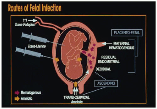
FIGURE 6-1 • Routes of fetal infection.


TABLE 6-6 PREDOMINANT PATHWAYS OF THE MAJOR FETAL AND NEONATAL INFECTION


Transplacental


Ascending


Postpartum


Bacteria


Listeriaa


Treponema pallidum


Mycobacterium tuberculosis


Borrelia


Campylobacter fetus (?)


Group B streptococci


Enteric bacillib


Haemophilus influenzae


Neisseria gonorrhea


Anaerobes


Actinomyces


Fusobacteria


Staphylococcia


Pseudomonasa


Nongroup B streptococcib


Viruses


Cytomegalovirusc


Herpes simplexc


Respiratory syncytial virus


Human immunodeficiency virus


Rubella


Mumps


Measles


Variola


Vaccinia


Poliovirus


ECHO


Hepatitis Bc


Western equine encephalitis


Human parvovirus


Varicella-zostera


Epstein-Barr virus


Coxsackie Ba


Protozoa


Toxoplasma


Plasmodium


Trypanosoma


Babesia


Fungi


Coccidioides


Candidac


Aspergillus


Torulopsis


Mycoplasmas


Mycoplasma hominis


Ureaplasma urealyticum


a Also utilize ascending route.

b Also utilize hematogenous route.

c Also acquired postnatally.


Perinatal infections commonly occur from exposure to maternal blood, body fluids, stool, or urine. The likelihood of infection varies significantly with the specific organism and various host factors (e.g., passively acquired antibody levels in the infant) and the IgG nadir in serum. Intrapartum transmission is more likely in a setting of prolonged labor combined with an infected maternal birth canal. Premature fetal inspiration results in perinatal pneumonia with high mortality. Postnatally acquired infections are transmitted most commonly through contact with caregivers (parents, relatives, visitors, and health care providers), the environment (medical equipment, other fomites), or breast milk (19). In most perinatal infections due to maternal transmission, the infant is exposed before illness is diagnosed in the mother and frequently occurs even before the mother becomes ill. Examples include maternally derived measles, enteroviral infection, chicken pox, and hepatitis.


BREAST MILK TRANSMISSION

Human milk protects against both specific pathogens and many illnesses (e.g., necrotizing enterocolitis, bacteremia, meningitis, respiratory tract illness, diarrheal disease, and
otitis media). Breast milk enhances nonpathogenic flora, decreases colonization with enteropathogens, aids development of the respiratory and intestinal mucosal barriers, provides secretory IgA and functioning immune cells (neutrophils, macrophages, T and B lymphocytes), and prevents gut inflammation and immunomodulation (20). Breast milk-associated infections include HIV-1, human T-lymphotropic virus-1 (HTLV-1), CMV, measles virus, and streptococci (21). Bacterial infections are rarely, if ever, transmitted to infants through breast milk. Although infections associated with breast milk are more commonly transmitted through other mechanisms, at least temporary cessation of breast-feeding has been recommended in certain maternal bacterial infections associated with Neisseria gonorrhoeae, Haemophilus influenzae, group B streptococci (GBS; Streptococcus agalactiae), staphylococci, Borrelia burgdorferi, Treponema pallidum, and Mycobacterium tuberculosis.


EVALUATION OF SUSPECTED FETAL INFECTION

Given the incredibly wide spectrum of fetal infections, pathologists must properly evaluate fetal and neonatal deaths. Infection should be suspected in newborns exhibiting any or all of the following: intrauterine growth restriction, failure to thrive, hydrops, jaundice, hepatosplenomegaly, skin rashes, hydrocephalus, microcephaly, eye lesions such as microphthalmia, chorioretinitis, and cataract. Wigglesworth has outlined a procedure for evaluating these infants by using serologic studies, radiology, bacteriologic studies including dark-field examination, and viral diagnostic studies including culture and electron microscopy (22). Nucleic acid amplification techniques have enabled the identification of infectious agents hitherto difficult or impossible to detect (23). It cannot be overemphasized that histopathologic examination constitutes only one facet of adequate autopsy examination in suspected prenatal or perinatal infection.


VIRAL INFECTIONS

Most significant viral infections in neonates or infants occur through transplacental or intrapartum transmission. The risk of transmission depends on whether the maternal infection is primary (e.g., HSV, HIV-1), secondary reactivation (e.g., HSV, CMV), or chronic (e.g., hepatitis B, HIV-1, HTLV-1). Fetal and neonatal viral infections are multifaceted. Many agents are teratogens and can affect the fetus or infant at any stage, leading to fetal death, malformation, self-limited or ongoing infection, and late sequelae. Table 6-7 summarizes the main features of fetal and neonatal viral infections, while Table 6-8 outlines the major pathologic features of commonly seen viral infections in infants and older children. Of the more commonly encountered viral illnesses in children, many exclusively involve the nervous system (e.g., poliomyelitis, rabies) and are discussed in Chapter 10. Respiratory tract pathogens are included in Chapter 12, and the hepatitis viruses in Chapter 15. Of the remainder, relatively few are encountered by the pathologist; many are opportunists in immunocompromised children. In general, these disseminated opportunistic viral infections resemble their perinatal infectious counterparts. For comprehensive information, the reader is referred to Feigin and Cherry’s textbook (1); only selected infections will be discussed here.


CYTOMEGALOVIRUS

CMV, the largest member of the family Herpesviridae, is encountered in all populations. A ubiquitous pathogen, CMV seroprevalence in adult populations ranges from 50% to 90%. It is the most common cause of congenital infection in the United States, with frequencies ranging from 0.2% to 2.2% of liveborn babies in the United States. Unlike congenital rubella and toxoplasmosis, intrauterine CMV transmission occurs in some women who are CMV seroimmune before pregnancy, at a much lower frequency than primary intrauterine infections. Approximately 1% of all congenitally infected infants excrete CMV in their urine within 3 weeks after birth; about 5% manifest perinatal disease at birth, and 15% develop late sequelae (24). More children may be affected by congenital CMV than by other perinatal diseases such as Down syndrome, fetal alcohol syndrome, and spina bifida. CMV is, therefore, one of the most common causes of birth defects and childhood disability.


Transmission

CMV infection may be transplacental, perinatal, or postnatal. Early, hematogenous gestational infections are the most devastating. Rarely, congenital CMV infection recurs in subsequent pregnancies. Primary CMV infection may rarely occur in the mother during delivery or lactation, and the lack of maternal antibody protection in these cases increases the risk for illness in the infant, although infections are rarely associated with clinical illness in full-term infants (25,26). Congenital and perinatal CMV immunity is complex; fetal protection cannot be assured with maternal immunity, since reinfection with a novel strain can occur (27). Infants who are susceptible (e.g., due to prematurity, seronegative mothers, or immunodeficiency) can have severe disease. CMV is also commonly reactivated in a setting of congenital or acquired immunodeficiency. Childcare centers also significantly transmit CMV, propagated by frequent mouthing of hands and toys. Approximately 20% to 40% of toddlers in day care shed the virus for years. These children function as an important infectious source for other children, parents, and daycare workers. After puberty, CMV infection is mainly sexually


transmitted. Virus is present in urine; oropharyngeal, cervical, and vaginal secretions; breast milk; semen; and tears and can be shed intermittently for years.


TABLE 6-7 VIRAL INFECTION IN THE FETUS AND NEONATE


Abortion


Stillbirth


Intrauterine Growth Restriction


Congenital Defects


Acute Perinatal Infection


Late Effects


Rubella


+


+


+


Cataract, retinopathy, sensorineural deafness, patent ductus arteriosus, pulmonary stenosis, VSD, microcephaly, cognitive impairment


Interstitial pneumonitis, cholestatic hepatitis with giant cell transformation, anemia, thrombocytopenia, myocarditis, immunopathy, osteoporosis, pancreatitis


Interstitial pulmonary fibrosis, hepatic fibrosis/cirrhosis, biliary atresia, arteriopathy with infarction, diabetes mellitus, chronic lymphocytic thyroiditis, panencephalitis, autism (?)


Cytomegalovirus


?


+


+


Microcephaly, hydrocephaly, microphthalmia


Necrotizing meningoencephalitis with arterial and periventricular calcification, hepatitis with giant cell transformation, cholangitis, inclusions in lung, renal tubules, rare pneumonitis and interstitial nephritis


Deafness, neurologic deficits, optic atrophy, noncirrhotic portal hypertension, and vascular and periventricular calcifications (brain)


Hypoganglionosis of bowel (see Pediatr Pathol 1984;2:85-102)


Herpes simplex


+


+


+


Microcephaly, hydranencephaly, microphthalmia


Hepatoadrenal necrosis, vesicular skin rash, vesicular/ulcerated stomatitis, esophagitis, necrotizing pneumonitis, chorioretinitis


Psychomotor retardation


Varicella-zoster


?


?


+


Limb hypoplasia, rudimentary digits, cutaneous scars in dermatome distribution, chorioretinitis, microphthalmia


Typical varicella, acute disseminated varicella with necrotizing cutaneous and visceral lesions


Blindness, psychomotor retardation


HIV


+


+


+


None


See Table 6-9A


See Table 6-9B


Parvovirus


+


+


Ocular defects


Anemia, hydrops, hepatic fibrosis, siderosis


?


Hepatitis B


Acute hepatitis, giant cell transformation, chronic active hepatitis, fulminant hepatitis


Cirrhosis, carrier state hepatocellular carcinoma


Hepatitis A


+


+


Rare


Mumps


+


+


Not proved


Perinatal parotitis (extremely rare)


Endocardial fibroelastosis (?)


Influenza


+


+


Unlikely


Rare influenza pneumonia, apnea


Vaccinia


+


+


+


Generalized vaccinia


Variola


+


+


?


?


Smallpox


Measles


+


+


?


Measles pneumonia


Polio


+


+


+


Paralytic polio


Paralysis


Echo


Meningitis, DIC


Coxsackie B


?


?


Disseminated disease, meningoencephalitis, myocarditis


Adenovirus


Pneumonia


RSV


Apnea, bronchiolitis, pneumonia


Asthma (?), COPD


DIC, disseminated intravascular coagulation; VSD, ventricular septal defect. +, occurs; -, does not occur.


TABLE 6-8 COMMON SYSTEMIC VIRAL INFECTIONS IN INFANTS AND CHILDREN


Virus


Localized or Self-Limited Disease


Disseminated or Serious Disease


Specific Inclusions


Comments


Adenoviruses


Acute respiratory illness (types 1, 2, 3, 4, 7, 21)


Laryngotracheitis, pneumonia


Types 1, 2, 4, 5, 7, 11


Hepatitis, massive hepatic necrosis


Pneumonia, hemorrhagic cystitis, gastroenteritis, meningoencephalitis


1. Large basophilic indistinctly demarcated intranuclear inclusion (smudge cells)


2. Smaller eosinophilic intranuclear with incomplete halo


Easily confused with disseminated HSV infection


Cytomegalovirus


Mononucleosis


Interstitial pneumonia


Gastroenteritis


Retinitis, encephalitis, glomerulonephritis


1. Large amphophilic or basophilic nuclear inclusions with distinct halo.


2. Smaller basophilic PAS-positive indistinct cytoplasmic inclusions


Disease in immunocompromised host is similar to neonatal pattern.


Herpes simplex


Localized oral, skin, or genital vesicular or ulcerated eruption, may be extensive


Hepatitis, hepatoadrenal necrosis, stomatitis, esophagitis, encephalitis, pneumonia


1. Type A eosinophilic nuclear inclusions with halo


2. Type B basophilic or amphophilic nuclear inclusions filling nucleus with peripheral chromatin rim, often multinucleate cells


Either type I or type II may disseminate: disseminated form resembles neonatal disease.


Varicella-zoster


Localized herpes zoster, acute varicella, generalized vesicular eruption


Disseminated zoster


Progressive disseminated varicella, pneumonia, meningoencephalitis, hepatitis


Multinuclear or mononuclear cells with nuclear type A inclusions, indistinguishable from HSV inclusions


Associated with Reye syndrome


Epstein-Barr virus


Mononucleosis


Fatal mononucleosis, hepatitis, myocarditis, immunodeficiency various hematologic phenotypes in X-linked lymphoproliferative syndrome


None


Implicated in oncogenesis, especially in X-linked lymphoproliferative syndrome, posttransplant lymphoproliferative syndrome


Rubeola


Uncomplicated primary measles, skin, conjunctiva, respiratory tract


Progressive measles


Cytoplasmic and nuclear inclusions in epithelial and Warthin-Finkeldey giant cells


Subacute sclerosing panencephalitis, late


Mumps


Parotitis


Orchitis/oophoritis


Meningitis, pancreatitis


Mastitis, nephritis, arthritis


None


Late sequelae include deafness and diabetes mellitus.


Coxsackievirus


Coxsackieviruses A; benign, self-limited febrile illness with respiratory disease


Coxsackieviruses B, myocarditis, meningoencephalitis


None


Echoviruses


Mild nonspecific febrile illness with respiratory disease


Hepatitis, hepatic necrosis, meningitis, adrenal and renal hemorrhage


None


Variola (smallpox)


Disease declared eradicated by WHO in 1980


Cytoplasmic Guarnieri bodies. Nuclear changes inconsistent


Vaccinia


Eczema vaccinatum


Disseminated vaccinia


Indistinguishable from smallpox


Hantavirus


?


Hantavirus pulmonary syndrome in adolescents


None


Noncarcinogenic pulmonary edema


HSV, herpes simplex virus; PAS, periodic acid-Schiff; WHO, World Health Organization.


Clinical Features

Transplacental transmission can result in congenital infection and neurologic sequelae. Perinatal and postnatal transmission usually fails to produce clinical disease except in extremely preterm infants (28). Around approximately 90% of infants born with congenital CMV infection do not exhibit clinical abnormalities at birth (so-called asymptomatic congenital CMV infection). Of the 40,000 children annually born with congenital CMV infection, approximately 10% to 15% exhibit clinical signs or symptoms. Infection involves multiple organ systems, particularly the reticuloendothelial and central nervous systems (CNS). Commonly observed physical signs in these infants include petechiae, jaundice, and hepatosplenomegaly (Figure 6-2). Neurologic abnormalities such as microcephaly and lethargy affect a significant proportion. Other physical signs include intrauterine growth restriction, chorioretinitis, optic atrophy, and seizures. Postnatally acquired infection results in neonatal sepsis with apnea, bradycardia, hepatitis, leukopenia, and prolonged thrombocytopenia. Older children with severe infection are typically immunodeficient. Disseminated CMV infection produces fever, leukopenia, thrombocytopenia, pneumonia, hepatitis, chorioretinitis, adrenalitis, and encephalitis. Infected infants may have the characteristic “blueberry muffin” lesions, a hemorrhagic purpura with mobile gray-blue skin papules that contain dermal extramedullary hematopoiesis. CNS lesions are irreversible and affect prognosis. Symptomatic liver disease commonly occurs, ranging from mild inclusion body cholangitis to severe cholestatic hepatitis (Figure 6-2). Noncirrhotic portal fibrosis with portal hypertension is rare but results in potentially lethal late sequela. Glomerulonephritis, ascites, and pulmonary hypoplasia are also described. A syndrome of hepatosplenomegaly, respiratory distress, a peculiar gray pallor, and atypical lymphocytosis occurs in multiple transfused low-birth-weight infants in this setting. Interstitial inclusion body pneumonitis (Figure 6-2) likely causes the high (24%) mortality rate.


Pathology

Morphologic hallmarks of CMV infection comprise cytomegaly with extremely large (25 to 40 µm) inclusion-bearing cells and both nuclear and cytoplasmic inclusions; often, the nucleolus is retained within the inclusion, appearing as an “accessory body” (29). A clear zone around the inclusion with chromatin margination renders an “owl’s eye” appearance. Inclusions vary from eosinophilic to deeply basophilic, depending on developmental stage. The inclusions are periodic acid-Schiff (PAS) and GMS positive, although immunostains are commonly used to specifically identify the virus. They are also CD15-positive, creating potential confusion with Hodgkin lymphoma. Potential virocytes include endothelial cells, epithelial cells (notably the biliary tree, pneumocytes, many exocrine cells, and renal tubular cells), fibroblasts, and histiocytes. Patchy, focal necrosis with mononuclear cells, occasionally neutrophils, and vascular and parenchymal calcifications characterizes the infection. Calcifications most commonly affect the developing brain. Giant cell transformation of hepatocytes is not a frequent feature.

Congenital neurologic and hematologic damage and developmental defects are evident at birth in 10% of

CMV-infected babies. During infancy, sequelae such as sensorineural deafness, psychomotor retardation, and cerebral palsy may develop even in infants lacking symptoms at birth. About 20% of all infected neonates suffer sequelae of a congenital CMV infection (30).


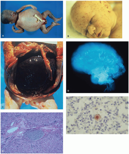
FIGURE 6-2 • Congenital CMV infection. A: Body with marked ascites. B: Face with petechiae as well as elsewhere. C: Abdominal cavity at autopsy showing hepatosplenomegaly. D: Skull x-ray with diffuse calcification secondary to necrosis. (See Malinger G, Lev D, Zahalka N, et al. Am J Neuroradiol 2003;24:28-32.) E: CMV hepatitis with inflammation around a bile duct and within the lobules. F: Lung: CMV immunostain with large nuclear inclusion.


Prognosis and Outcome

Mortality rate among symptomatic children is now less than 5%. Of the symptomatic children who survive infancy, most will suffer mild to severe psychomotor and perceptual handicaps, and approximately one half will develop sensorineural hearing loss, cognitive impairment, and microcephaly (28). Predictors of adverse neurologic outcome include microcephaly, chorioretinitis, presence of other neonatal or neurologic abnormalities, and cranial abnormalities on CT scans. Petechiae and intrauterine growth restriction independently predict hearing loss. Children asymptomatic at birth have a better long-term prognosis, but approximately 10% develop sensorineural hearing loss, often bilateral. Neurologic complications occur in asymptomatic congenital CMV infection less frequently than in symptomatic infection. This inability to predict outcome in infants at risk for the development of hearing loss and other sequelae necessitates monitoring and follow-up of all children with congenital CMV infection (32).


HERPES SIMPLEX VIRUS (HSV)

First described as hepatoadrenal necrosis by Haas in 1935 (33), HSV types 1 and 2 cause severe perinatal infections and, less frequently, prenatal and postnatal infections. HSV-2 predominates, and about 20% of infections are caused by HSV-1 (34). The risk to the infant is highest at the time of delivery in mothers with primary genital herpes, but fetal infection may occur in the absence of visible maternal lesions.


Transmission

Most HSV infections in infants are acquired during passage through an infected birth canal. Maternal skin infections and even nipple lesions, as well as paternal disease, pose a threat to the infant. Severe fetal intrauterine infection can also occur as a consequence of either primary or recurrent maternal infection. HSV antigen in endometrium, decidua, and placenta suggests possible transplacental passage. Case reports demonstrate HSV infections in infants caused by maternal HSV-positive breast lesions, and conversely, virus from primary infantile gingivostomatitis may infect the mother’s breast.


Clinical Features

Neonatal infections manifest in the first week of life. Although the neonatal form of the disease may be relatively benign, the majority of cases result in death from disseminated disease with meningoencephalitis (50%) or serious neurologic impairment (30%). The clinical presentation is variable and diagnosis may be extremely difficult; seizures, cyanosis, shock, and bleeding diatheses are common manifestations. Since no specific sign or symptom is diagnostic, the diagnosis should be strongly considered in the presence of HSV risk factors, atypical sepsis, unexplained acute hepatitis, or focal seizure activity. Neonatal HSV infection may be either disseminated or relatively localized. In general, the younger the patient at presentation, the more disseminated the lesions. Infants with subclinical encephalitis usually have skin and mucous membrane lesions (Figure 6-3A). Conversely, at least one-third of newborns with disseminated HSV do not have detectable skin or mucous membrane lesions at the time of presentation (34). In older infants and children, clinical presentations include orolabial and genital herpes, herpes gladiatorum, herpetic whitlow, eczema herpeticum, and ocular herpes. Herpes encephalitis in older children is an emergency that requires a high index of suspicion to diagnose (35).


Pathology

The pathologic hallmark of disseminated HSV are patchy and focal well-demarcated punctate areas of yellow-tan to hemorrhagic coagulative necrosis with little cellular inflammatory reaction at the periphery of irregular zones of necrosis (Figure 6-3B, C) (34). There are two types of inclusions (Figure 6-3D). Early infectious inclusions (Cowdry type B) stain variably (usually amphophilic, sometimes basophilic), are homogeneous and glassy, occupy the entire nucleus, and
push the chromatin to the nuclear membrane. The later inclusions (Cowdry type A) are smaller, deeply eosinophilic, round, or polygonal and separated from the nuclear membrane by a clear halo. Multinucleated cells are more likely to contain Cowdry type B inclusions. Type A inclusions reflect excess viral capsid material following extrusion of encapsidated viral DNA. Seventy-five to eighty percent of cases of disseminated HSV involve liver (Figure 6-3B, C) and adrenal glands. Lesions may also be seen in the lung, brain, spleen, bone marrow, and GIT. Care must be exercised in the evaluation of necrotizing and ulcerated skin or mucous membrane lesions; HSV inclusions can usually be found at the periphery of such lesions, as secondary bacterial or yeast infection may obscure the underlying viral lesion.


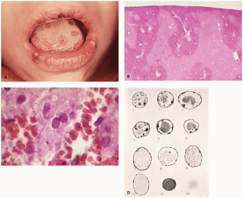
FIGURE 6-3 • Herpes simplex infection. A: Herpetic stomatitis. B: Liver (low power) with multifocal areas of coagulative necrosis. C: Liver (high power) with smooth nuclear inclusions usually at the interface between the necrotic and viable parenchyma. D: Pictorial representation of inclusions—camera lucida drawings by E. Piotti; each nucleus corresponds to the types of inclusions seen in the first reported case of HSV infection. (With permission from Singer DB. Pathology of neonatal Herpes simplex virus infection. Perspect Pediatr Pathol 1981;6:243-278.)


Prognosis and Outcome

Neonatal herpes is a potentially devastating illness, with 80% mortality without treatment. Even with therapy, the mortality rate for disseminated disease remains very high (50%). About 25% of survivors may have neurologic defects and/or blindness. Maternal therapy or prophylactic infant treatment, in certain situations, may decrease shedding, hasten clinical resolution, and protect the patient.

The consequences of neonatal HSV infection can be severe (37). Disease may be localized to skin, eye, and mouth (SEM disease), involve the CNS, or disseminate to multiple organs. Most survivors in the latter two categories have neurologic sequelae. Neonatal herpes may occur in the absence of skin lesions; if infection is suspected, swabs of the oropharynx, conjunctiva, rectum, skin lesions, mucosal lesions, urine, and cerebrospinal fluid (CSF) should be promptly submitted for laboratory studies (38).


HUMAN PARVOVIRUS INFECTION

Parvovirus B19, better known as a cause of erythema infectiosum (fifth disease), is a single-stranded DNA virus of the Erythrovirus genus that primarily targets erythroid precursors in the bone marrow. Because the erythrocyte P antigen is the cellular receptor for the virus, individuals lacking this antigen are resistant to infection.


Transmission

Transmission occurs through contact with respiratory droplets, saliva, and, less commonly, blood and urine. Seroprevalence data show that peak parvovirus infections occur in school-age children. Modes of entry into bloodstream and placenta are not clearly known.


Clinical Features

Intrauterine infection (39) results in fetal anemia, a pronounced leukoerythroblastic reaction, and hepatitis with excessive iron deposition. Parvovirus is a major cause of disease and possibly accounts for up to 16% of cases of “idiopathic” nonimmune hydrops (40). In Anand’s series (39), two of six affected pregnancies resulted in fetal hydrops and death, whereas the other four infants were normal. Fetal and perinatal presentations other than hydrops have been described. Ocular lesions include microphthalmia, aphakia, and dysplasia of sclera, anterior segment, and retina. Liveborn infants can present with a lethal constellation of anemia, petechial rash, purpuric “blueberry muffin” lesions, and severe liver disease with hepatic fibrosis and siderosis that mimic the syndrome of neonatal hemochromatosis (41).

Although postnatal infection is frequently asymptomatic, the best known clinical illness is immune-mediated erythema infectiosum or fifth disease. Fifth disease is a highly contagious pediatric illness with a slapped cheek appearance and lacy erythematous exanthem on the face, trunk, and proximal limbs in children. Adults manifest arthralgias and arthritis. Severe consequences occur most often in individuals with hemoglobinopathy, red blood cell abnormalities, or immunodeficiency. Erythroid abnormalities include pure red cell aplasia and aplastic anemia (especially aplastic crisis in a setting of chronic hemolytic anemia). Vasculitis and hemophagocytic syndrome can also occur. In some series, parvovirus has been the agent most frequently associated with myocarditis, but the significance of this finding has been questioned in an autopsy study.


Pathology

The bone marrow shows variable degrees of erythroid hypoplasia. Morphologic abnormalities in red cell precursors include giant pronormoblasts with vacuoles and multiple nucleoli. Distinctive eosinophilic intranuclear inclusions occur in erythroid lineage cells. By DNA hybridization and electron microscopy, inclusions contain B19 virus (39), and the virus is readily detected in tissue with immunohistochemical and PCR techniques (40). Histologic studies on very young fetuses show ocular malformations and generalized intense inflammatory reactions. The first clue to the infection may be infected red cells in the fetal capillaries of the placenta.


RUBELLA

Rubella is caused by a single-stranded RNA virus. Originally described by Gregg as a classic triad of cataracts, deafness, and congenital heart disease (42), the “expanded rubella syndrome” is rare in countries where rubella vaccination is the norm. However, congenital rubella syndrome (CRS) is unfortunately not just a subject of historical interest. Existence of both a nonimmune population of women of childbearing age and the many survivors of the 1964 to 1965 epidemic attest to the continued importance of this disease. Investigation of adult individuals enables delineation of late effects of congenital infection (43).


Transmission

Rubella virus is capable of infecting the fetus at any time during gestation. Virus reaches the fetus in emboli of necrotic placental tissue and then affects the fetus by at least three mechanisms: (a) inhibition of cell growth, (b) cytolysis, and (c) compromise of blood supply (43). These factors consequently incite necrosis, inflammation, and scarring in virtually limitless combinations and permutations. Unlike CMV, maternal antibodies to rubella virus protect the fetus from infection. Postnatal and childhood infections are transmitted through inhalation of droplets of nasopharyngeal secretions. Per the CDC, endemic rubella and CRS have been
eliminated from the United States through 2011, but international importation continues (44).


Clinical Features

The incidence and pattern of fetal rubella vary strikingly with gestational age at the time of maternal viremia. Congenital heart defects result from infection in the first trimester of gestation, deafness and neurologic deficits follow infection through the 4th gestational month, and retinopathy ensues through the 5th gestational month. Infection late in gestation more likely produces inflammatory and destructive lesions, without evidence of malformation. The probability of the fetus suffering significant damage decreases from 80% to 90% in first trimester to near 0% beyond 20 weeks. In either case, the virus is recoverable for months to years after birth. With the possible exception of microcephaly, most CNS abnormalities result from meningoencephalitis and/or necrosis. Necrosis, presumably ischemic, is related to the vascular lesions seen in a majority of cases. True developmental malformation is rare (43). A late-onset chronic progressive panencephalitis with neuropathologic changes similar to subacute sclerosing panencephalitis (SSPE) occurs in the second decade of life in some survivors of CRS. These changes include meningeal and perivascular infiltrates of lymphocytes and plasma cells with glial nodules, predominantly in the white matter. Rubella virus has been recovered from these late lesions. Deafness in CRS is related both to CNS damage causing central auditory imperception and to inflammation and scarring in the cochlea. CRS and Down syndrome are considered to be the main causes of combined deafness and blindness in patients over 18 years of age, whereas CHARGE syndrome is the main cause in children (45). Disseminated sequelae may relate to vascular spread and cytopathic effects on endothelial cells. Deafness, cardiovascular and neurologic damage, and retinopathy are rare if infection occurs beyond the second trimester, suggesting a protective role of maternal antibodies (46).

Postnatal rubella presents as a prodrome followed by a characteristic postauricular lymphadenopathy. Fine maculopapular rash appears 1 to 5 days later, starting in the face and spreading to limbs and trunk and lasting about 3 days. Complications are infrequent but include immune manifestations such as arthritis, encephalitis, Guillain-Barré syndrome, and thrombocytopenia. Surveillance of postnatal and congenital infection is an essential component of CRS prevention, since rubella is difficult to diagnose on clinical grounds alone. Laboratory differentiation of rubella from other exanthems, such as measles, parvovirus B19, human herpes virus 6, enteroviruses, and various endemic arboviruses, is essential. Current testing includes RT-PCR and sequencing for diagnosis and epidemiologic surveillance, and oral fluid tests for detection of rubella-specific IgG and IgM salivary antibody responses (47).


Pathology

A wide spectrum of cardiovascular disease is seen in CRS. In addition to the characteristic patent ductus arteriosus, lesions most commonly include pulmonary artery branch stenosis, myocarditis, and systemic arterial hypoplasia and stenosis. Valvular sclerosis has been frequent in some series (43). The arterial lesion of CRS is distinctive and possibly unique: fibromuscular intimal proliferation, devoid of inflammatory change, leads to patchy and focal vascular stenosis. The media and adventitia are usually not disrupted, and there is no calcification (except in the brain). Chronic meningeal inflammation, perivascular lymphocytic infiltrates, gliosis, and mineralization of cerebral arterioles may occur. Transient bone lesions consist of focal osteopenia and growth inhibition. Metaphyseal changes reminiscent of syphilis, that is, longitudinal radiologic striations, occur in one-half of the patients. Interstitial pneumonitis is seen in up to 75% of CRS infants and may persist for up to a year after birth. Alterations of the lymphoreticular system are variable; both precocious germinal centers (from viral antigenic stimulus) and lymphoid depletion are encountered. Histiocytic proliferation and erythrophagocytosis may be seen. Hepatic changes include cholestatic hepatitis, giant cell transformation, necrosis, extramedullary hematopoiesis, and fibrosis; cirrhosis may ensue. On occasion, bile duct proliferation mimics extrahepatic biliary atresia; true biliary atresia has been reported anecdotally. Eye changes include cataracts, lens necrosis, ciliary body inflammation, iridocyclitis, and retinitis. Interstitial nephritis and chronic lymphocytic thyroiditis have also been described. The placenta may show villitis, villous stromal necrosis and sclerosis, and vascular endothelial lesions (48). Due to its short and benign course, no specific histopathologic features characterize postnatal rubella infection.


Prognosis and Outcome

Characteristically, CRS sequelae affect the eyes (cataracts or retinopathy) and hearing (sensorineural deafness). Multiorgan involvement may include myocarditis, hepatitis, cytopenia, meningoencephalitis, and visceromegaly. Cardiac teratogenic effects include patent ductus arteriosus, pulmonary artery stenosis, and supra-aortic stenosis. Long-term effects of intrauterine rubella infection include an increase in the prevalence of diabetes, thyroid disorders, early menopause, and osteoporosis (49). HLA haplotypes that are associated with autoimmune disorders occur in increased frequency. Approximately 20% of CRS survivors develop diabetes mellitus, usually in the second or third decade. Persistent viral infection has been implicated as the virus may be recovered from the pancreas and lymphocytic infiltration of the pancreas may be seen at postmortem exam. Postnatal growth retardation probably results from active virus, as evidenced by its prolonged shedding in nasopharyngeal secretions.


VARICELLA ZOSTER VIRUS

VZV (human herpes virus 3) is a double-stranded DNA virus. The existence of a fetal varicella syndrome was suggested in 1974, although the first case had been reported years earlier. Several studies describe the defects associated with fetal varicella infection, and a specific neonatal syndrome is associated with maternal varicella (but not by maternal zoster) occurring during the first half of pregnancy (50).


Transmission

Congenital VZV infections are transmitted transplacentally. The risk of embryopathy with maternal infection in the first 20 weeks of gestation is estimated at 0.4% to 2%. Intrauterine insult occurring between 8 and 20 weeks of gestation results in a fetal disease with distinctive herpes zoster-like dermatome distribution. Although the virus has not been isolated from affected fetuses or newborns, virus specific IgM has been demonstrated in affected fetus and VZV DNA sequences have been recovered from the placenta. Neonatal varicella infection follows in utero near-term exposure or postnatal contact with the mother or others (household or nursery). Infants delivered of mothers who were infected more than 5 days before delivery fared best, presumably because there was time for production and transfer of maternal antibody. Perinatal infection can be severe when the mother presents with chicken pox exanthem from 5 days before to 2 days after delivery, since maternal transplacental viremia occurs without sufficient time for maternal-fetal antibody transfer. Postnatal transmission occurs via respiratory droplets and contact or aerosolization of virus from varicella or zoster skin lesions. Infection peaks in winter and spring.


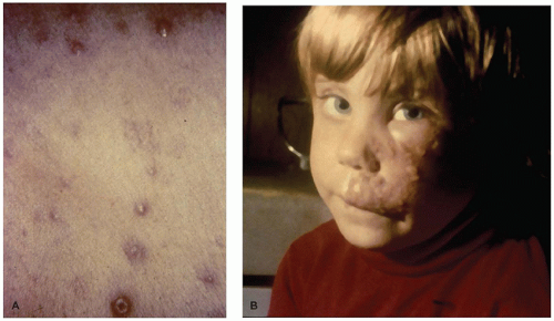
FIGURE 6-4 • Varicella-zoster virus infection. A: Neonatal varicella with skin lesions. B: Herpes zoster in an older child.


Clinical Features

Fetal varicella results in multiple defects of skin, limbs, eyes, and brain, giving an impression of a sudden devastating, but self-limited, herpes zoster-like in utero illness (50). All patients exhibit cicatricial skin lesions corresponding to the affected dermatome. There is frequent associated hypoplasia of the underlying bone and soft tissue. Hypoplastic limbs, many seriously deformed by scarring, occur in 80% of cases. Occasional calcification of the liver suggests dissemination, and viral-like inclusions can be seen in the lung. CNS involvement may take the form of necrotizing encephalitis with calcification. Microphthalmia, severe chorioretinitis with scarring, and cataract lead to blindness. Neurologic abnormalities frequently correspond anatomically to the afflicted dermatome and include limb paresis, microcephaly, Horner syndrome, cranial nerve palsies, and cortical atrophy.

Neonatal varicella in the newborn may be limited to the skin (Figure 6-4) or disseminate widely; disseminated disease carries a very high mortality rate, largely due to varicella pneumonia. Older children develop a prodrome for 2 to 3 days, followed by a transient scarlatiniform rash that may precede or accompany the characteristic varicelliform rash, which appears on the trunk and spreads out as crops of 1- to 4-mm maculopapular lesions. These progress to clear fluid-filled vesicles (“dew drop on a rose petal”) and pustules, with accompanying distressing pruritis. Lesions of various stages are seen in a given patient, and the patient remains infectious until all the lesions have crusted. Excoriation may leave shallow pink depressions that lead to scars when complicated by secondary bacterial infection. Vesicles may also develop on mucous membranes and leave multiple small ulcers. Rarely, there may be septic shock, hemolytic-uremic
syndrome, necrotizing pneumonia, encephalitis, hepatitis, and/or Reye syndrome (51).

VZV infects sensory nerves, migrates to sensory ganglia during acute infections, and remains latent there, to cause later herpes zoster (Figure 6-4). Involvement of nonneuronal satellite cells, which interface with multiple neurons, may allow the virus to involve large geographic areas (e.g., an entire dermatome) during reactivation. A prodrome of pain, itching, burning, and paresthesia may precede the characteristic zosteriform eruption by 4 to 5 days, as may constitutional symptoms such as headache, fever, and malaise. Lesions may continue to develop within the dermatome over a week and last for 2 to 3 weeks but may last longer in debilitated and immunodeficient patients. Ulcers, scaling, hyperpigmentation, and secondary bacterial infection with resultant scarring may complicate the clinical picture. Herpes zoster is uncommon in childhood.


Pathology

Chicken pox is a clinical diagnosis. However, Tzanck smears of vesicular or pustular fluid (of varicella or zoster) allows rapid identification of VSZ-infected cells by demonstrating intranuclear inclusions and giant cells similar to those seen in HSV infection. Skin biopsies also show features similar to HSV, that is, ballooning degeneration progressing to acantholytic intraepidermal vesicle sometimes involving adnexa. Unlike HSV infection, dermal leukocytoclastic vasculitis with occasional hemorrhage may occur. Inclusions begin as faint basophilic intranuclear bodies with peripheral chromatin condensation, later becoming eosinophilic with a surrounding halo. An immunostain is available for specific identification. Disseminated VZV may induce hemorrhagic necrosis in a variety of organs, with little or no inflammation or eosinophilic intranuclear inclusions. Pulmonary lesions consist of an interstitial mononuclear infiltrate, edema, hemorrhage, and hyaline membranes, all apparent within focal, sharply defined centrilobular areas of necrosis (51).


Prognosis and Outcome

Chicken pox is almost always self-limited. Death from chicken pox is distinctly unusual (12 deaths/100,000 cases), except in immunodeficient patients in whom pneumonia, meningoencephalitis, and hepatitis may develop (52). Less frequently, other systemic manifestations may occur, including nephritis, myocarditis, arthritis, myositis, uveitis, orchitis, and idiopathic thrombocytopenic purpura (53). Reye syndrome has been described following treatment with salicylates, and aspirin is contraindicated in patients with chicken pox.


HUMAN IMMUNODEFICIENCY VIRUS (HIV)

The first cases of pediatric acquired immunodeficiency syndrome (AIDS) were reported in 1982-1983, soon after recognition of adult disease. Over 90% of cases of childhood HIV infection are acquired by vertical transmission. HIV/AIDS in children resembles adult disease in both its primary viral cytopathic effect on the lymphoid and nervous systems and its secondary immunodeficiency effects including opportunistic infections and neoplasia. Adult and pediatric HIV infection also differs with respect to diagnostic methods, effects on the response capability of the developing immune system, etiologies of secondary infections, and type and distribution of pathologic lesions. HIV-2 causes clinical disease similar to HIV-1 but with a significantly slower progression to immune suppression. In West African infants, there is infrequent HIV-2 vertical transmission and no cases of late postnatal seroconversion (54). HIV-2 transmission through breast milk occurs less commonly than for HIV-1, but the risk and possible factors contributing to transmission have not been adequately quantified. Recent volumes have detailed the epidemiology, immunopathogenesis, molecular biology, and clinicopathologic aspects of pediatric AIDS (55,56,57). The CDC surveillance case definitions for HIV and AIDS are shown in Tables 6-9A, 6-9B, and 6-10 (58).


Transmission

Vertical transmission is the most common mode of acquisition of pediatric HIV. Transmission of virus by breast-feeding, sexual abuse, and heterosexual or homosexual relationships accounts for most of the remainder. The risk of transmission through transfusion of blood or blood products has been almost eliminated. Vertical transmission can take place in utero, intrapartum, or postpartum, through breast-feeding. To some extent, the resultant infections can be distinguished on the basis of culturable virus or HIV genome in cord and infant blood. The best predictor of transmission risk is maternal viral burden, as measured by maternal plasma HIV-1 RNA level. Levels under 500 copies per milliliter imply minimal risk of perinatal transmission (59). Treatment of HIV-infected mothers with effective antiviral agents has significantly decreased the rate of vertical transmission. Obstetric interventions such as caesarian section or shortening of postrupture transmembrane exposure may also decrease the rate of perinatal infection (60). Coinfections appear to increase the risk of mother-to-child HIV transmission such as chorioamnionitis, maternal genital infection (e.g., HSV), systemic infections (e.g., hepatitis B), mastitis, and infant infections (e.g., periodontitis, oral candidiasis, and diarrhea) (61). Despite their immature immune system and prolonged repetitive exposure to breast milk, most infants born to HIV-infected women escape infection. In infected infants, the course and response to treatment depend upon the timing (in utero, intrapartum, or during breast-feeding) and potentially the route of infection, probably


due to immunologic factors. Immune factors may be responsible for the adverse effects even in infants who escape actual infection (62).


TABLE 6-9A 2008 SURVEILLANCE CASE DEFINITION FOR HIV INFECTION AMONG CHILDREN AGED <18 MONTHSa


Criteria for definitive or presumptive HIV infection


Child born to an HIV-infected mother and laboratory criterion or at least one other criteria met


Laboratory criterion for definitive HIV infection


Positive results on two separate specimens (not including cord blood) using HIV virologic (nonantibody) tests (HIV nucleic acid detection is method of choice)


Laboratory criterion for presumptive HIV infection


Criterion for definitively HIV infected not met, and


Positive result on one specimen (not including cord blood) using HIV virologic tests AND no subsequent negative results from HIV virologic or antibody tests


Other criteria (for cases that do not meet above laboratory criteria)


HIV infection diagnosed by a physician or qualified medical care provider based on the laboratory criteria and documented in a medical record. Oral reports of prior laboratory test results are not acceptable.


or


When test results regarding HIV infection status are not available, documentation of a condition that meets the criteria in the 1987 pediatric surveillance case definition for AIDS


Criteria for uninfected with HIV, definitive or presumptive


Child born to an HIV-infected mother is either definitively or presumptively uninfected with HIV if (1) the criteria for definitive or presumptive HIV infection are not met and (2) at least one of the following laboratory criteria or other criteria are met.


Laboratory criteria for uninfected with HIV, definitive


At least two negative HIV DNA or RNA virologic tests from separate specimens, both of which were obtained at age ≥1 mo and one of which was obtained at age ≥4 mo


or


At least two negative HIV antibody tests from separate specimens obtained at age ≥6 mo


and


No other laboratory or clinical evidence of HIV infectionb


Laboratory criteria for uninfected with HIV, presumptive


Two negative RNA or DNA virologic tests, from separate specimens, both of which were obtained at age ≥2 wk and one of which was obtained at age ≥4 weeks


or


One negative RNA or a DNA virologic test from a specimen obtained at age ≥8 weeks


or


One negative HIV antibody test from a specimen obtained at age ≥6 mo


or


One positive HIV virologic test followed by at least two negative tests from separate specimens, one of which is a virologic test from a specimen obtained at age ≥8 wk or an HIV antibody test from a specimen obtained at age ≥6 mo


and


No other laboratory or clinical evidence of HIV infectionb


Other criteria (for cases that do not meet above laboratory criteria)


Determination of uninfected with HIV by a physician or qualified medical care provider based on the laboratory criteria and who has noted the HIV diagnostic test results in the medical record. Oral reports of prior laboratory test results are not acceptable


and


No other laboratory or clinical evidence of HIV infectionb


Criteria for indeterminate HIV infection


Child born to an HIV-infected mother if the criteria for infected with HIV and uninfected with HIV are not met


aThese guidelines are intended for public health surveillance only and are not a guide for clinical diagnosis.

b No positive results from virologic tests (if tests were performed) and no AIDS-defining condition for which no other underlying condition indicative of immunosuppression exists (see Table 6-10).


Modified from Schneider E, Whitmore S, Glynn KM, et al.; Centers for Disease Control and Prevention. Revised surveillance case definitions for HIV infection among adults, adolescents, and children aged <18 mo and for HIV infection and AIDS among children aged 18 mo to <13 years—United States, 2008. MMWR 2008;57(RR-10):1-12.


TABLE 6-9B 2008 SURVEILLANCE CASE DEFINITION FOR HIV INFECTION AMONG CHILDREN AGED 18 MONTHS TO <13 YEARSa


Criteria for HIV infection


At least one of laboratory criteria or the other criterion should be met.


Laboratory criteria


Positive result from a screening test for HIV antibody (e.g., reactive EIA), confirmed by a positive result from a supplemental test for HIV antibody (e.g., Western blot or indirect immunofluorescence assay)


or


Positive result or a detectable quantity by an HIV virologic (nonantibody) testsb


Other criterion (for cases that do not meet laboratory criteria)


HIV infection diagnosed by a physician or qualified medical care provider based on the laboratory criteria and documented in a medical record. Oral reports of prior laboratory test results are not acceptable.


Criteria for AIDS


Children aged 18 mo to <13 y are categorized for surveillance purposes as having AIDS if the criteria for HIV infection are met and at least one of the AIDS-defining conditions has been documented.


aThese guidelines are intended for public health surveillance only and are not a guide for clinical diagnosis. The 2008 laboratory criteria for reportable HIV infection among persons aged 18 mo to <13 y exclude confirmation of HIV infection through the diagnosis of AIDS-defining conditions alone (see Table 6-10). Laboratory-confirmed evidence of HIV infection is now required for all reported cases of HIV infection among children aged 18 mo to <13 y.

b For HIV screening among children aged 18 mo to <13 y infected through exposure other than perinatal exposure, HIV virologic (nonantibody) tests should not be used in lieu of approved HIV antibody screening tests. A negative result (i.e., undetectable or nonreactive) by an HIV virologic test (e.g., viral RNA nucleic acid test) does not rule out the diagnosis of HIV infection.


TABLE 6-10 AIDS-DEFINING ILLNESSES IN THE PEDIATRIC AGE GROUP


Bacterial infections, multiple or recurrenta


Candidiasis of bronchi, trachea, or lungs


Candidiasis of esophagusb


Cervical cancer, invasivec


Coccidioidomycosis, disseminated or extrapulmonary


Cryptococcosis, extrapulmonary


Cryptosporidiosis, chronic intestinal (>1-month duration)


Cytomegalovirus disease (other than liver, spleen, or nodes), onset at age >1 mo


Cytomegalovirus retinitis (with loss of vision)b


Encephalopathy, HIV related


Herpes simplex: chronic ulcers (>1-month duration) or bronchitis, pneumonitis, or esophagitis (onset at age >1 mo)


Histoplasmosis, disseminated or extrapulmonary


Isosporiasis, chronic intestinal (>1-month duration)


Kaposi sarcomab


Lymphoid interstitial pneumonia or pulmonary lymphoid hyperplasia complexa,b


Lymphoma, Burkitt (or equivalent term)


Lymphoma, immunoblastic (or equivalent term)


Lymphoma, primary, of brain


Mycobacterium avium complex or M. kansasii, disseminated or extrapulmonaryb


M. tuberculosis of any site, pulmonary,b,c disseminated,b or extrapulmonaryb


Mycobacterium, other species or unidentified species, disseminatedb or extrapulmonaryb


Pneumocystis jiroveci pneumoniab


Pneumonia, recurrentb,c


Progressive multifocal leukoencephalopathy


Salmonella septicemia, recurrent


Toxoplasmosis of brain, onset at age >1 mob


Wasting syndrome attributed to HIV


a Only among children aged <13 y. (CDC. 1994 Revised classification system for human immunodeficiency virus infection in children <13 years of age. MMWR 1994;43[No. RR-12].)

b Condition that might be diagnosed presumptively.

c Only among adults and adolescents aged ≥13 y. (CDC. 1993 Revised classification system for HIV infection and expanded surveillance case definition for AIDS among adolescents and adults. MMWR 1992;41[No. RR-17].)


Source: Schneider E, Whitmore S, Glynn KM, et al.; Centers for Disease Control and Prevention. Revised surveillance case definitions for HIV infection among adults, adolescents, and children aged <18 mo and for HIV infection and AIDS among children aged 18 mo to <13 years—United States, 2008. MMWR 2008;57(RR-10):1-12.


Breast-feeding by an HIV-1-positive mother increases transmission risk by 4% to 22% over the risk for prior prenatal and perinatal transmission. However, the lack of acceptable, feasible, affordable, sustainable, and safe (AFASS) water for breast milk alternatives complicates infant feeding practices in less developed nations. Unless AFASS criteria are satisfied, current WHO/UNICEF guidelines recommend exclusive breast-feeding for all infants for at least the first 6 months because of reduced infant mortality among exclusively breast-fed, HIV-exposed infants (63). Conversely, the avoidance of breast-feeding in maternal HIV-1 infection is an important component of preventing mother-to-child transmission in the United States and other developed countries. Issues related to breast milk HIV-1 transmission include the increased risk of primary maternal HIV-1 infection during lactation, the health of the HIV-1-infected mother, the presence of virus, potentially immunologically protective factors and factors that enhance HIV-1 transmission in breast milk, and possible interventions that prevent or limit HIV-1 transmission through breast milk (64). These interventions include early weaning, education, and support to decrease the occurrence of mastitis or nipple lesions, antiretroviral therapy for the mother or infant, treating the human milk to decrease the viral burden (ultraviolet light, freezing, and thawing), and stimulating the infant’s immune defenses with active or passive immunization.


Clinical Features

Clinical manifestations include hepatosplenomegaly, lymphadenopathy, failure to thrive, fever of unknown origin (FUO), chronic diarrhea, various infections, parotitis, chronic otitis media, lymphoid interstitial pneumonitis (LIP), HIV nephropathy, HIV encephalopathy, HIV cardiomyopathy, idiopathic thrombocytopenia purpura, and lymphoma. Age-specific data suggest that HIV manifestation changes with the child’s age, and these may further vary based on geographic location (65,66,67). HIV encephalopathy, HIV cardiomyopathy, idiopathic thrombocytopenia purpura, and lymphoma may occur later than other manifestations.

There is great variation in rapidity of onset, age of onset, and rate of progression in pediatric AIDS. In perinatally infected infants, the onset of symptomatic disease occurs at 6 to 8 months of age, as compared with a mean of about 18 months in transfusion-acquired pediatric AIDS (and years in adults). This extremely rapid progression undoubtedly reflects early disruption of differentiation in the developing cellular immune system that results from HIV-induced destruction of CD4 lymphocytes before the establishment of a fully developed immunologic response. There is also marked variation in the rate of progression of HIV in pediatric patients once they are symptomatic. Some perinatally infected children have onset of disease in the first year of life characteristically with Pneumocystis jiroveci pneumonia (PJP; formerly Pneumocystis carinii pneumonia, PCP), HIV encephalopathy, and recurrent severe bacterial infections. Another group is characterized by onset after the first year and a more indolent and chronic course of mucosal candidiasis, LIP, and cardiovascular disease. The reason(s) for these differences are, as yet, unclear. Children with AIDS do not show the marked degree of lymphopenia seen in adults but are more likely to have hyperglobulinemia. Cutaneous anergy is seen in infants. Severe bacterial infections are extremely common in pediatric AIDS, occurring in over 80% of affected children. Among pediatric opportunistic infections, candidiasis is the most frequent (Figure 6-5), beginning as oral thrush and affecting the entire GIT; PJP is the most frequent fatal infection in infancy (Figure 6-5). Other common opportunistic pathogens, depending on geographic distribution and/or population, include CMV (Figure 6-5), Mycobacterium avium/intracellulare, tuberculosis (TB), aspergillosis, cryptococcosis, cryptosporidiosis, histoplasmosis, HSV, adenoviral pneumonia, measles, and respiratory syncytial virus (RSV). About 25% of children with AIDS develop a lymphoproliferative syndrome with generalized lymphadenopathy and splenomegaly.

Children with HIV demonstrate lower motor, cognitive, and adaptive functioning compared to uninfected children. Risk factors that may negatively affect the development of infected children include neurologic abnormalities, progression of the disease, and poor environmental factors (68). Anemia, also a very common complication of pediatric HIV infection, is associated with a poor prognosis. Failure of erythropoiesis may be the most important mechanism for anemia (69). Survival in children with AIDS is in general shorter than survival in HIV-infected adults.

Clinical manifestations in children living with HIV/AIDS differ from those in adults, due to a poorly developed immunity that allows greater dissemination throughout various organs. In developing countries, HIV-infected children have an increased frequency of malnutrition and common childhood infections such as otitis, pneumonia, gastroenteritis, and TB. Symptoms common to many treatable conditions, such as recurrent fever, diarrhea, and generalized dermatitis, tend to be more persistent and severe and often do not respond as well to treatment (70).

Only gold members can continue reading. Log In or Register to continue

Stay updated, free articles. Join our Telegram channel

Sep 23, 2016 | Posted by in PATHOLOGY & LABORATORY MEDICINE | Comments Off on Congenital and Acquired Systemic Infectious Diseases

Full access? Get Clinical Tree

Get Clinical Tree app for offline access