29
Chest Wall and Mediastinum
The chest wall and mediastinum are parts of the thorax. The thorax has a bony and cartilaginous skeleton and contains the principal organs of respiration and circulation. The 12 thoracic vertebrae make up the posterior wall of the thorax. The 12 pairs of ribs, along with their costal cartilages and intercostal muscles, form the lateral and anterolateral wall. In the midline, the sternum is the anterior wall. The thorax is approximately conical in shape with a relatively narrow superior inlet (superior thoracic aperture) and a broader inferior outlet (inferior thoracic aperture).
The thoracic cavity is divided into the right and left pleural cavities by the mediastinum (Fig. 29.1). The mediastinum is bounded anteriorly by the sternum and posteriorly by the vertebral column; its floor is formed by the diaphragm. Superiorly, it is continuous with structures of the neck through the superior thoracic aperture, which forms the boundary of the superior mediastinum.
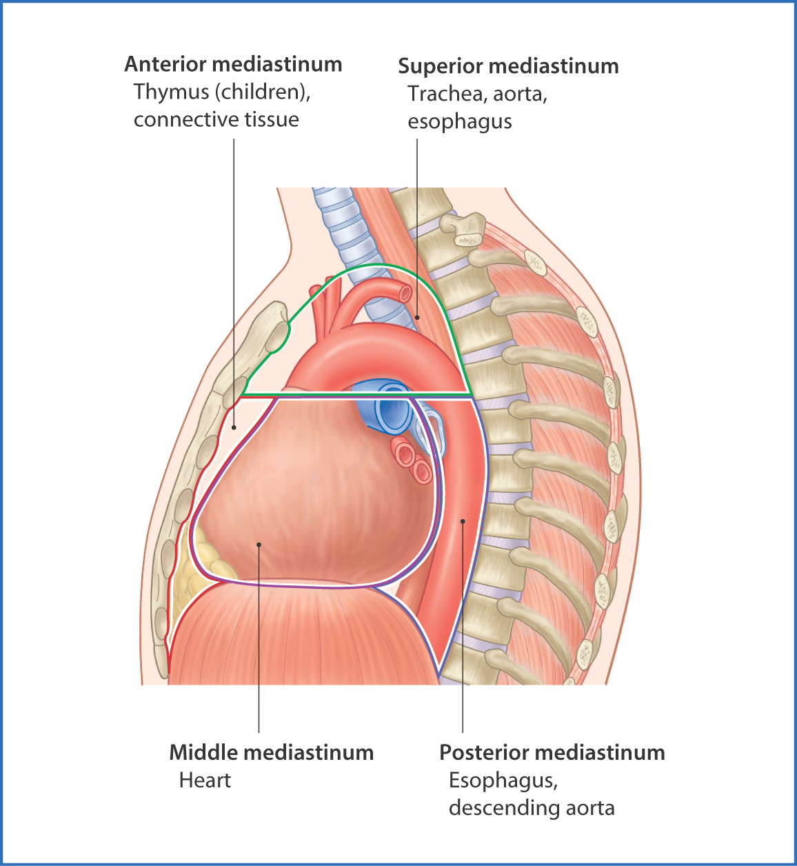
FIGURE 29.1 Subdivisions of the mediastinum and their contents.
Muscles
The muscles of the thorax (Table 29.1) are arranged in three layers:
- The external intercostal muscles (Fig. 29.2) form the external layer and are contained within the 11 intercostal spaces; the muscles extend from the tubercle of the ribs to the costochondral junction.
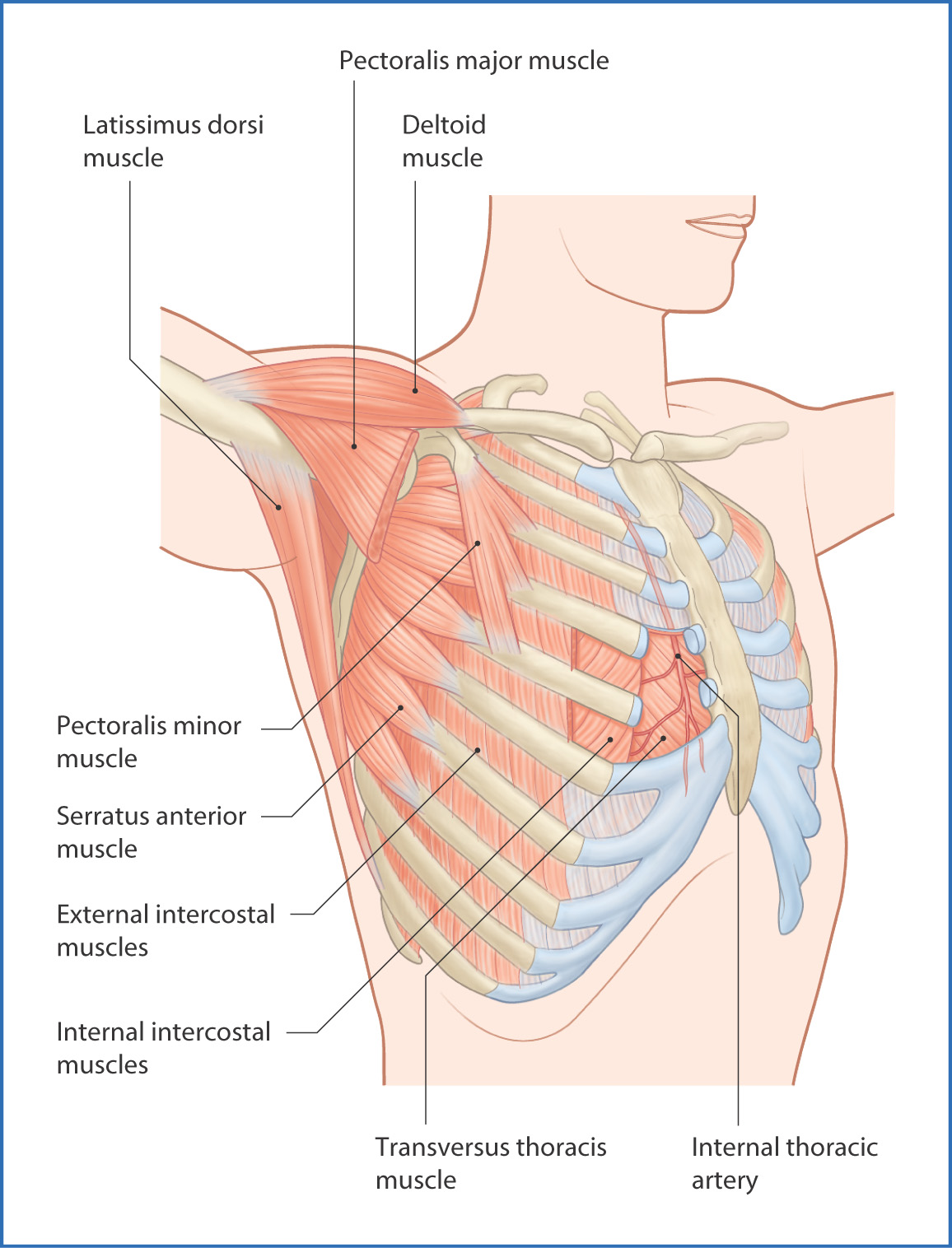
FIGURE 29.2 Muscles of the chest wall.
- The internal intercostal muscles form the middle layer and occupy the 11 intercostal spaces from the border of the sternum to the angle of the ribs.
- The innermost intercostal, subcostales, and transversus thoracis muscles form the internal layer of the chest wall muscles.
TABLE 29.1 Muscles of the Chest Wall
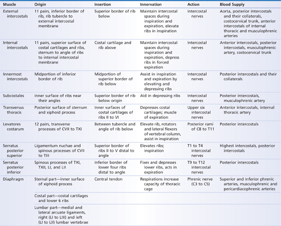
These muscles pull on the ribs to enable the changes in thoracic volume needed for breathing and help maintain the rigidity of the chest wall.
The levatores costarum muscles arise from the transverse processes of vertebrae CVII to TXI and insert into the external surface of the rib below. They are innervated by the posterior rami of the spinal nerves and, as their name suggests, elevate the ribs.
Nerves
The anterior rami of thoracic spinal nerves TI to TXI supply the overlapping segmental innervation of the skin and tissues of the chest wall. Spinal nerve TXII forms the subcostal nerve. Collectively, these nerves are called the intercostal nerves. They pass laterally from their origin at the spinal cord into the costal groove of each rib, and they continue anteriorly around the chest wall and run between the internal and the innermost intercostal muscles. Anteriorly, they terminate as cutaneous branches that provide sensation to the skin of the chest wall dermatomes (see Fig. 26.5).
Arteries
The internal thoracic artery (Fig. 29.3) is a branch of the subclavian artery. It descends toward the abdomen posterior to the first six costal cartilages and
- supplies blood to the upper part of the anterior chest wall
- gives off anterior intercostal arteries to the first six intercostal spaces
- divides at the sixth intercostal space into two terminal branches—the superior epigastric artery, which supplies the lower intercostal spaces and the anterior abdominal wall, and the musculophrenic artery, which supplies the peripheral part of the diaphragm
- gives off anterior intercostal arteries to the first six intercostal spaces
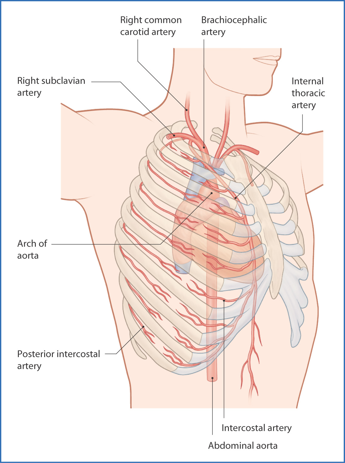
FIGURE 29.3 Arterial supply to the chest wall.
The first two posterior intercostal spaces receive their blood supply from the supreme intercostal artery, a branch of the costocervical trunk from the subclavian artery. The remaining nine posterior intercostal spaces are supplied by the posterior intercostal arteries, which are branches of the thoracic aorta.
Veins and Lymphatics
Stay updated, free articles. Join our Telegram channel

Full access? Get Clinical Tree


