• Elevated serum IgG4 (not invariably present, and not diagnostic of autoimmune pancreatitis)
More common in type 1 than type 2 AIP
• Steroid therapy is usually very effective
Recurrence reported in 6-26%
• Often mimics pancreatic adenocarcinoma clinically and radiographically
Macroscopic
• Enlarged, firm pancreas ± mass lesion; may mimic adenocarcinoma
Usually head is most prominently involved
• Stenosis of pancreatic duct and intrapancreatic common bile duct are common
Microscopic
• Dense, lymphoplasmacytic infiltration centered around main and interlobular pancreatic ducts
• Periductal, lobular, and perilobular fibrosis
• Obliterative phlebitis and venulitis
• 2 main types
Type 1: Lobular and interlobular distribution, obliterative phlebitis, numerous IgG4(+) plasma cells
Type 2: Duct-centric distribution, granulocytic epithelial lesions, only rare IgG(+) plasma cells
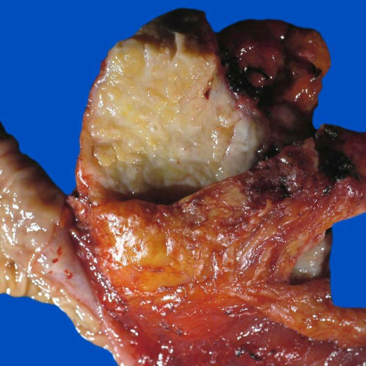
Surgical Specimen This pancreatic resection from a case of type 1 autoimmune pancreatitis (AIP) shows a dense, infiltrative, fibrotic process in the head of the pancreas.
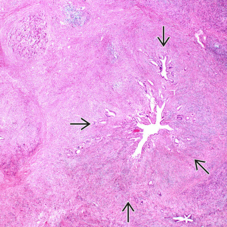
Periductal Inflammation, Type 1 Autoimmune Pancreatitis Low-power view of type 1 AIP shows marked chronic inflammation surrounding a large duct and involving the periductal stroma .
Phlebitis, Type 1 Autoimmune Pancreatitis This vein has been infiltrated by inflammatory cells, with resultant edema and destruction of the wall of the vessel. Note the surrounding fibrosis.
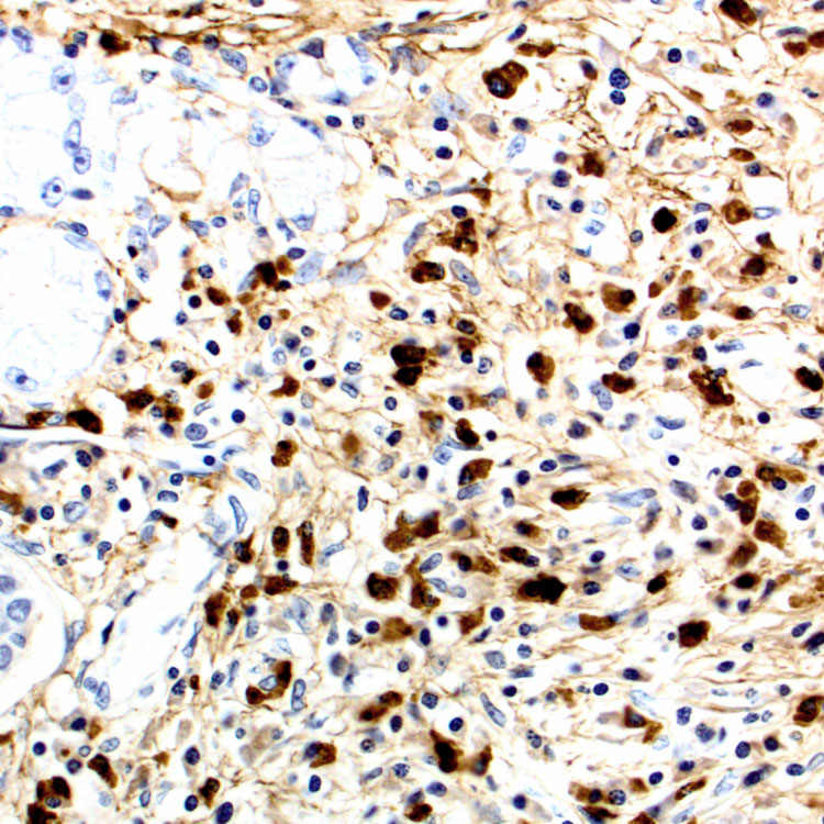
IgG4 Immunostain, Type 1 IgG4 stain in type 1 AIP shows a large number of IgG4(+) plasma cells (> 10/HPF) in the periductal stroma.
 Similar fibroinflammatory process often affects other organs such as bile ducts, salivary glands, retroperitoneum, and lymph nodes


 .
.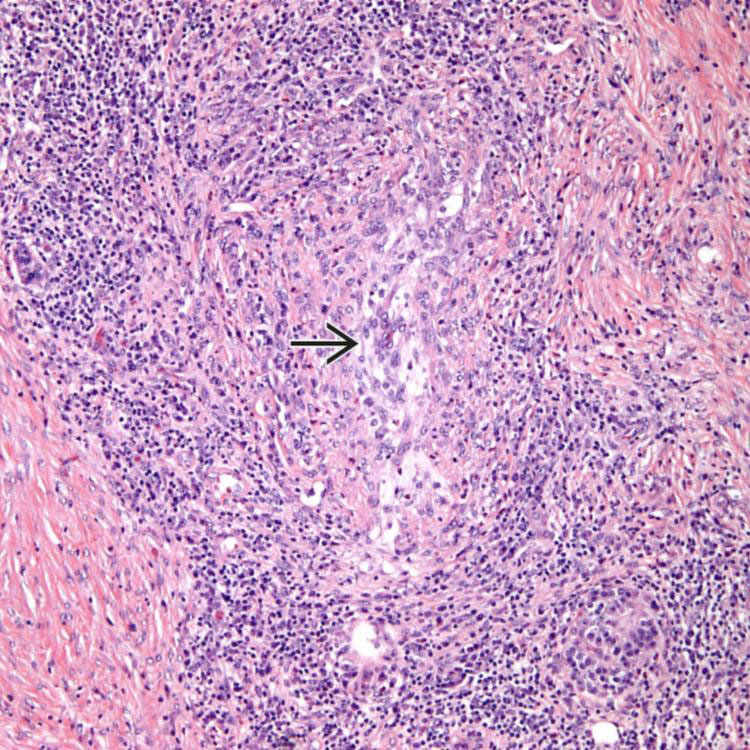
 has been infiltrated by inflammatory cells, with resultant edema and destruction of the wall of the vessel. Note the surrounding fibrosis.