Appendix at the confluence of the three teniae. (With permission from Fischer JE, Bland KI, Callery MP, et al., eds. Mastery of Surgery. 5th ed. Philadelphia, PA: Lippincott Williams & Wilkins; 2006.)
The Appendix
•Arises at the confluence of the three teniae
•Following the anterior tenia is a useful technique to find the appendix in a difficult operation
•Symptoms of appendicitis are related to the position of the appendix (anterior, retrocecal, pelvic, retroileal)
•The blood supply is from the appendicular artery, a branch of the ileocolic artery (from the superior mesenteric artery)
•Innervated by the autonomic nervous system
•The early pain of appendicitis is visceral or vague periumbilical pain
•The focused RLQ pain occurs only after the appendiceal inflammation irritates the parietal peritoneum and activates the somatic pain fibers
•Luminal obstruction is the inciting event and is most commonly due to lymphoid hyperplasia or a fecalith
•Tumors can also cause luminal obstruction (incidence of tumors is 0.1% to 1.0%)
•Obstruction leads to bacterial overgrowth and compromise of lymphatic, venous, and arterial flow, which result in ischemia, necrosis, and perforation
The most common location for a perforation is the antimesenteric border of the middle third of the appendix because it has the poorest blood supply.
A 24-year-old woman presents with a one-day history of new periumbilical pain that radiates to the right lower quadrant. A diagnosis of appendicitis is confirmed by ultrasound (US). What is the likelihood that she will also have leukocytosis?
Approximately 90% of patients with acute appendicitis have a WBC greater than 10,000.

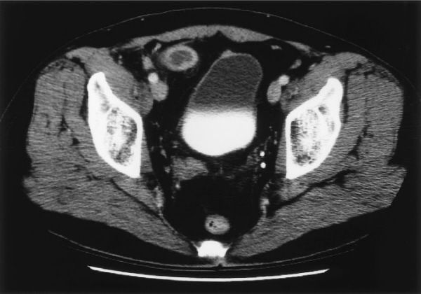
Acute appendicitis seen on computed tomography (CT) scan. (With permission from Mulholland MW, Lillemoe KD, Doherty GM, Maier RV, Upchurch GR, eds. Greenfield’s Surgery. 4th ed. Philadelphia, PA: Lippincott Williams & Wilkins; 2005.)
Acute Appendicitis
•The “classic” presentation is periumbilical pain radiating to the right lower quadrant accompanied by:
•Anorexia (usually first)
•Abdominal pain (usually precedes nausea)
•Low-grade fever and nausea are present in only 50% of patients
•Symptom duration of greater than 24 to 36 hours is uncommon in non-perforated appendicitis
•Menstrual and sexual histories as well as pelvic and rectal exams are imperative
•The most common finding on physical exam is focal abdominal tenderness with signs of peritoneal irritation
•Common signs of appendicitis include:
•Dunphy: Increased pain with coughing
•Rovsing: Left lower quadrant palpation induces right lower quadrant pain
•Obturator: Pain on internal rotation of the right hip
•Iliopsoas: Pain on extension of the right hip
•Urinalysis often reveals minimal white cells, red cells, and bacteria
•β-HCG must be checked in female patients to rule out an ectopic pregnancy
A WBC greater than 20,000 suggests a perforation, which is also true for the workup of many other gastrointestinal diseases.
A 32-year-old woman with suspected acute appendicitis has an abdominal CT. What findings on CT are consistent with a diagnosis of appendicitis?
CT findings consistent with appendicitis are appendiceal diameter of >6 mm, right lower quadrant fat stranding, lack of filling of the appendix with contrast, and a fecalith (non-specific).
Ultrasonography and Computed Tomography in the Evaluation of Acute Appendicitis
•A clinical diagnosis of acute appendicitis can obviate the need for any imaging workup, particularly in a male patient
•US is useful but operator-dependent
•Sonographic criteria for appendicitis include a non-compressible tubular structure, diameter >6 mm, and presence of a fecalith or complex mass
•US (transabdominal and transvaginal) is particularly useful in women of menstrual age to evaluate other pathologic conditions
•Non-visualization of the appendix does not rule out appendicitis
•CT is less operator-dependent
•Oral and/or rectal contrast can improve the yield
•Criteria for appendicitis include a thickened appendix >6 mm in diameter, wall thickness >1.5 mm, periappendiceal inflammation, and the presence of a phlegmon, fecalith, or abscess
•A CT scan can also suggest conditions other than appendicitis
A 79-year-old man presents with a recent history of right lower quadrant pain over McBurney’s point, which is now generalized. He is noted to have temperature of 101.8°F and mild tachycardia. His CBC reveals a WBC of 28,000, a hematocrit of 22%, and a platelet count of 285,000. What is the most likely diagnosis?
Perforated cecal cancer can present with anemia and signs and symptoms of appendicitis.
Colon Perforation
•Signs and symptoms of appendicitis with concomitant anemia suggest colon cancer
•A history of diverticular symptoms suggests perforated diverticulitis
•Resection of the involved segment of colon is the standard management
•An ostomy may need to be created
Perforated cecal cancer should be suspected in any elderly patient with anemia and signs of appendicitis.
A 24-year-old man is diagnosed in the emergency department to have acute appendicitis. What is the next step in management?
Stay updated, free articles. Join our Telegram channel

Full access? Get Clinical Tree


