Slipped capital femoral epiphysis.
A mother notices her 6-year-old son ambulating with a limp. He occasionally complains of hip pain and will rest to relieve the pain. Radiographs show flattening of the femoral head. What is the most likely diagnosis?
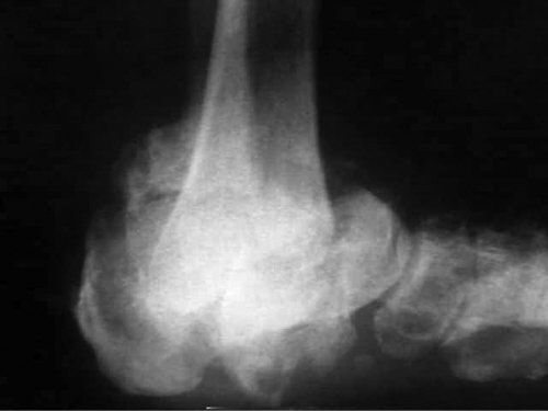
Leg-Calve-Perthes disease commonly occurs in boys 4 to 8 years of age.
Leg-Calve-Perthes Disease
•Avascular necrosis of the proximal femoral epiphysis
•Acute onset of hip pain with a limp and loss of hip motion
•X-ray shows flattening of the femoral head
•Easily confused with septic arthritis
•Treatment is maintenance of range of motion with limited exercise
•Femoral head will remodel without sequelae most of the time
A 14-year-old male soccer player presents with anterior knee pain. On physical exam, he is tender over his tibial tuberosity. What is the most likely diagnosis?
Osgood-Schlatter disease is one of the most common causes of knee pain in the adolescent and is caused by traction apophysitis of the tibial tuberosity.
Osgood-Schlatter Disease
•Tibial tubercle apophysitis caused by traction injury from patellar tendon
•Symptoms: Pain in front of knee
•Diagnosis: X-ray with irregular shape or fragmenting of the tibial tubercle
•Treatment
•Mild symptoms: Activity limitation
•Severe symptoms: Cast for 6 weeks followed by activity limitation
A 49-year-old man with poorly controlled diabetes presents with painful flatfoot in his right lower extremity. X-rays demonstrate the classic “bag of bones” appearance indicating multiple unhealed fractures. What is the most likely diagnosis?
A Charcot foot is a common problem among diabetics and is caused by the associated neuropathy.
Charcot Joint
•Joint destruction caused by an inability to sense the required distribution of weight at the affected joint
•Associated with underlying neurologic disorders
•Diabetes is the most common cause of neuropathy
Charcot ankle.
An 89-year-old female trips and falls over her bed at home. She presents to the emergency room with a laceration on her eye, questionable loss of consciousness, and a flexed and externally rotated right leg. Further workup reveals a negative head CT scan and a right femoral neck fracture. What is the appropriate thromboprophylaxis for this patient?
Low molecular weight heparin or low-dose unfractionated heparin.
Deep Vein Thrombosis
•50% of deep vein thrombosis have been prevented by the effective use of thromboprophylaxis
•If surgery is to be delayed, preoperative anticoagulation is recommended
•Either low-dose molecular weight heparin (LMWH) or unfractionated heparin can be used
•Postoperative prophylaxis should be initiated with fondaparinux, LMWH, warfarin, or low-dose unfractionated heparin for 10 to 35 days after surgery
•Aspirin alone is not considered effective prophylaxis
What muscles make up the rotator cuff?
The acronym SITS describes the muscles.
Rotator Cuff Muscles
•S: Supraspinatus
•I: Infraspinatus
•T: Teres minor
•S: Subscapularis
A 31-year-old mother of two children notices her right hand falls asleep whenever she is performing any overhead activities. EMG/NCV studies are normal. What are two tests to diagnose thoracic outlet syndrome (TOS)?
An Adson maneuver or a Wright test may reproduce symptoms of thoracic outlet syndrome.
Tests for Thoracic Outlet Syndrome
•Adson maneuver
•Elevation of first rib by placing shoulder in slight abduction and extension
•Contraction of the scalene muscles and rotation of the head to the affected side to stretch/extend the neck
•When the patient inhales while in this position, diminished pulse or reproduction of symptoms indicates presence of TOS
•Wright test
•External rotation, abduction, and extension of arm with the neck rotated away causes a diminished pulse and reproduction of symptoms
What ligaments are disrupted in an acromioclavicular separation?
The coracoclavicular and acromioclavicular ligaments.
•The most common site is the middle third of the clavicle
•Treatment is a sling and gentle range of motion (as pain allows)
•Main risk with this fracture is vascular impingement
•97% heal without any other intervention
•If <2 cm of shortening or compromise of overlying skin, recommend surgical treatment
Proximal Humerus Fractures
•Most commonly located at the surgical neck
•Treatment is closed reduction and immobilization
•An anatomic neck fracture (above the tuberosities) is associated with avascular necrosis
What tendon is involved in “tennis elbow” (lateral epicondylitis)?
The extensor carpi radialis brevis.
A 32-year-old man develops wrist pain after falling on his outstretched hand. He has tenderness over the anatomic snuffbox. What is the most likely diagnosis?
A scaphoid fracture is classically associated with pain in the anatomic snuff box of the wrist.
Scaphoid Fracture
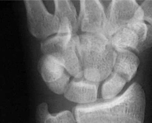
Scaphoid fracture.
•May not be detected on initial X-rays
•A bone scan or MRI may be necessary for diagnosis
•Treatment is with a long arm thumb “spica” cast
•Repeat an X-ray in 3 weeks
•Fracture of the distal radius
•Usually happens after a fall on an outstretched hand
•Treatment: Closed reduction
Scaphoid fractures are associated with a high incidence of avascular necrosis because the blood supply enters the scaphoid distally.
A 12-year-old girl falls off a swing set onto her right arm. X-rays show a fracture of her proximal ulna. What should also be noted on the X-ray?
A dislocated radial head is associated with a proximal ulnar fracture.
Monteggia Fracture
•A well-described fracture of the proximal ulna with radial head dislocation
•Caused by a fall on an outstretched hand
•Treatment: Open reduction internal fixation
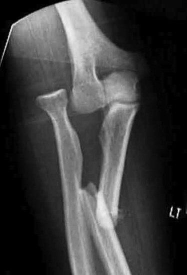
Monteggia fracture.
What is the most common cause of failure of a carpal tunnel release?
An incomplete release.
Carpal Tunnel Syndrome
•Entrapment of the median nerve at the wrist (carpal canal)
•Usually idiopathic but can be caused by a distal radius fracture
•Positive Tinel sign and positive Phalen maneuver
•Initial treatment with splinting, NSAIDs, and possible carpal tunnel injection
Median Nerve Innervation
•First and second lumbricals
•Thenar muscles except ulnar head of the flexor digitorum brevis (FDB)
•Skin of palmar 3½ fingers
Ulnar Nerve Innervation
•Hypothenar muscles
•All interossei muscles
•Third and fourth lumbricals and ulnar FDB
•Sensory to all of fifth finger, ½ of fourth finger, and dorsal hand
Radial Nerve Innervation
•Wrist extension, finger extension, thumb extension, triceps (no hand muscles)
•Sensory to first 3½ fingers on dorsal side
•Saturday night palsy
•Wrist drop, decreased sensation of dorsum of hand, and posterolateral forearm
•Due to laying with arm over a chair for an extended period
A 35-year-old laborer notes that upon waking from sleep his ring finger is locked in flexion. He also notes a tender nodule at the base of his ring finger. What is the most likely diagnosis?
He has a trigger finger.
Stenosing Tenosynovitis (“Trigger finger”)
•Caused by the passage of a swollen flexor tendon sheath beneath the A1 pulley
•Finger can lock in either flexion or extension
•Conservative treatment is with NSAIDs and injection
•If conservative treatment fails, then release the A1 pulley
A 50-year-old secretary is having difficulty in opening jars due to wrist pain. She has tenderness over her radial wrist. What test is used to diagnose de Quervain’s disease?
A Finkelstein test is performed by placing the thumb in a fist and deviating the wrist to the ulnar side. This test exacerbates the tenosynovitis of the first dorsal compartment of the hand (abductor pollicis longus and extensor pollicis brevis).
Stay updated, free articles. Join our Telegram channel

Full access? Get Clinical Tree


