and John E. Skandalakis1
(1)
Centers for Surgical Anatomy and Technique, Emory University School of Medicine Piedmont Hospital, Atlanta, GA, USA
Abstract
Five typical locations of the appendix, in order of frequency, are retrocecal-retrocolic, free or fixed; pelvic or descending; subcecal, passing downward and to the right; ileocecal, passing upward and to the left, anterior to the ileum; and ileocecal, posterior to the ileum. Congenital absence of the appendix is too rare to be considered seriously, but an apparent absence may be the result of intussusception.
The incision for open appendectomy is usually made over McBurney’s point. To expose a deeply buried retrocecal appendix, it may be necessary to incise the posterior peritoneum lateral to the cecum. During laparoscopic appendectomy, the patient should have an orogastric tube placed to decompress the stomach. Use of a Foley catheter for bladder decompression helps to avert iatrogenic trocar injury to the bladder.
Anatomy
Relations and Positions of the Appendix
The appendix arises from the posteromedial side of the cecum about 1.7 cm from the end of the ileum. The cecum is related posteriorly to the iliopsoas muscle and the lumbar plexus of nerves. Anteriorly it is related to the abdominal wall, the greater omentum, or coils of ileum. The base of the appendix is located at the union of the teniae. For all practical purposes, the anterior tenia ends at the appendiceal origin.
Five typical locations of the appendix, in order of frequency, are:
Retrocecal-retrocolic, free or fixed
Pelvic or descending
Subcecal, passing downward and to the right
Ileocecal, passing upward and to the left, anterior to the ileum
Ileocecal, posterior to the ileum
Studies have found that the first two positions are the most common, but with significant variations.
Mesentery
The mesentery of the appendix is embryologically derived from the posterior side of the mesentery of the terminal ileum. The mesentery attaches to the cecum as well as to the proximal appendix. It contains the appendicular artery.
Vascular System of the Appendix
Arterial Supply
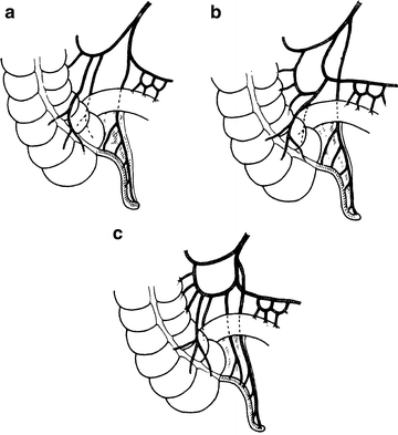
The appendicular artery arises from the ileocolic artery, an ileal branch, or from a cecal artery. Although the appendicular artery is usually singular (Fig. 11.1), duplication is often seen. In addition to the typical appendicular artery, the base of the appendix may be supplied by a small branch of the anterior or posterior cecal artery.


Figure 11.1.
Blood supply to the appendix. (a, b) Usual type with a single appendicular artery. (c) Paired appendicular arteries (By permission of JE Skandalakis, SW Gray, and JR Rowe. Anatomical Complications in General Surgery. New York: McGraw-Hill, 1983).
Venous Supply
The appendicular artery and the appendicular vein are enveloped by the mesentery of the appendix. The vein joins the cecal veins to become the ileocolic vein, which is a tributary of the right colic vein.
Lymphatic Drainage
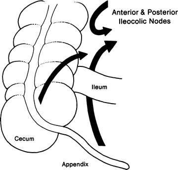
Lymphatic drainage from the ileocecal region is through a chain of nodes on the appendicular, ileocolic, and superior mesenteric arteries through which the lymph passes to reach the celiac lymph nodes and the cisterna chyli (Fig. 11.2). Some studies describe a secondary drainage (which passes anterior to the pancreas) to subpyloric nodes.


Figure 11.2.
Lymphatic drainage of the appendix (By permission of JE Skandalakis, SW Gray, and JR Rowe. Anatomical Complications in General Surgery. New York: McGraw-Hill, 1983).
Technique
Appendectomy
The incision for appendectomy is usually made over McBurney’s point. It is made at right angles to a line between the anterior superior iliac spine and the umbilicus at 2/3 the distance from the umbilicus; 1/3 of the incision should be above the line, and 2/3 should be below.
Stay updated, free articles. Join our Telegram channel

Full access? Get Clinical Tree


