Antifungal Drugs


Antifungal Drugs


Overview


Fungal Infections


Fungal infections can be divided into three groups: systemic mycoses, subcutaneous mycoses, and superficial mycoses.


Systemic mycoses can cause signs and symptoms of soft tissue infection, urinary tract infection, pneumonia, meningitis, or septicemia. The diseases can be chronic and indolent or invasive and life-threatening. The systemic mycoses are most commonly caused by members of the genera Aspergillus, Blastomyces, Candida, Coccidioides, Cryptococcus, and Histoplasma. Some infections (e.g., blastomycosis, coccidioidomycosis, and histoplasmosis) that are endemic to certain geographic regions are found in both immunocompetent and immunocompromised individuals. Other infections (e.g., aspergillosis, candidiasis, cryptococcosis, and mucormycosis) are more likely to occur in immunocompromised or debilitated patients, such as those receiving immunosuppressive drugs, those with indwelling catheters or prostheses, or those with human immunodeficiency virus (HIV) infection, diabetes, or chronic renal, hepatic, or cardiac diseases. These conditions either suppress cellular immunity or facilitate colonization and infection by fungi. For example, there has been an increased incidence of invasive infections caused by Aspergillus, Scedosporium, and Fusarium species in recipients of hematopoietic stem cell transplants in recent decades.


Subcutaneous mycoses are often caused by puncture wounds contaminated with soil fungi. Examples of these infections are chromomycosis, pseudallescheriasis, and sporotrichosis.


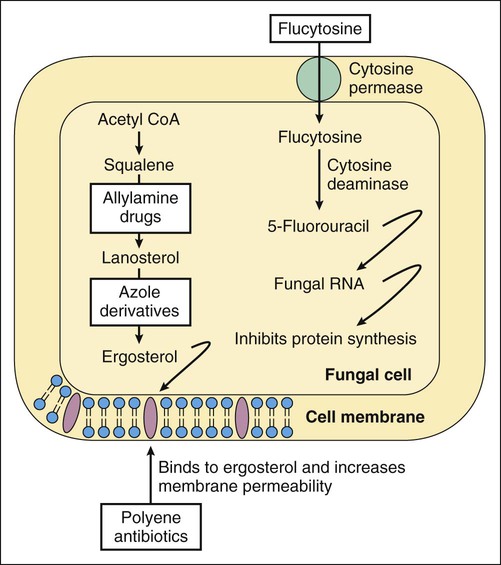
Superficial mycoses are infections of the nails, skin, and mucous membranes, and are usually caused by dermatophytes or yeasts. The most common dermatophytes are Epidermophyton, Microsporum, and Trichophyton species. Dermatophyte infections of the nails are referred to as tinea unguium or onychomycosis. Other dermatophyte infections include tinea pedis (athlete’s foot), tinea capitis (ringworm of the scalp; Box 42-1), tinea corporis (ringworm of the body), and tinea cruris (jock itch). These infections usually manifest as a rash with pruritus (itching) and erythema. Ringworm is described as an annular (ring-shaped), scaling rash with a clear center.


Box 42-1   A Case of Flaking Skin and Hair Loss


Case Discussion


Tinea capitis is a dermatophyte infection of skin and hair on the scalp that is most common in prepubertal children, with the highest frequency at 3 to 7 years of age. Trichophyton tonsurans has been the most common fungus responsible for this infection in North America for several decades, but numerous other dermatophytes may cause the infection, including species that are primarily transmitted by humans (T. tonsurans and Trichophyton violaceum) or animals (Microsporum canis). The clinical presentation is variable, depending on the causative agent and other factors, but the diagnosis can be established by microscopic examination of skin and hair samples mixed with potassium hydroxide. Fungal culture is the most reliable diagnostic method, but results take several weeks to obtain. Griseofulvin is the standard therapy for tinea capitis, but higher doses must be used for successful treatment—for example, 10 to 15 mg of ultramicrosized griseofulvin per kilogram per day or 20 to 25 mg of the microsized form per kilogram per day for 6 to 8 weeks. Infections that do not respond to griseofulvin may be treated with fluconazole, itraconazole, or terbinafine.


The most common yeasts causing superficial mycoses are Candida albicans and other Candida species. Affected patients may have thrush (oral candidiasis), vaginal candidiasis, or Candida infections of the axilla, groin, and gluteal folds (including diaper rash in infants). Less common yeasts causing superficial mycoses include Malassezia furfur (also called Pityrosporum orbiculare) and Malassezia ovalis (also called Pityrosporum ovale). M. furfur causes tinea versicolor or pityriasis versicolor, a skin infection characterized by hypopigmented and hyperpigmented macules, typically in the shoulder girdle area. P. ovale and M. furfur cause seborrheic dermatitis, characterized by scaling and erythema on the ears, eyebrows, nose, and chest.


Clinical Uses and Mechanisms of Antifungal Drugs
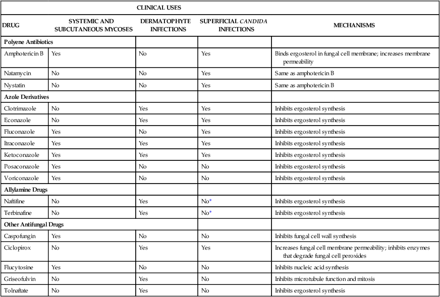

Fungi are eukaryotic organisms whose growth is not inhibited by antibacterial or antiviral drugs. Drugs that are selectively toxic to fungi have been discovered, however, and they are used to treat fungal infections in humans and animals.


As shown in Table 42-1, drugs used in the treatment of systemic and subcutaneous mycoses include a polyene antibiotic (amphotericin B), several azole derivatives (fluconazole, itraconazole, ketoconazole, and voriconazole), an echinocandin drug (caspofungin), and flucytosine. The other drugs listed are used in the treatment of superficial mycoses. Amphotericin B tends to be used for treating severe mycoses, whereas the azoles are used for less severe infections. Newer antifungal agents (e.g., voriconazole and caspofungin) can be used to treat invasive Candida and Aspergillus infections. Flucytosine is usually administered in combination with amphotericin B for the treatment of systemic Cryptococcus or Candida infections.


Many antifungal drugs act by impairing plasma membrane function in fungal cells. The selective toxicity of these drugs is a result of the difference in the sterols found in fungal and mammalian cell membranes. Fungal cell membranes contain ergosterol, whereas mammalian cell membranes contain cholesterol. Some antifungal drugs bind to ergosterol and thereby increase plasma membrane permeability, whereas other drugs inhibit the synthesis of ergosterol (Fig. 42-1; see Table 42-1).


Polyene antibiotics selectively bind to ergosterol in fungal membranes. This action increases fungal plasma membrane permeability and allows the cytoplasmic contents to escape from the cell. The polyene drugs can also bind to cholesterol in mammalian cells, and this may account for their ability to damage renal cell membranes and cause toxicity.


The mechanism of action of ciclopirox is uncertain. Some studies found that it increases fungal cell membrane permeability by inhibiting amino acid transport into fungal cells and altering membrane structure. Another study suggested that it chelates polyvalent cations (Fe3+, Al3+) and thereby inhibits metal-dependent enzymes responsible for degradation of peroxides in fungal cells.


The allylamine drugs and the azole derivatives block distinct steps in ergosterol biosynthesis, but these groups of drugs have little effect on cholesterol biosynthesis in humans. Allylamine drugs such as terbinafine inhibit squalene epoxidase, which converts squalene to squalene-2,3-oxide, the immediate precursor of lanosterol. The azoles such as fluconazole inhibit 14α-demethylase, a cytochrome P450 enzyme that converts lanosterol to ergosterol.


The echinocandin drugs (e.g., caspofungin) represent a new class of antifungal agents that inhibit the synthesis of a fungal cell wall component, β-(1,3)-D-glucan. The fungal cell wall surrounds the plasma membrane and normally protects the cell from osmotic and mechanical stress.


Flucytosine, a pyrimidine antimetabolite, is the only antifungal drug that affects nucleic acid. Flucytosine is converted to 5-fluorouracil (5-FU) in fungal cells by cytosine deaminase, an enzyme not found in mammalian cells. 5-FU is then incorporated into fungal RNA, and this inhibits fungal protein synthesis.


Griseofulvin acts by binding to fungal microtubules and thereby inhibiting microtubule function and mitosis. The mechanism of action of tolnaftate is uncertain, but it appears to inhibit squalene epoxidase in a manner similar to allylamine drugs such as terbinafine.

< div class='tao-gold-member'>

Stay updated, free articles. Join our Telegram channel

Jul 23, 2016 | Posted by in PHARMACY | Comments Off on Antifungal Drugs

Full access? Get Clinical Tree

Get Clinical Tree app for offline access