32
Anterolateral Abdominal Wall and Groin
The abdomen is the part of the trunk that lies between the diaphragm and pelvis. The abdominal and pelvic cavities are continuous, and the abdominal cavity is lined by a serous membrane (peritoneum, see Chapters 33 and 34) and contains most of the organs of the digestive system.
From superficial to deep, the layers covering the anterior and lateral aspects of the abdomen are skin, subcutaneous tissue, muscles or fascia, extraperitoneal tissue, and peritoneum (Table 32.1). This multilayered musculofascial organization contributes to the anatomy of the groin. During embryologic descent of the male gonads, these layers are drawn inferiorly and contribute to formation of the spermatic cord and scrotum.
TABLE 32.1 Anterolateral Abdominal Wall—Layers and Derivatives
Anterior Abdominal Wall Layers | Scrotal Layers | Inguinal Canal |
1 Skin | Skin |
|
2 Superficial fascia | Dartos fascia and muscle |
|
3 External abdominal oblique muscle/aponeurosis | External spermatic fascia | Superficial inguinal ring, anterior wall, floor—inguinal and lacunar ligaments |
4 Internal abdominal oblique muscle/aponeurosis | Cremaster muscle | Roof and posterior wall via conjoint tendon |
5 Transversus abdominis muscle | Cremaster muscle |
|
6 Transversalis fascia | Internal spermatic fascia | Deep inguinal ring and posterior wall |
7 Extraperitoneal connective tissue/fat | Fat |
|
8 Peritoneum | Tunica vaginalis |
|
Structural support for the abdominal region is provided superiorly by the inferior margin of the rib cage, anteriorly by the muscles of the anterior abdominal wall, inferiorly by the pelvic girdle and pelvic diaphragm (see Chapter 36), and posteriorly by the vertebral column, ribs, and back muscles.
Muscles
The anterior abdominal wall contains the rectus abdominis muscle, which is a long muscle in the midline that flexes the trunk and extends from the inferior rib cage to the pelvic inlet. The rectus abdominis is covered by several layers of fascia derived from three anterolateral abdominal muscles (Fig. 32.1).
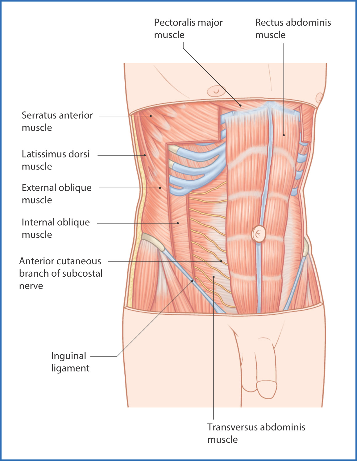
FIGURE 32.1 Anterior abdominal wall muscles.
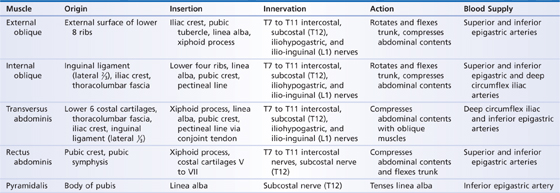
The anterolateral abdominal wall is composed of three sheet-like muscles (Table 32.2): an outer external oblique, a middle internal oblique, and an inner transversus abdominis muscle (see Fig. 32.1). Anteriorly, these muscles become aponeurotic, fuse, and form a sheath around the rectus abdominis muscle. They rotate and flex the trunk anteriorly and laterally and assist in expiration, coughing, vomiting, and defecation.
TABLE 32.2 Muscles of the Anterolateral Abdominal Wall
The rectus sheath (Table 32.3), formed by the anterolateral abdominal muscle aponeurosis, contains the superior and inferior epigastric vessels, the segmental thoraco-abdominal nerves (T7 to T11), the subcostal (T12) nerves, and lymphatic vessels.
TABLE 32.3 Components of the Rectus Sheath
Level | Anterior Wall | Posterior Wall |
Above costal margin | Aponeurosis of external oblique muscle | Costal cartilages |
Costal margin to lower | Aponeurosis of external oblique muscle, anterior lamina of internal oblique muscle |
 of sheath


