46
Ankle and Foot Joints
The ankle joint is a freely moving hinge joint between the leg and the foot. It is formed by the lower ends of the tibia and fibula and the trochlea of the talus. The tibia and the fibula form a socket that is wider anteriorly than posteriorly, and the talus moves in the socket during dorsiflexion and plantar flexion of the ankle. The joint capsule of the ankle is thickened on each side by ligaments (Figs. 46.1 and 46.2).
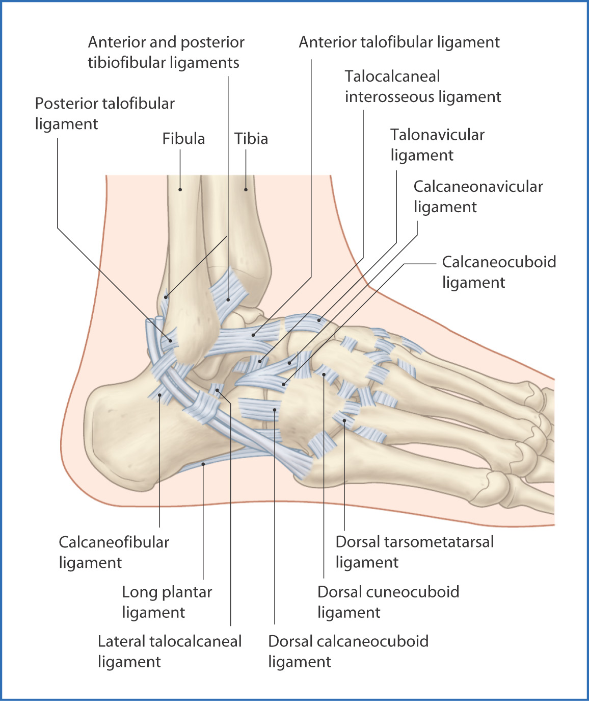
FIGURE 46.1 Lateral view of the ankle ligaments.
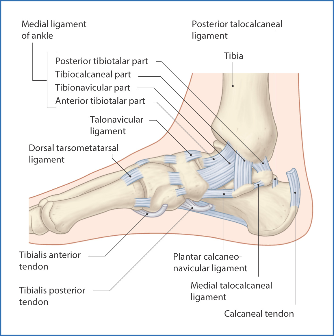
FIGURE 46.2 Medial view of the ankle ligaments.
The foot consists of individual bones supported by ligaments and muscles (Fig. 46.3). All joints between the bones in the foot are synovial. Together, the bones and joints create an arch that supports the body’s weight, serves as a shock absorber, and acts as a lever to propel the body forward.
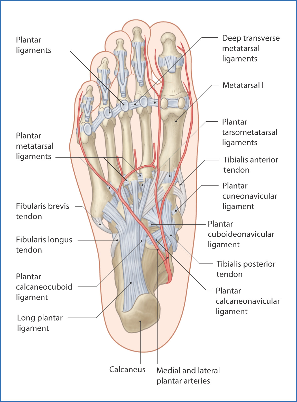
FIGURE 46.3 Plantar surface showing the ligaments and arterial supply.
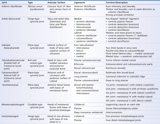
The most important joints in the foot are the subtalar, talocalcaneonavicular, and calcaneocuboid joints. The last two form the transverse tarsal joint. The subtalar joint is where the talus rests on the calcaneus and where most of the inversion and eversion movements of the foot occur. It has a joint capsule reinforced by ligaments. The remaining joints are listed in Table 46.1.
TABLE 46.1 Joints of the Ankle and Foot
Ligaments and Fascia
Because of the angle at the junction of the leg and foot, the tendons of the extrinsic foot muscles need to be bound down by fascial bands (retinacula). On the lateral side of the ankle, the fibularis muscle tendons are kept in place by the superior fibular retinaculum, which joins the lateral malleolus to the calcaneus. The inferior fibular retinaculum between the dorsum of the foot and the lateral aspect of the calcaneus prevents displacement of these tendons.
On the medial side of the ankle, the flexor retinaculum (between the medial malleolus and the calcaneus, plantar aponeurosis, and adjacent bony prominences of the foot) helps maintain the position of the flexor tendons from the posterior compartment of the leg.
On the front of the ankle, two extensor retinacula prevent the extensor muscle tendons from bowing during extension (dorsiflexion) of the foot and toes. The superior extensor retinaculum is attached to the anterior borders of the fibula and tibia. The Y-shaped inferior extensor retinaculum has its lateral base on the upper part of the calcaneus, and its arms are attached to the medial malleolus and the medial side of the plantar aponeurosis.
The lateral ligament is composed of three separate bands that arise from the lateral malleolus and insert onto the neck of the talus (anterior talofibular ligament), the calcaneus (calcaneofibular ligament), and the lateral tubercle of the talus (posterior talofibular ligament). The lateral ligament is weaker than the medial ligament and prevents inversion of the foot.
The foot has numerous dorsal and plantar ligaments (Figs. 46.3 and 46.4), but three ligaments maintain the important longitudinal arch of the foot:
- The plantar calcaneonavicular ligament (spring ligament) connects the sustentaculum tali of the calcaneus to the navicular bone and strengthens the longitudinal arch of the foot.
- The long plantar ligament extends from the calcaneus to the tuberosity of the cuboid bone—some fibers extend to the bases of the metatarsals and form a tunnel for the tendon of the fibularis longus muscle.
- The plantar calcaneocuboid ligament (short plantar ligament) is deep to the long plantar ligament and extends from the calcaneus to the inferior surface of the cuboid.
- The long plantar ligament extends from the calcaneus to the tuberosity of the cuboid bone—some fibers extend to the bases of the metatarsals and form a tunnel for the tendon of the fibularis longus muscle.
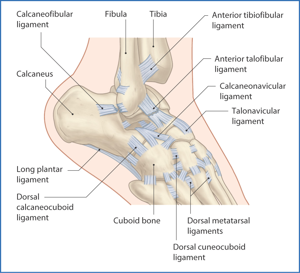
FIGURE 46.4 Dorsal ligaments of the foot.
Structures around the Ankle Joint
The ankle joint has no intrinsic muscles. Instead, muscle tendons from the three compartments of the leg extend across the ankle to the foot. These structures provide additional support to the ankle joint and facilitate movement.
On the medial side of the ankle joint, just posterior to the medial malleolus, are (from anterior to posterior)
- the tendons of the tibialis posterior and flexor digitorum longus muscles
- the posterior tibial vein and artery and the tibial nerve
- the tendon of the flexor hallucis longus muscle
- the calcaneal tendon, which is the most posterior structure at the ankle
- the posterior tibial vein and artery and the tibial nerve
Stay updated, free articles. Join our Telegram channel

Full access? Get Clinical Tree


