Solid, epithelioid, and spindled areas may be present
• Immunopositive for vascular markers: CD31, CD34, factor VIII
• Nuclear staining for transcription factors FLI-1 and ERG helps in confirming endothelial differentiation
Ancillary Tests
• High Ki-67 (> 10%), diffuse p53 and diffuse MYC staining favors angiosarcoma over benign vascular tumors
Top Differential Diagnoses
• Epithelioid hemangioendothelioma
• Carcinoma
• Other sarcomas
• Hepatic small vessel neoplasm
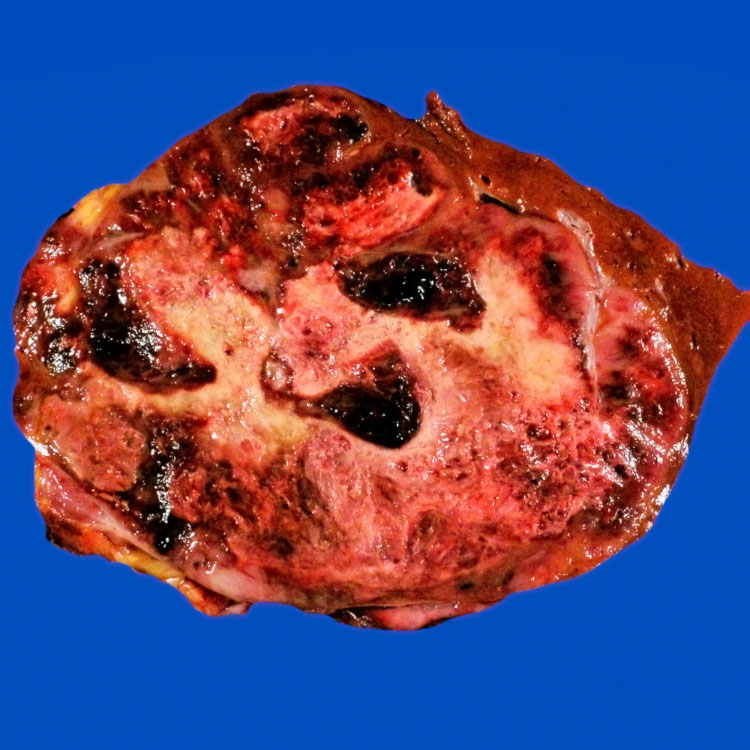
Hemorrhagic Cystic Tumor This cross section from a partial hepatectomy for angiosarcoma shows numerous cystic, blood-filled spaces. [Courtesy C. Trower, PA (ASCP) and A. Folpe, MD.]
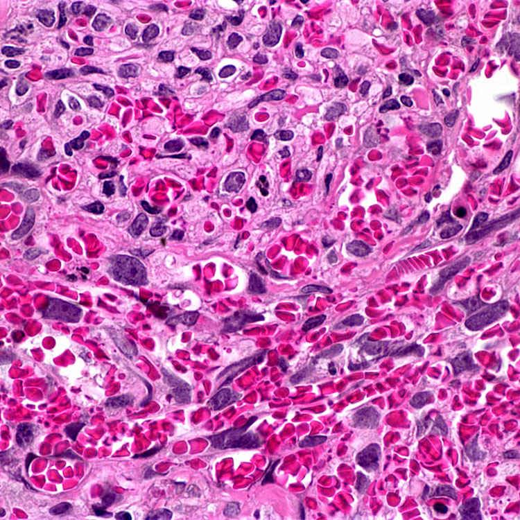
Spindled Tumor Cells This angiosarcoma has a spindled growth pattern. The tumor cells are admixed with red blood cells and forming vascular lumina in some places. The normal hepatic architecture is no longer apparent.
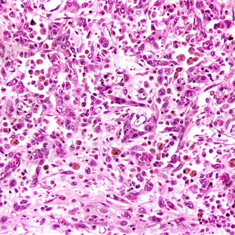
Atypical Epithelioid Cells Markedly atypical neoplastic endothelial cells line poorly formed vascular spaces. Note that the normal hepatic architecture has been destroyed.
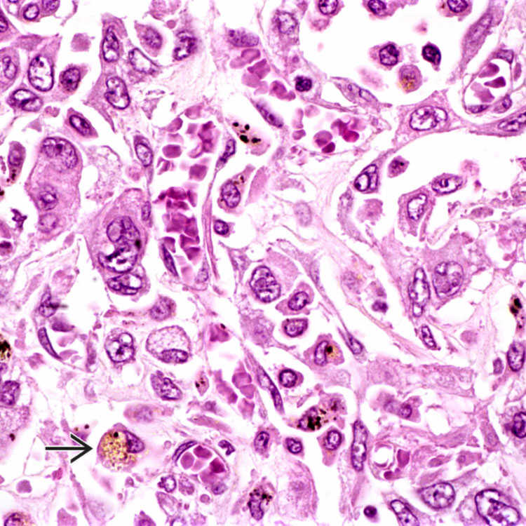
Marked Cytologic Atypia This angiosarcoma shows markedly atypical endothelial cells with enlarged, bizarre nuclei. The tumor cells show scattered vascular differentiation. Note the admixed red blood cells and hemosiderin-laden macrophages .

 .
.


