AJCC T4 Carcinomas (Chest Wall or Skin Involvement)
TERMINOLOGY
Definitions
Carcinomas with skin or chest wall involvement classified as T4 by AJCC/UICC
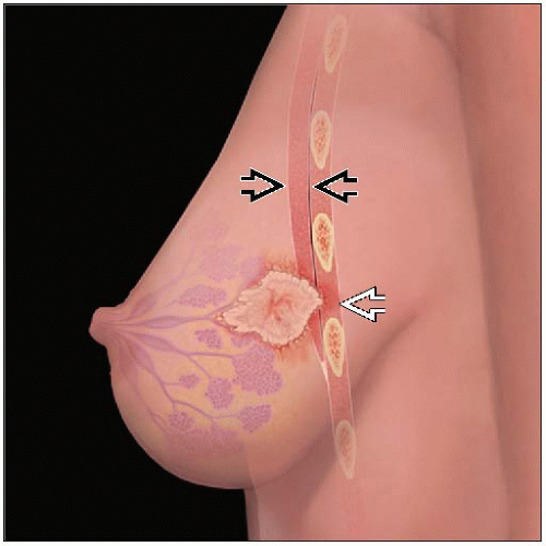
Chest wall involvement (T4a)
Carcinoma invading into skeletal muscle of chest wall beyond pectoralis muscle
Skin involvement (T4b)
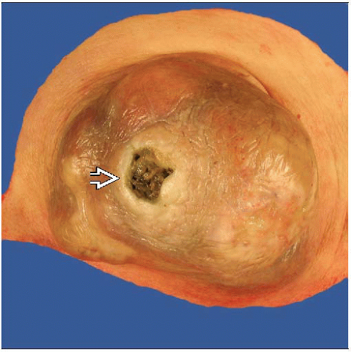
Invasion into skin with ulceration
Satellite skin nodules
Skin edema or peau d’orange (“skin of an orange”): Due to tethering of the skin by Cooper’s ligaments causing pitting of the swollen skin
Inflammatory breast carcinoma (T4d)
Superficial dermal lymphatic involvement with erythema and swelling (peau d’orange)
Classified as T4d by AJCC/UICC
Term “locally advanced breast carcinoma”
Includes all T4 tumors and some large T3 carcinomas (AJCC stages IIIA and IIIB)
ETIOLOGY/PATHOGENESIS
Etiology
Incidence of carcinomas with noninflammatory chest wall and skin invasion (T4a, b, c) increases with age
Delays in diagnosis in older patients may allow carcinomas to reach larger sizes
Do not have biologic features of increased aggressiveness
Incidence of inflammatory carcinomas (T4d) increases to age 50 and then levels off
More likely to be poorly differentiated and hormone receptor negative
Likely have specific biological profile that promotes early lymph-vascular dissemination
EPIDEMIOLOGY
Incidence
Inflammatory carcinomas: 1-5% of breast carcinomas
T4b carcinomas: Approximately 1% of carcinomas
T4a and T4c carcinomas exceedingly rare (< 1%)
Carcinomas with skin involvement but not classified as T4 make up 2-3% of carcinomas
Age Range
T4a, b, c: Mean age is 10 years older than usual for women with breast carcinoma (i.e., 70s rather than 60s)
T4d: Occurs in women at younger mean age (40s to 50s)
CLINICAL IMPLICATIONS
Prognosis
Patients whose prognosis was so poor that surgery was not beneficial were originally classified as having T4 cancers
Majority of these patients had very large bulky local disease
With current chemotherapy and radiotherapy, patients with carcinomas that respond to treatment may become candidates for surgery
Smaller carcinomas with only skin ulceration have better prognosis than other T4 carcinomas
MACROSCOPIC FINDINGS
Specimen Handling
Skin
If skin invasion, carcinoma will be fixed to skin
Take sections of carcinoma closest to skin and any skin ulceration
If satellite skin lesions are present, take sections to demonstrate there is no direct connection with main carcinoma
Chest wall
Deep margin of specimens should be examined to determine if muscle is present
In majority of cases, any muscle present will be pectoralis (chest wall invasion does not include this muscle)
Take sections to demonstrate relationship of carcinoma to muscle
If true chest wall invasion, ribs and serratus muscle will be resected
Bones should be examined for direct invasion
Inflammatory carcinoma
Majority of patients will have received neoadjuvant chemotherapy
May be difficult to determine location and extent as these carcinomas are typically diffusely invasive
If clip marks prior biopsy site, this should be found and sampled
In cases without gross or radiologic correlate, sections taken every cm across specimen, including samples of skin, may be necessary
MICROSCOPIC FINDINGS
T4a: Extension to Chest Wall
Carcinoma must invade into serratus muscle between ribs
Correlation with radiologic findings may be necessary to demonstrate true chest wall invasion
Specimens will typically contain portions of ribs
Ribs should be radiographed and sectioned to determine if carcinoma has invaded into bone
Stay updated, free articles. Join our Telegram channel

Full access? Get Clinical Tree


