Infected hepatocytes may be slightly enlarged and multinucleated

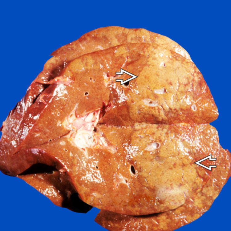
This liver specimen from an autopsy case shows large, irregular, variably sized, yellow-tan foci of necrosis
 .
.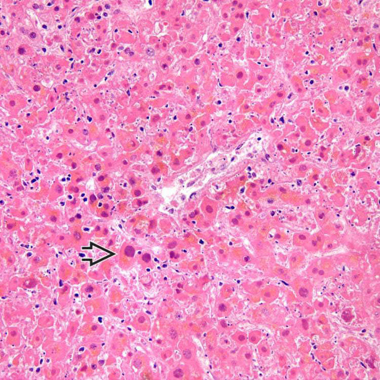
Large zones of necrosis with minimal inflammation, as seen here, are typical of adenovirus infection. Dark, smudgy nuclear inclusions (“smudge cells”) are visible even at low power
 .
.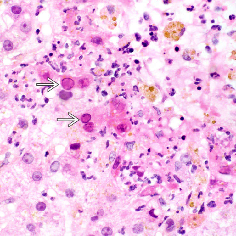
Hyperchromatic, smudgy nuclei, or “smudge cells” with chromatin margination
 are characteristic of adenovirus infection. They are seen here within hepatocytes at the periphery of a necrotic focus. Note that the necrotic hepatocytes are largely dropped out in this case, accompanied by mild neutrophilic infiltrates.
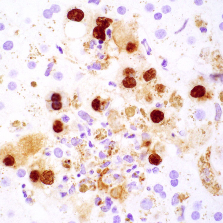
Immunohistochemical stain for adenovirus highlights infected hepatocytes with intense nuclear, and some cytoplasmic, reactivity.
CLINICAL ISSUES
Presentation


• Fulminant hepatitis typically occurs in immunocompromised or transplant patients


Stay updated, free articles. Join our Telegram channel
Full access? Get Clinical Tree
