Acute Pancreatitis

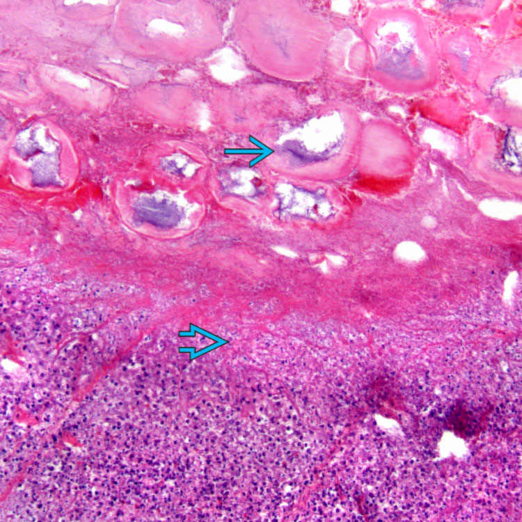
 Large areas of fat necrosis along with variable parenchymal necrosis


image Saponification

image Hemorrhage, vascular thrombosis


• Mild acute pancreatitis is most often clinical (rather than morphologic) diagnosis
image Spotty peripancreatic or perilobular fat necrosis and interstitial acute inflammation


Top Differential Diagnoses


• Chronic pancreatitis
image Fibrosis, chronic inflammation, lacks inflammatory component

• Autoimmune pancreatitis
image Prominent lymphoplasmacytic infiltrate and storiform fibrosis

image
Acute Pancreatitis With Saponification
Pancreatic necrosis image and saponification of peripancreatic fat with calcification image are shown in a patient with acute pancreatitis.


image
Necrosis and Acute Inflammation
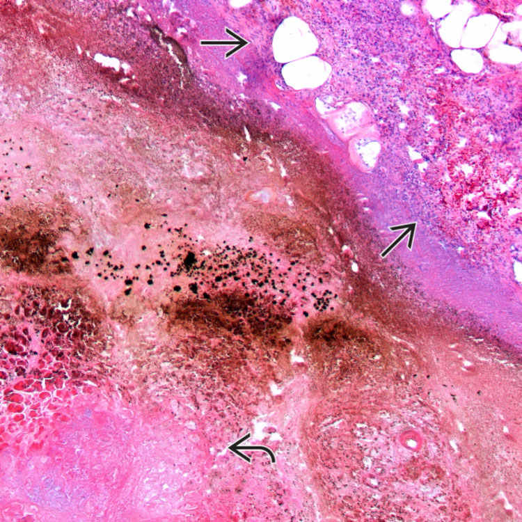
Trypsinogen activation is a key step in the development of acute pancreatitis. The release of digestive enzymes results in fat necrosis with acute inflammation image and pancreatic parenchymal necrosis image .

image
Hemorrhage and Fat Necrosis
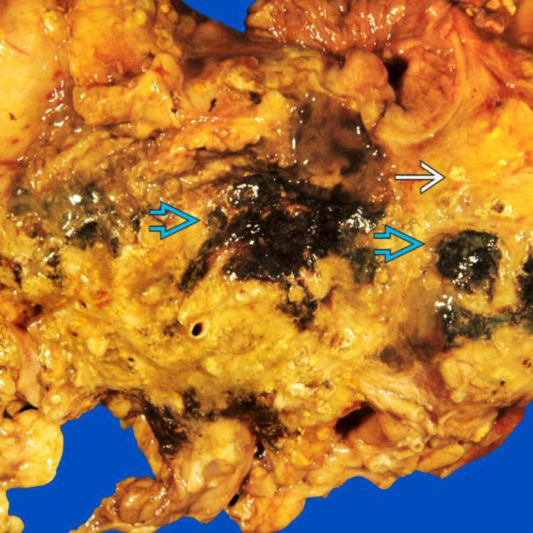
Gross examination reveals chalky white foci of fat necrosis image and black areas of hemorrhage image .

image
Acute Pancreatitis on CT
This CT shows severe acute necrotizing pancreatitis with heterogeneous and diminished enhancement of the pancreas image .


TERMINOLOGY


Definitions


• Acute inflammatory process involving pancreas


ETIOLOGY/PATHOGENESIS


Mechanical


• Gallstones, biliary sludge, periampullary diverticulum, neoplasms, duodenal stricture or obstruction
image Gallstones most common cause, accounting for 35-60% of cases


Toxic


• Ethanol, methanol, scorpion venom, organophosphate poisoning
image Alcohol is 2nd most common cause of acute pancreatitis overall


Trauma


• Blunt or penetrating abdominal injury, iatrogenic injury during procedure

Apr 20, 2017 | Posted by in PATHOLOGY & LABORATORY MEDICINE | Comments Off on Acute Pancreatitis

Full access? Get Clinical Tree

Get Clinical Tree app for offline access