• Lining cells are positive for trypsin &/or lipase
Top Differential Diagnoses
• Serous cystadenoma
Also lined by single layer of cuboidal epithelium
• Retention cyst
Lacks morphological or immunohistochemical evidence of acinar cell differentiation
• Intraductal papillary-mucinous neoplasm and mucinous cystic neoplasm
Neoplastic cysts lined by mucinous epithelium
• Acinar cell cystadenocarcinoma and acinar cell carcinoma
Both lesions show sheets of neoplastic cells with nuclear atypia and many mitoses
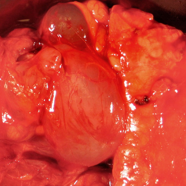
Gross Photo of Multiple Unilocular Cysts This acinar cell cystadenoma shows multiple unilocular, thin-walled cysts. The cysts are filled with clear fluid and lack a mural nodule.
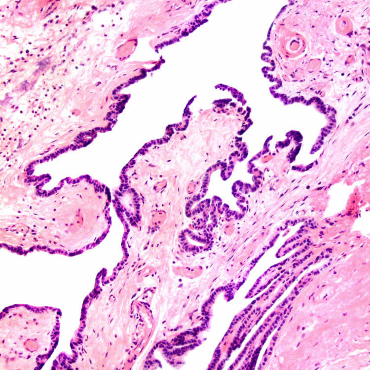
Low Magnification Acinar cell cystadenomas are lined by a single layer of cuboidal epithelium and could easily be mistaken for a serous cystadenomas.
Higher Magnification Incipient acinar structures are seen on this H&E section. Some of the cells lining these structures show apical eosinophilic granules . These granules are PAS positive and diastase resistant and represent zymogen granules.
Immunohistochemical Stain for Trypsin The apical cytoplasmic compartment of the cyst lining cells is positive for trypsin .
TERMINOLOGY
Definitions
• Benign pancreatic cyst lined by cells with acinar cell differentiation
CLINICAL ISSUES
Epidemiology
• Age
Young to middle-aged
Only gold members can continue reading. Log In or Register to continue


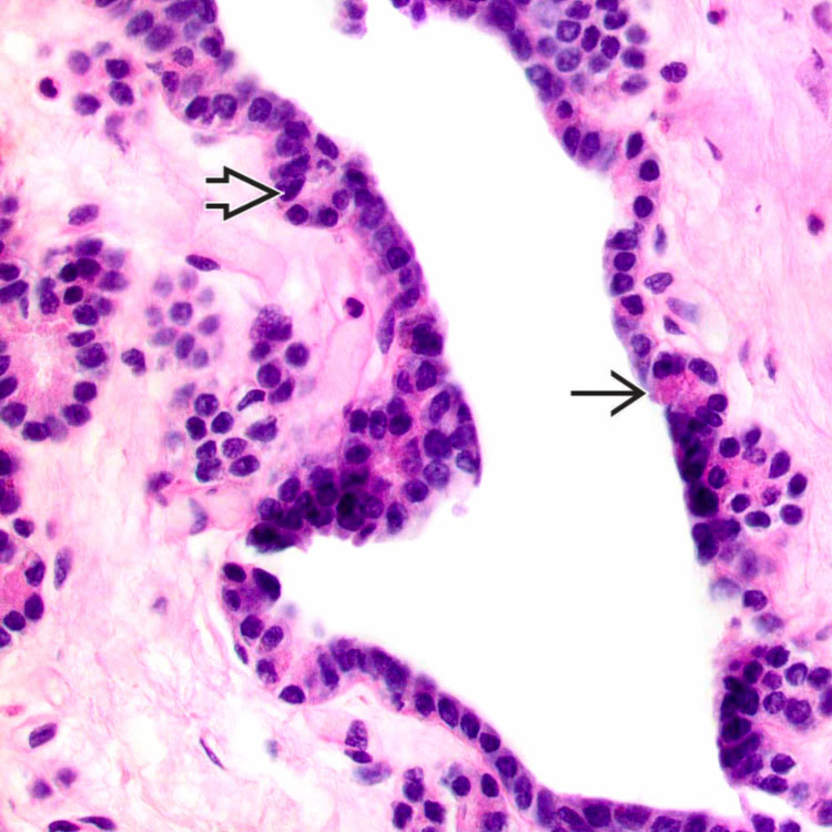
 are seen on this H&E section. Some of the cells lining these structures show apical eosinophilic granules
  . These granules are PAS positive and diastase resistant and represent zymogen granules.
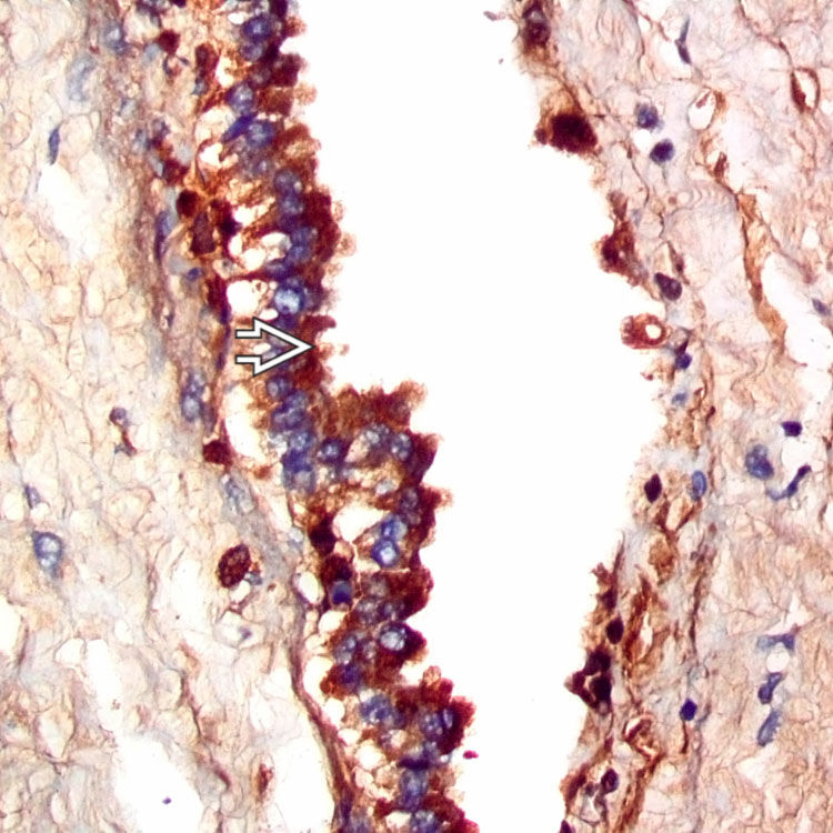
 .
.


