and John E. Skandalakis1
(1)
Centers for Surgical Anatomy and Technique, Emory University School of Medicine Piedmont Hospital, Atlanta, GA, USA
Abstract
The surgical anatomy of the anterior and posterior (lumbar) abdominal wall, umbilical region, and groin is presented in detail. The laparoscopic anatomy of the inguinal area, including its layers, fossae, and spaces, along with an updated section on repair, is of great importance for contemporary surgeons. The principles of proper abdominal incisions and the surgical anatomy of the major types of incisions are illustrated.
The etiology, pathogenesis, evaluation, and operating room strategy for treating incisional hernias of the abdominal wall are enumerated. A new section details component separation. Non-mesh and mesh repairs for various hernias (epigastric, umbilical, spigelian, indirect inguinal, sliding indirect inguinal, direct inguinal, external supravesical, femoral, lumbar) are presented with step-by-step details, as is repair of hydrocele.
Anatomy
General Description of the Anterior Abdominal Wall
The anterior abdominal wall can be thought of as having two parts: anterolateral and midline. The anterolateral portion is composed of the external oblique, internal oblique, and transversus abdominis muscles (often referred to as “the three flat muscles of the anterior abdominal wall”). These muscles are arranged such that their fibers are roughly parallel as they approach their insertion on the rectus sheath.
The midline (middle) portion is composed of the rectus abdominis and pyramidal muscles. The rectus muscle is enclosed in a stout sheath formed by the bilaminar aponeuroses of the three flat muscles, which divide and pass anteriorly and posteriorly around it. The sheath attaches medially to the linea alba (Fig. 4.1), which is formed by decussation. In 10–20 % of the subjects, the pyramidal muscle is absent on one or both sides, but when present its insertion into the linea alba is a landmark for an accurate midline incision.
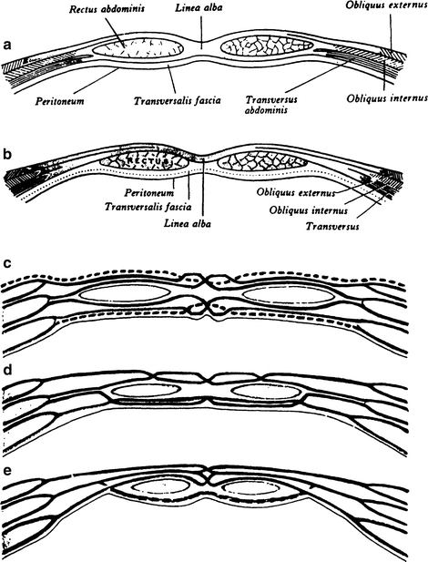
Figure 4.1.
(a) Transverse sections through the anterior abdominal wall, traditional view: immediately above the umbilicus. (b) Below the arcuate line. (c–e) Schematic transverse sections through the ventral abdominal wall showing bilaminar aponeuroses, external oblique, internal oblique, transversus abdominis, and sites of linear decussation that compacted from linea alba (By permission of PL Williams, R Warwick, and M Dyson, et al. (eds.), Gray‘s Anatomy, 37th ed. Edinburgh: Churchill Livingstone, 1989).
The following array shows some comparisons between the structures of the upper three-quarters of the midline portion of the abdominal wall and the lower one-quarter:
Upper midline | Lower midline |
|---|---|
Linea alba well developed | Linea alba poorly developed |
Right and left recti well separated | Right and left recti close together |
Anterior and posterior layers of rectus sheath present | Only anterior layer of rectus sheath present |
Aponeurosis of external oblique weak or absent | Aponeurosis of external oblique strong and well developed |
Umbilical Region
The variations of the anatomy of the umbilical region may be appreciated from the drawing presenting the relations of the umbilical ring to the linea alba, round ligament, urachus (median umbilical ligament), and umbilical fascia (Fig. 4.2).
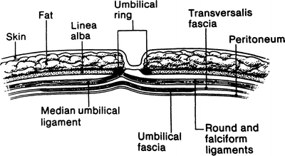
Figure 4.2.
Diagrammatic sagittal section through a normal umbilicus showing the relation of the umbilical ring to the linea alba, the round ligament, the urachus, and the umbilical and transversalis fasciae. Note the absence of subcutaneous fat over the umbilical ring (By permission of JE Skandalakis, SW Gray, AR Mansberger, et al. Hernia: Surgical Anatomy and Technique. New York: McGraw-Hill, 1989).
Figures 4.3 and 4.4 present variations of the umbilical fascia in relation to several anatomical entities. Note that in Fig. 4.4a, the fascia covers the umbilical ring in toto.
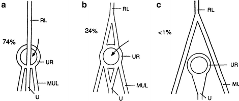
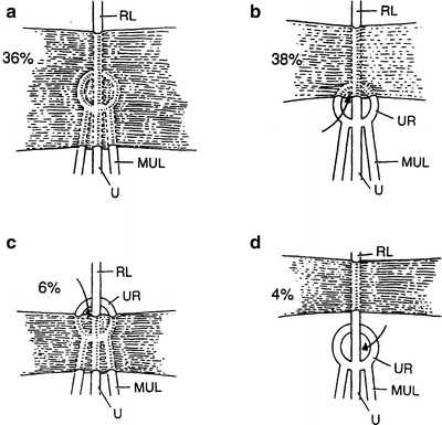
Figure 4.3.
Variations in the disposition of the umbilical ligaments as seen from the posterior (peritoneal) surface of the body wall. Arrows indicate (a) usual relations (74 %) of the umbilical ring (UR), the round ligament (RL), the urachus (U), and the medial umbilical ligaments (MUL). The round ligament crosses the umbilical ring to insert on its inferior margin. (b) Less common configuration (24 %). The round ligament splits and is attached to the superior margin of the umbilical ring. (c) Rare configuration (less than 1 %). The round ligament branches before reaching the umbilical ring. Each branch continues with the medial umbilical ligament without attaching to the umbilical ring (modified by permission of R Orda and H Nathan. Int Surg 58(7):454–464, 1973).
Figure 4.4.
Variations in the presence and form of the insertion of the umbilical fascia as seen from the posterior (peritoneal) surface of the body wall. (a) The thickened transversalis fascia forms the umbilical fascia covering the umbilical ring (36 %). Arrows indicate (b) the umbilical fascia covers only the superior portion of the umbilical ring (38 %); (c) the umbilical fascia covers only the inferior portion of the umbilical ring (6 %); (d) though present, the umbilical fascia does not underlie the umbilical ring (4 %). (No figure: fascia is entirely absent in 16 %). (modified by permission of R Orda and H Nathan. Int Surg 58(7): 454–464, 1973).
Layers of the Lower Anterior Body Wall
In the inguinal region, the layers of the abdominal wall are:
Skin.
Subcutaneous fasciae (Camper and Scarpa) containing fat (superficial fascia).
Innominate fascia (Gallaudet). It is not always recognized as a distinct entity. Its absence is of no surgical importance. It is responsible for the formation of the external spermatic fascia.
External oblique aponeurosis, including the inguinal, lacunar, and reflected inguinal ligaments.
Spermatic cord.
Transversus abdominis muscle and aponeurosis, internal oblique muscle, falx inguinalis (Henle), and the conjoined tendon (when present).
Anterior lamina of transversalis fascia.
Posterior lamina of transversalis fascia.
Preperitoneal connective tissue with fat.
Peritoneum.
Fossae of the Lower Anterior Abdominal Wall (Fig. 4.5)
Figure 4.5.
Diagram of the fossae of the anterior abdominal wall and their relation to the sites of groin hernias: (A) umbilicus, (B) median umbilical ligament (obliterated urachus), (C) medial umbilical ligament (obliterated umbilical arteries), (D) lateral umbilical ligament containing inferior (deep) epigastric arteries, and (E) falciform ligament. Sites of possible hernias: (1) lateral fossa (indirect inguinal hernia), (2) medial fossa (direct inguinal hernia), (3) supravesical fossa (supravesical hernia), and (4) femoral ring (femoral hernia) (By permission of JS Rowe, JE Skandalakis, and SW Gray. Am Surg 39(5):269–270; 1973).
The inner (posterior) surface of the anterior body wall above the inguinal ligament and below the umbilicus is divided into three shallow fossae on either side of a low ridge formed in the midline by the median umbilical ligament, the obliterated urachus. Each of these fossae is a potential site for a hernia. From lateral to medial, these fossae are:
The lateral fossa, bounded medially by the inferior epigastric arteries. It contains the internal inguinal ring, the site of indirect inguinal hernia.
The medial fossa, between the inferior epigastric artery and the medial umbilical ligament (remnant of the umbilical artery). It is the site of direct inguinal hernia.
The supravesical fossa, between the medial and the median umbilical ligaments. It is the site of external supravesical hernia.
Anatomical Entities of the Groin
Superficial Fascia (Fig. 4.6)
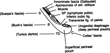
Figure 4.6.
Diagram of the relations of the superficial fasciae of the inguinal area showing the formation of the superficial perineal pouch (By permission of JE Skandalakis, SW Gray, AR Mansberger, et al. Hernia: Surgical Anatomy and Technique. New York: McGraw-Hill, 1989).
This fascia (described here only for the male) is divided into a superficial part (Camper) and a deep part (Scarpa). The superficial part extends upward on the abdominal wall and downward over the penis, scrotum, perineum, thigh, and buttocks. The deep part extends from the abdominal wall to the penis (Buck’s fascia), the scrotum (dartos), and the perineum (Colles’ fascia).
Usually two spaces are formed: the superficial perineal cleft and the superficial perineal pouch. The cleft is situated between Colles’ fascia and the muscle fascia that covers the muscles of the superficial perineal pouch. The pouch is defined by the perineal membrane, the external perineal fascia (Gallaudet), and the ischiopubic rami.
Aponeurosis of the External Oblique Muscle (Fig. 4.6)
Below the arcuate line (Douglas), the aponeurosis of the external oblique muscle joins with the aponeuroses of the internal oblique and transversus abdominis muscles to form the anterior layer of the rectus sheath. This aponeurosis forms or contributes to three anatomical entities in the inguinal canal:
Inguinal ligament (Poupart)
Lacunar ligament (Gimbernat)
Reflected inguinal ligament (Colles)
(Included sometimes is the pectineal ligament (Cooper), which is also formed from tendinous fibers of the internal oblique, transversus, and pectineus muscles.)
Inguinal Ligament (Poupart) (Fig. 4.7)
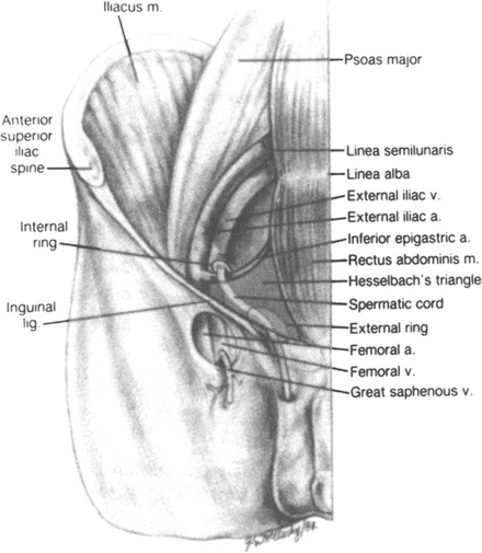
Figure 4.7.
Hesselbach triangle, site of direct inguinal hernia. Medial border of triangle is related to the lateral border of the rectus abdominis, site of supravesical hernia (By permission of SW Gray, JE Skandalakis, and DA McClusky. Atlas of Surgical Anatomy for General Surgeons. Baltimore: Williams & Wilkins, 1985).
This is the thickened lower part of the external oblique aponeurosis from the anterior superior iliac spine laterally to the superior ramus of the pubic tubercle. The middle one-third has a free edge. The lateral two-thirds is attached strongly to the underlying iliopsoas fascia.
Lacunar Ligament (Gimbernat) (Fig. 4.8)
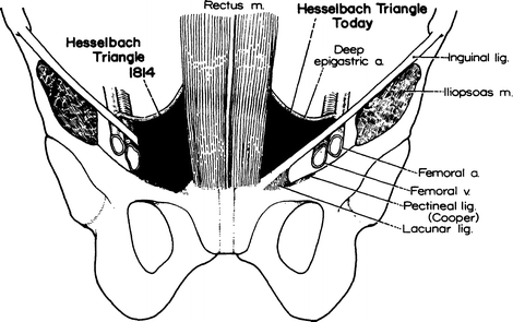
Figure 4.8.
Left: triangle described by Hesselbach (1814). Right: the slightly smaller triangle accepted today (By permission of JE Skandalakis, SW Gray, and JR Rowe. Anatomical Complications in General Surgery. New York: McGraw-Hill, 1983).
This is the most inferior portion of the inguinal ligament and is formed from external oblique tendon fibers arising at the anterior superior iliac spine. Its fibers recurve through an angle of less than 45° before attaching to the pectineal ligament. Occasionally it forms the medial border of the femoral canal.
Pectineal Ligament (Cooper) (Fig. 4.8)
This is a thick, strong tendinous band formed principally by tendinous fibers of the lacunar ligament and aponeurotic fibers of the internal oblique, transversus abdominis, and pectineus muscles and, with variation, the inguinal falx. It is fixed to the periosteum of the superior pubic ramus and, laterally, the periosteum of the ilium. The tendinous fibers are lined internally by transversalis fascia.
Conjoined Area (Fig. 4.9)
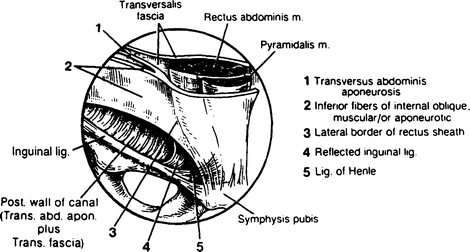
Figure 4.9.
The “conjoined area” (By permission of JE Skandalakis, GL Colborn, SW Gray, et al. Contemp Surg 38(1):20–34, 1991).
By definition, this is the fusion of fibers of the internal oblique aponeurosis with similar fibers from the aponeurosis of the transversus abdominis muscle, just as they insert on the pubic tubercle, the pectineal ligament, and the superior ramus of the pubis.
The above configuration is rarely encountered; published data suggest that it will be found in 5 % of individuals or fewer. We have proposed the term conjoined area. This has obvious practical application to the region containing the falx inguinalis (Henle ligament), the transversus abdominis aponeurosis, the inferomedial fibers of the internal oblique muscle or aponeurosis, the reflected inguinal ligament, and the lateral border of the rectus sheath.
Arch of the Transversus Abdominis
The inferior portion of the transversus abdominis, the transversus arch, becomes increasingly less muscular and more aponeurotic as it approaches the rectus sheath. Close to the internal ring, it is covered by the much more muscular arch of the internal oblique muscle. Remember that in the inguinal canal region, the internal oblique is muscular. The transversus abdominis is aponeurotic.
Falx Inguinalis (Henle Ligament) (Fig. 4.9)
Henle ligament is the lateral, vertical expansion of the rectus sheath that inserts on the pecten of the pubis. It is present in 30–50 % of individuals and is fused with the transversus abdominis aponeurosis and transversalis fascia.
Interfoveolar Ligament (Hesselbach)
This is not a true ligament. It is a thickening of the transversalis fascia at the medial side of the internal ring. It lies anterior to the inferior epigastric vessels.
Reflected Inguinal Ligament (Colles’) (Fig. 4.9)
This is formed by aponeurotic fibers from the inferior crus of the external ring that extend to the linea alba.
Iliopubic Tract (Fig. 4.10)
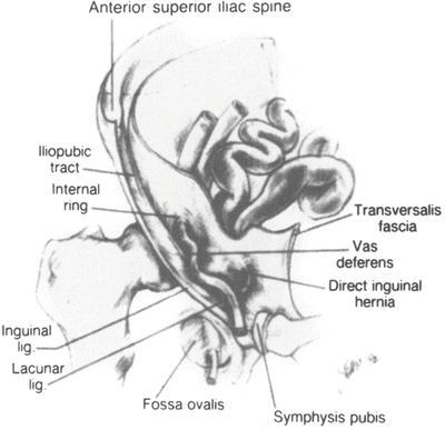
Figure 4.10.
Site of direct inguinal hernia. Note the internal ring and the iliopubic tract (By permission of SW Gray, JE Skandalakis, and DA McClusky. Atlas of Surgical Anatomy for General Surgeons. Baltimore: Williams & Wilkins, 1985).
This is an aponeurotic band extending from the iliopectineal arch to the superior ramus of the pubis. It forms part of the deep musculoaponeurotic layer together with the transversus abdominis muscle and aponeurosis and the transversalis fascia.
The tract passes medially, contributing to the inferior border of the internal ring. It crosses the femoral vessels to form the anterior margin of the femoral sheath, together with the transversalis fascia. The tract curves around the medial surface of the femoral sheath to attach to the pectineal ligament. It can be confused with the inguinal ligament.
Transversalis Fascia (Fig. 4.11)
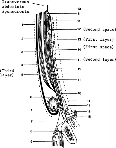
Figure 4.11.
Highly diagrammatic representation of the layers and spaces of the inguinal area and the space of Bogros: (1) innominate fascia; (2) external oblique aponeurosis; (3) internal oblique muscle; (4) transversus abdominis muscle and its aponeurosis; (5) transversalis fascia anterior; (6) external spermatic fascia; (7) Cooper ligament; (8) pubic bone; (9) pectineus muscle; (10) transversalis fascia; (11) transversalis fascia posterior lamina; (12) vessels; (13) peritoneum; (14) home (space) of the prosthesis, space of Bogros; (15) preperitoneal fat; (16) transversus abdominis aponeurosis and anterior lamina of transversalis fascia; (17) femoral artery; and (18) femoral vein (drawn by RC Read. From Skandalakis JE, Colborn GL, Androulakis JA, Skandalakis LJ, and Pemberton LB. Embryologic and anatomic basis of inguinal herniorrhaphy. Surg Clin North Am 73(4):799–836, 1993, with permission).
Although the name transversalis fascia may be restricted to the internal fascia lining the transversus abdominis muscle, it is often applied to the entire connective tissue sheath lining the abdominal cavity. In the latter sense, it is a fascial layer covering the muscles, aponeuroses, ligaments, and bones. In the inguinal area the transversalis fascia is bilaminar, enveloping the inferior epigastric vessels.
Related to the transversalis fascia is the space of Bogros, which is, for all practical purposes, a lateral extension of the retropubic space of Retzius. It is located just beneath the posterior lamina of the transversalis fascia out in front of the peritoneum (Fig. 4.11). The space of Bogros is used for the location of prostheses during the repair of inguinal hernias.
Figure 4.12 shows normal relations of the transversalis fascia in the lateral and lower parts of the abdominal wall.
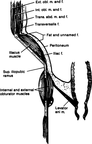
Figure 4.12.
Diagram of the normal relations of the transversalis fascia in the lateral and lower parts of the abdominal wall (By permission of EW Lampe. Transversalis fascia. In: Nyhus LM, and Condon RL. Hernia, 3rd ed. Philadelphia: Lippincott, 1989).
Iliopectineal Arch
This is a medial thickening of the iliopsoas fascia deep to the inguinal ligament. The surgeon does not directly use this arch, but it is important as the junction of a number of structures of the groin. These structures are:
The insertion of fibers of the external oblique aponeurosis.
The insertion of fibers of the inguinal ligament.
The origin of part of the internal oblique muscle.
The origin of part of the transversus abdominis muscle.
Part of the lateral attachment of the iliopubic tract. It contributes also to the lateral wall of the femoral sheath.
Hesselbach Triangle (Fig. 4.8)
As described by Hesselbach in 1814, the base of the triangle was formed by the pubic pecten and the pectineal ligament. The boundaries of this triangle as usually described today are:
Superolateral: the inferior (deep) epigastric vessels
Medial: the rectus sheath (lateral border)
Inferior (or, the base): the inguinal ligament
This is smaller than that described by Hesselbach in 1814. Most direct inguinal hernias occur in this area.
Inguinal Canal
The inguinal canal in the adult is an oblique rift in the lower part of the anterior abdominal wall. It measures approximately 4 cm in length. It is located 2–4 cm above the inguinal ligament, between the opening of the external (superficial) and internal (deep) inguinal rings.
The external, or superficial, inguinal ring is a triangular opening in the aponeurosis of the external oblique muscle. The superior and inferior crura form the margins of the ring: the superior crura are composed of the aponeurosis of the external oblique itself, and the inferior crura are formed by the inguinal ligament.
The internal, or deep, inguinal ring is a normal defect in the transversalis fascia shaped like an inverted V or U. Its arms—anterior and posterior—are a special thickening of the transversalis fascia, forming a sling. Its inferior border is formed by another thickening of the transversalis fascia—the iliopubic tract—which is not always very aponeurotic. The ring’s position in the transversalis fascia corresponds to the middle of the inguinal ligament.
The contents of the inguinal canal for males and females are as follows.
Male
The spermatic cord contains a matrix of connective tissue continuous with the preperitoneal connective tissue. The cord consists of:
Ductus deferens
Three arteries: the internal spermatic (testicular), deferential, and external spermatic (cremasteric)
One venous plexus (pampiniform)
Genital branch of the genitofemoral nerve
Ilioinguinal nerve
Sympathetic fibers from the hypogastric plexus
Three layers of fascia: the external spermatic fascia, a continuation of the innominate fascia; the middle, cremasteric layer, continuous with the internal oblique muscle fibers and muscle fascia; and the internal spermatic fascia, an extension of the transversalis fascia
Female
Round ligament of the uterus
Genital branch of the genitofemoral nerve
Cremasteric vessels
Ilioinguinal nerve
Coverings as described for the male, although usually less distinct
The anterior wall of the inguinal canal is formed by the aponeurosis of the external oblique muscle (Fig. 4.13) and by participation of the internal oblique muscle more laterally. The superior wall (“roof”) of the canal is formed by the arching lower borders of the internal oblique and transversus abdominis muscles and their aponeuroses. The inferior wall is formed by the inguinal and lacunar ligaments. From both anatomical and surgical standpoints, the posterior wall is the most important wall of the inguinal canal. The posterior wall (“floor”) is formed primarily by fusion of the aponeurosis of the transversus abdominis muscle and the transversalis fascia in three-fourths of subjects, yielding a strong wall, and by only the transversalis fascia in the remainder—yielding a weak wall (Figs. 4.14 and 4.15). Medially the posterior wall is reinforced by the internal oblique aponeurosis.
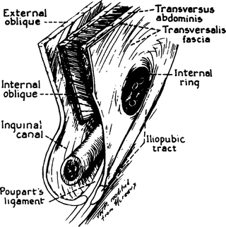

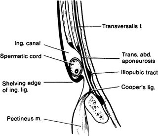
Figure 4.13.
Parasagittal section through right mid-inguinal region, illustrating separation of the musculoaponeurotic lamina into anterior and posterior inguinal walls (By permission of LM Nyhus. The preperitoneal approach and iliopubic tract repair of inguinal hernia. In: Nyhus LM, and Condon RL. Hernia, 3rd ed. Philadelphia: Lippincott, 1989).
Figure 4.14.
Diagrammatic cross section of a “strong” posterior inguinal canal wall. Notice the thickness and extent of the aponeurosis of the transversus abdominis muscle (By permission of EW Lampe. Transversalis fascia. In: Nyhus LM, and Condon RL. Hernia, 3rd ed. Philadelphia: Lippincott, 1989).
Figure 4.15.
Diagrammatic cross section of a “weak” posterior inguinal canal wall. Compare with Fig. 4.14 (By permission of EW Lampe. Transversalis fascia. In: Nyhus LM, and Condon RL. Hernia, 3rd ed. Philadelphia: Lippincott, 1989).
Fruchaud viewed hernias not by their clinical presentation, but rather by their origin within the groin, in an area he termed the myopectineal orifice (Fig. 4.16). This area is bounded superiorly by the arch of the internal oblique muscle and transversus abdominis muscle, laterally by the iliopsoas muscle, medially by the lateral border of the rectus muscle and its anterior lamina, and inferiorly by the pubic pecten. The inguinal ligament spans and divides this framework.
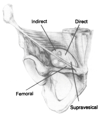
Figure 4.16.
Anterior view of Fruchaud’s myopectineal orifice (modified by permission of GE Wantz. Atlas of Hernia Surgery. New York: Raven Press, 1991, p. 4).
Surgical Ellipse (Fig. 4.17)
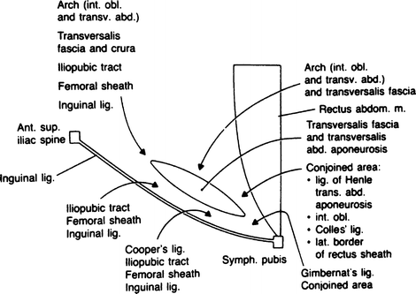
Figure 4.17.
Highly diagrammatic presentation of the surgical ellipse (By permission of JE Skandalakis, SW Gray, AR Mansberger, et al. Hernia: Surgical Anatomy and Technique. New York: McGraw-Hill, 1989).
With the patient in the supine position, the surgeon is dealing with the following anatomical areas and entities of the inguinal region, which are incorporated into an elliptical area: floor of the inguinal canal, superior medial edge (above), inferior lateral edge (below), medial apex, and lateral apex.
The floor (posterior wall) of the ellipse is formed by the transversus abdominis aponeurosis and the transversalis fascia. If the posterior wall is intact, its integrity prevents herniation (Figs. 4.14 and 4.15): none of the four types of inguinal hernia (indirect, direct, external supravesical, femoral) will develop.
The superior medial (upper) edge of the ellipse is formed by the conjoined area and the arch (internal oblique muscle and transversus abdominis muscle with their aponeuroses).
The inferior lateral (lower) edge is formed by the inguinal ligament, iliopubic tract, femoral sheath, and Cooper ligament. The iliopubic tract, femoral sheath, Cooper ligament, and occasionally the inguinal ligament are used for repair.
The medial apex—close to the symphysis pubis—is formed by Gimbernat ligament below and the conjoined area above.
The lateral apex—at the internal ring—is formed by the arch (internal oblique and transversus abdominis muscles and aponeuroses), transversalis fascia and crura, iliopubic tract, femoral sheath, and inguinal ligament.
Remember:
✔The transversalis fascia and transversus abdominis aponeurosis together are “good stuff” for repair of inguinofemoral herniation.
Femoral Canal and Its Sheath
The femoral canal—within the groin area, below the inguinal ligament—is 1.25–2 cm long and occupies the most medial compartment of the femoral sheath. The femoral sheath is formed anteriorly and medially by the transversalis fascia and some transversus aponeurotic fibers, posteriorly by the pectineus and psoas fasciae, and laterally by the iliacus fascia. The sheath forms three compartments, the most medial of which is the femoral canal, through which a femoral hernia may pass. The boundaries are:
Lateral: a connective tissue septum and the femoral vein
Posterior: the pectineal ligament (Cooper)
Anterior: the iliopubic tract or the inguinal ligament or both
Medial: the aponeurotic insertion of the transversus abdominis muscle and transversalis fascia or, rarely, the lacunar ligament
Blood Supply of the Anterior Abdominal Wall
Arterial Supply
Where there have been no previous incisions, the blood supply to the abdominal wall creates no problem. Where scars are present, the surgeon should be familiar with the blood supply to avoid necrosis from ischemia to specific areas. If possible, the surgeon should proceed through an existing scar.
The lower anterolateral abdominal wall is supplied by three branches of the femoral artery. They are, from lateral to medial, the superficial circumflex iliac artery (Fig. 4.18), the superficial epigastric artery, and the superficial external pudendal artery. Branches of these arteries travel toward the umbilicus in the subcutaneous connective tissues. The superficial epigastric artery anastomoses with the contralateral artery, and all three arteries have anastomoses with the deep arteries.
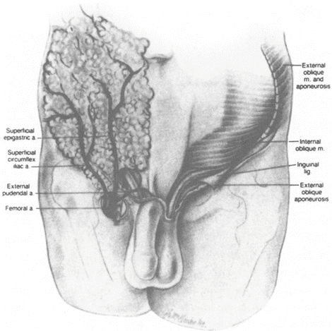
Figure 4.18.
The skin of the lower abdominal wall has been removed to show the superficial branches of the femoral artery (By permission of SW Gray, JE Skandalakis, and DA McClusky. Atlas of Surgical Anatomy for General Surgeons. Baltimore: Williams & Wilkins, 1985).
The deep arteries lie between the transversus abdominis and the internal oblique muscles. They are the posterior intercostal arteries 10 and 11, the anterior branch of the subcostal artery, the anterior branches of the four lumbar arteries, and the deep circumflex iliac artery.
The rectus sheath is supplied by the superior epigastric artery, which arises from the internal thoracic artery, and the inferior epigastric artery, which arises from the external iliac artery just above the inguinal ligament.
The superior epigastric artery enters the upper end of the rectus sheath, deep to the rectus muscle. Musculocutaneous branches pierce the anterior rectus sheath to supply the overlying skin. The perforating arteries are closer to the lateral border of the rectus than to the linea alba. Creating an incision too far laterally will result in bleeding from the several perforating arteries; cutting the musculocutaneous nerves will cause muscle paralysis.
The inferior epigastric artery arises in the preperitoneal connective tissue. It enters the sheath at or below the level of the semicircular line of Douglas, passing between the rectus muscle and the posterior layer of the sheath.
Venous Drainage
The veins follow the arteries.
Nerve Supply of the Anterior Abdominal Wall
Both the anterolateral portion of the abdominal wall and the rectus abdominis muscle are supplied by anterior rami of the 7th to the 12th thoracic nerves and the first lumbar nerve (Fig. 4.19). A branch—the lateral cutaneous ramus—arises from each anterior ramus and pierces the outer two flat muscles, innervating the external oblique and forming the lateral cutaneous nerve. The anterior rami of the last six thoracic nerves enter the posterior layer of the rectus sheath, innervating the rectus muscle and sending perforating branches through the anterior layer of the sheath to form the anterior cutaneous nerves. The first lumbar nerve forms an anterior cutaneous nerve without passing through the sheath. These relationships are shown diagrammatically in Fig. 4.19. Rectus muscle paralysis, with weakening of the abdominal wall, will result from section of more than one of these nerves.
Figure 4.19.
The course of the anterior ramus of segmental nerves in the anterior body wall. (a) 7th to 12th thoracic nerves. (b) First lumbar nerve (By permission of JE Skandalakis, SW Gray, and JR Rowe. Anatomical Complications in General Surgery. New York: McGraw-Hill, 1983).
The nerves of the lower abdominal wall area are illustrated in Fig. 4.20.
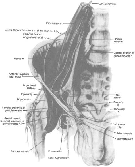
Figure 4.20.
Nerves of the inguinal region with which the surgeon should be familiar with (By permission of SW Gray, JE Skandalakis, and DA McClusky. Atlas of Surgical Anatomy for General Surgeons, Baltimore: Williams & Wilkins, 1985).
Panorama of Laparoscopic Cadaveric Anatomy of the Inguinal Area (Figs. 4.21, 4.22, 4.23, 4.24, 4.25, 4.26, 4.27, 4.28, 4.29, 4.30, 4.31, 4.32, 4.33, 4.34, 4.35, and 4.36)
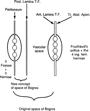
Figure 4.21.
Highly diagrammatic presentation of laparoscopic anatomy of the inguinal area demonstrating layers, fossae, and spaces (From Colborn GL, Skandalakis JE. Laparoscopic cadaveric anatomy of the inguinal area. Prob Gen Surg 12(1):13–20, 1995, with permission).
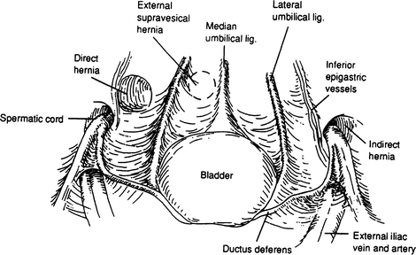
Figure 4.22.
Laparoscopic topographic anatomy of the inguinal region. In men, the spermatic vessels join the vas deferens to form the spermatic cord. The presence of a fascial defect lateral or medial to the inferior epigastric vessels defines an indirect or a direct hernia, respectively (modified by permission from JH Peters, and AE Ortega. Laparoscopic inguinal hernia repair. In: Hunter JG, and Sackier JM, eds. Minimally Invasive Surgery. New York: McGraw-Hill, 1993, p. 300).
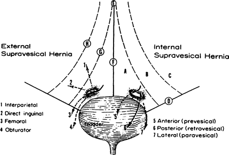
Figure 4.23.
The bladder and anterior abdominal wall viewed posteriorly. Possible pathways of external supravesical hernias are shown on the left and those of internal supravesical hernias are shown on the right. (A) Supravesical fossa with mouth of supravesical hernia. (B) Medial fossa. (C) Lateral fossa. (D) Inguinal ligament. (E) Umbilicus. (F) Middle umbilical ligament (obliterated urachus). (G) Lateral umbilical ligament (obliterated umbilical artery). (H) Inferior (deep) epigastric artery (By permission of PN Skandalakis, LJ Skandalakis, SW Gray, and JE Skandalakis. Supravesical hernia. In: Nyhus LM, and Condon RE, eds. Hernia, 4th ed. Philadelphia: Lippincott, 1995, p. 401).
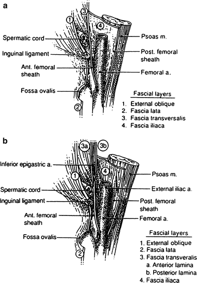
Figure 4.24.
(a) Highly diagrammatic drawing of the transversalis fascia and femoral sheath (old concept). (b) Highly diagrammatic drawing of the transversalis fascia and femoral sheath (new concept), emphasizing the bilaminar nature of the transversalis fascia in the inguinal area (By permission of JE Skandalakis, GL Colborn, JA Androulakis, et al. Embryologic and anatomic basis of inguinal herniorrhaphy. Surg Clin North Am 73:817, 1993).
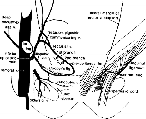
Figure 4.25.
The deep inguinal vasculature within the space of Bogros (By permission of R Bendavid. The space of Bogros and the deep inguinal venous circulation. Surg Gynecol Obstet 174:355–358, 1992).
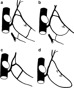
Figure 4.26.
(a–d) Variations in the vasculature of the deep inguinal venous system (By permission of R Bendavid. The space of Bogros and the deep inguinal venous circulation. Surg Gynecol Obstet 174: 355–358, 1992).
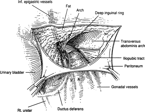
Figure 4.27.
After incising and retracting the peritoneum, the inferior epigastric vessels (the most superficial anatomical entities) will be seen. The arch and the iliopubic tract may or may not be seen (From Colborn GL, and Skandalakis JE. Laparoscopic cadaveric anatomy of the inguinal area. Prob Gen Surg 12(1):13–20, 1995, with permission).
Figure 4.28.
Posterior view of myopectineal orifice of Fruchaud. The boundaries are as follows: superior, the arch; lateral, iliopsoas muscle; medial, lateral edge of rectus abdominis muscle; and inferior, pubic pecten (By permission of GE Wantz and Lippincott-Raven. Atlas of Hernia Surgery. New York: Raven Press, 1991).
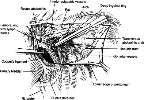
Figure 4.29.
With more cleaning the arch, iliopubic tract, and ligament of Cooper can be seen (From Colborn GL, and Skandalakis JE. Laparoscopic cadaveric anatomy of the inguinal area. Prob Gen Surg 12(1):13–20, 1995, with permission).
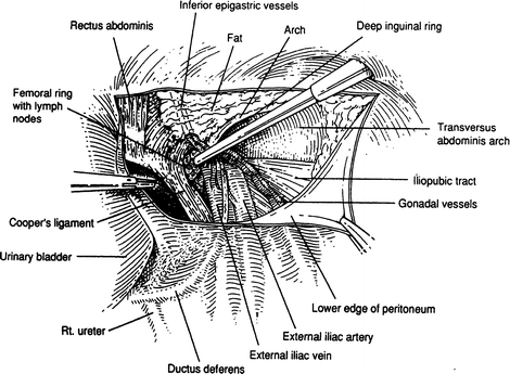
Figure 4.30.
With further cleaning, more entities will be seen: the spermatic cord and the iliac vessels (From colborn GL, and Skandalakis JE. Laparoscopic cadaveric anatomy of the inguinal area. Prob Gen Surg 12(1):13–20, 1995, with permission).
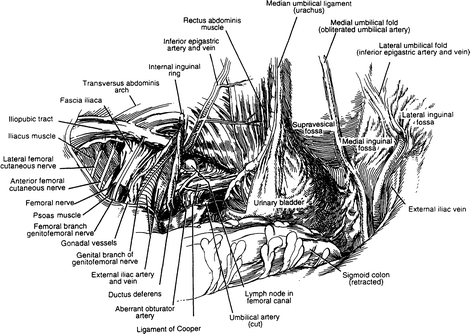
Figure 4.31.
Panoramic laparoscopic view of the anatomical entities of the inguinal area (From Colborn GL, and Skandalakis JE. Laparoscopic cadaveric anatomy of the inguinal area. Prob Gen Surg 12(1):13–20, 1995, with permission).
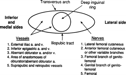
Figure 4.32.
Highly diagrammatic presentation of the vessels and nerves related to the iliopubic tract (From Colborn GL, and Skandalakis JE. Laparoscopic cadaveric anatomy of the inguinal area. Prob Gen Surg 12(1):13–20, 1995, with permission).
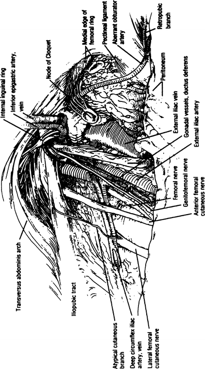
Figure 4.33.
Another panoramic laparoscopic view of the anatomy of the inguinal area (By permission of WG Brick, GL Colbom, TR Gadacz, et al. Crucial anatomic lessons for laparoscopic herniorrhaphy. Am Surg 61:175, 1995).
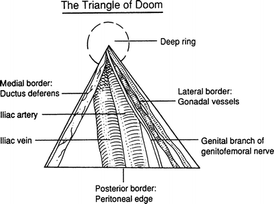
Figure 4.34.
The triangle of doom (From Colborn GL, and Skandalakis JE. Laparoscopic cadaveric anatomy of the inguinal area. Prob Gen Surg 12(1):13–20, 1995, with permission).
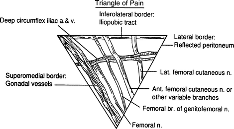
Figure 4.35.
The triangle of pain (From Colborn GL, and Skandalakis JE. Laparoscopic cadaveric anatomy of the inguinal area. Prob Gen Surg 12(1):13–20, 1995, with permission).
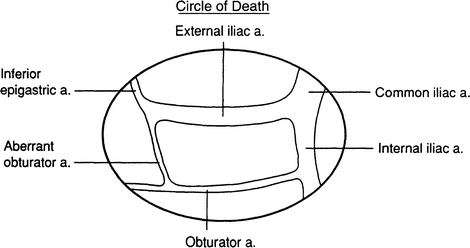
Figure 4.36.
The circle of death (From Colborn GL, and Skandalakis JE. Laparoscopic cadaveric anatomy of the inguinal area. Prob Gen Surg 12(1):13–20, 1995, with permission).
General Description of the Posterior (Lumbar) Body Wall
The lumbar area of the posterior body wall is bounded:
Superiorly: by the 12th rib
Inferiorly: by the crest of the ilium
Posteriorly: by the erector spinae (sacrospinalis) muscles
Anteriorly: by the posterior border of the external oblique muscle
In this area, the body wall is composed of the following layers of muscle and fascia:
Thick, tough skin.
Superficial fascia: two layers of fibrous tissue with fat between them.
A superficial muscle layer composed of the latissimus dorsi muscle posterolaterally and the external oblique muscle anterolaterally.
Thoracolumbar fascia containing three layers: posterior, middle, and anterior. The posterior and middle layers envelop the sacrospinalis muscle, and the middle anterior layer envelops the quadratus lumborum. Another characteristic of the middle layer of the thoracolumbar fascia is its lateral continuation to the transversus abdominis aponeurosis by fusion of all three layers. Therefore, the transversus abdominis aponeurosis should be accepted on faith as part of the thoracolumbar fascia.
A middle muscular layer of the sacrospinalis, internal oblique, and serratus posterior inferior muscles.
The deep muscular layer, composed of the quadratus lumborum and psoas muscles.
Transversalis fascia.
Preperitoneal fat.
Peritoneum.
Within this area, two triangles may be described: the inferior lumbar triangle (Petit) and the superior lumbar triangle (Grynfeltt).
The base of the inferior lumbar triangle is the iliac crest. The anterior (abdominal) boundary is the posterior border of the external oblique muscle. The posterior (lumbar) boundary is the anterior border of the latissimus dorsi muscle. The floor of the triangle is formed by the internal oblique muscle with contributions from the transversus abdominis muscle and the posterior lamina of the thoracolumbar fascia. The triangle is covered by superficial fascia and skin.
The base of the superior lumbar triangle is the 12th rib and the serratus posterior inferior muscle. The anterior (abdominal) boundary is the posterior border of the internal oblique muscle; the posterior (lumbar) boundary is the anterior border of the sacrospinalis muscle. The floor of the triangle is formed by the aponeurosis of the transversus abdominis muscle arising by fusion of the layers of the thoracolumbar fascia. The roof of the triangle is formed by the external oblique and latissimus dorsi muscles.
Stay updated, free articles. Join our Telegram channel

Full access? Get Clinical Tree


