TABLE 20–2 Unconventional Virus (Prion) Diseasesa
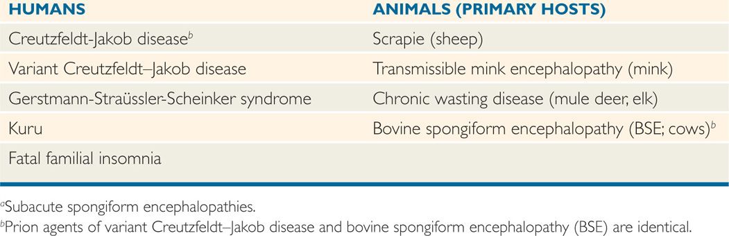
TABLE 20–3 Biologic and Physical Properties of Prions
Chronic progressive pathology without remission or recovery
No inflammatory response
No alteration in pathogenesis by immunosuppression or immunopotentiation
Estimated diameter of 5 to 100 nm
No virion-like structures visible by electron microscopy
Transmissible to experimental animals
No interferon production or interference by conventional viruses
Unusual resistance to ultraviolet irradiation, alcohol, formalin, boiling, proteases, and nucleases
Can be inactivated by prolonged exposure to steam autoclaving or 1N or 2N NaOH
Progressive neurologic diseases in humans and animals
Most persistent viral infections involve well-differentiated cells, such as lymphocytes and neuronal cells. They can be classified as (1) diseases associated with “conventional” viral agents that possess nucleic acid genomes and protein capsids and/or envelopes induce immune responses and can be grown in cell culture systems; and (2) diseases associated with “unconventional” agents that are small, filterable infectious agents, known as “prions,” which are transmissible to certain experimental animals, but do not contain nucleic acids, do not appear to be associated with immune or inflammatory responses by the host, and have not been cultivated in cell culture.
Include conventional viruses and unconventional agents
“Prions” do not produce immune or inflammatory responses
Persistence of conventional viruses can result from infection of a nonpermissive cell in the host with restrictive cytolytic effects, preservation of viral nucleic acid in infected host’s cells, and mutations that interfere with or severely limit viral replication or antigenicity.
Persistence can be due to a variety of mechanisms
DISEASES ASSOCIATED WITH CONVENTIONAL AGENTS
The following conditions are the major persistent infections caused by conventional viral agents. They are summarized in Table 20-1.
 Subacute Sclerosing Panencephalitis
Subacute sclerosing panencephalitis (SSPE) is discussed in Chapter 10. It is a rare chronic measles virus infection of children that usually appears 2 to 10 years after measles virus infection and produces progressive neurologic disease characterized by an insidious onset of personality change, progressive intellectual deterioration, and both motor and autonomic nervous system dysfunctions.
Persistence of measles virus after acute childhood infection
 Progressive Postrubella Panencephalitis
Even more rarely, a degenerative neurologic disorder similar to SSPE is associated with persistent rubella virus infection of the CNS. This condition is seen most often in adolescents who have had the congenital rubella syndrome. Rubella virus has been isolated from brain tissue in these patients using cocultivation techniques.
Can be a late sequela of congenital rubella infection
 Progressive Multifocal Leukoencephalopathy
Progressive multifocal leukoencephalopathy (PML) is a subacute, degenerative disease of the brain found primarily in adults with (1) immunosuppressive diseases, especially acquired immunodeficiency syndrome (AIDS) and hematologic malignancies; or (2) diseases requiring therapy with immunosuppressive agents. PML is due to a polyomavirus (JC virus) and is considered in Chapter 19.
Progressive neurologic disease of severely immunocompromised persons
 Persistent Enterovirus Infection
Persons with congenital or severe acquired immunodeficiency, especially those with agammaglobulinemia, may develop a chronic CNS infection due to an echovirus or other enterovirus. Headache, confusion, lethargy, seizures, and cerebrospinal fluid (CSF) pleocytosis are common manifestations. The virus can be isolated from the CSF. Clinical improvement may be achieved by the administration of human hyperimmune globulin to the infecting virus type. Relapse, however, occurs when therapy is discontinued, indicating persistence of virus despite the therapy.
Associated with humoral immunodeficiencies
Temporary improvement with virus type-specific hyperimmune globulin
AIDS DEMENTIA COMPLEX
Human immunodeficiency virus (HIV) causes a persistent infection of the CNS in many patients with symptomatic AIDS known as AIDS dementia complex (ADC) or HIV-associated dementia (HAD). The virus does not directly infect the nerve cells but the virus produced by perivascular macrophages and/or microglia may produce a bystander effect causing inflammation that may damage brain and spinal cord. The clinical course may vary from a mild sub-acute illness (early stage of HIV infection) to severe progressive dementia (late stages of HIV infection). HAD primarily occurs with more advanced HIV infection and symptoms include encephalitis, behavioral changes, and a gradual decline in cognitive function. HAD is more common is HIV-infected infants than infected adults. For more on HIV/AIDS, see Chapter 18.
Late stages of AIDS
HUMAN DISEASES CAUSED BY UNCONVENTIONAL AGENTS: SUBACUTE SPONGIFORM ENCEPHALOPATHIES
A group of progressive degenerative diseases of the CNS has been shown to be caused by infectious agents with unusual physical and chemical properties, which are now known as prions. The Nobel Prize in Medicine for 1997 was awarded to Stanley Prusiner for his work in identifying the role of prions in disease. Prions cause bovine spongiform encephalopathy (BSE) in cattle, scrapie in sheep, and five fatal CNS diseases in humans (Table 20-2). Prions can be the etiologic agents of inherited, communicable, or sporadic diseases. The pathogenesis of these illnesses is not well understood, but the pathologic and clinical features are similar. Varying degrees of neuronal loss and astrocyte proliferation occur. The diseases are known as “spongiform” encephalopathies or transmissible spongiform encephalopathies (TSE) because of the vacuolar changes in the cortex and cerebellum (Figures 20–1 and 20–2). The incubation periods for these diseases are months to years, and their courses are protracted and inevitably fatal.
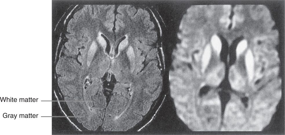
FIGURE 20–1. Appearance of brain with spongiform encephalopathy. (Left) Normal brain. (Right) Brain infected with a prion. Note the sponge-like appearance. (Reproduced with permission from Nester EW: Microbiology: A Human Perspective, 6th edition. 2009.)
Stay updated, free articles. Join our Telegram channel

Full access? Get Clinical Tree


