and John E. Skandalakis1
(1)
Centers for Surgical Anatomy and Technique, Emory University School of Medicine Piedmont Hospital, Atlanta, GA, USA
Abstract
The surgical anatomy of the deep fascia, axilla, lobules and ducts, vascular system, and lymphatic drainage of the breast is provided. The muscles and nerves involved in mastectomy (including their nerve supply) are presented in detail. Step-by-step techniques are given for breast biopsy, modified radical mastectomy, and lumpectomy with sentinel lymph node biopsy and axillary dissection. Instructions from the surgeon to the pathologist are enumerated.
Anatomy
General Description of the Breast
The adult female breast is located within the superficial fascia of the anterior chest wall. The base of the breast extends from the second rib superiorly to the sixth or seventh rib inferiorly and from the sternal border medially to the midaxillary line laterally. Two-thirds of the base of the breast lies anterior to the pectoralis major muscle; the remainder lies anterior to the serratus anterior muscle. A small part may lie over the aponeurosis of the external oblique muscle.
In about 95 % of women, there is a prolongation of the upper lateral quadrant toward the axilla. This tail (of Spence) of breast tissue enters a hiatus (of Langer) in the deep fascia of the medial axillary wall. This is the only breast tissue found normally beneath the deep fascia.
Deep Fascia
The deep pectoral fascia envelops the pectoralis major muscle, and the clavipectoral fascia envelops the pectoralis minor and part of the subclavius muscles. The axillary fascia lying across the base of the axillary pyramidal space is an extension of the pectoralis major fascia and continues as the fascia of the latissimus dorsi. It forms the dome of the axilla (Fig. 3.1a). Where the axillary vessels and nerves to the arm pass through the fascia, they take with them a tubular fascial sleeve, the axillary sheath.
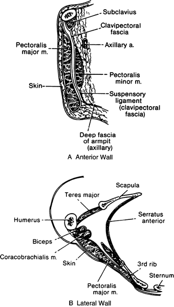
Figure 3.1.
Diagram of the walls of the axilla (modified by permission of JV Basmajian and CE Slonecker. Grant’s Method of Anatomy, 11th ed. Baltimore: Williams & Wilkins, 1989).
The clavipectoral fascia (Fig. 3.1a) can be thought of as consisting of five parts:
The attachment to the clavicle and the envelope of the subclavius muscle
That part between the subclavius and pectoralis minor muscles, referred to by some as “the costocoracoid membrane”
The thickened lateral band between the first rib and the coracoid process, the costocoracoid ligament
The pectoralis minor envelope
The suspensory ligament of the axilla attaching to the axillary fascia
Axilla
The axilla is shaped like a pyramid with an apex, a base, and four walls. The apex is a triangular space bordered by the clavicle, the upper border of the scapula, and the first rib. The base consists of the axillary fascia beneath the skin of the axillary fossa. The anterior wall is composed of three muscles—the pectoralis major, the pectoralis minor, and the subclavius—and the clavipectoral fascia, which envelops the muscles and fills the spaces between them. The posterior wall is formed by the scapula and three muscles: the subscapularis, the latissimus dorsi, and the teres major. The medial wall consists of the lateral chest wall, with the second to sixth ribs, and the serratus anterior muscle. The lateral wall is the narrowest of the walls, being formed by the bicipital groove of the humerus (Fig. 3.1b).
The axilla contains lymph nodes (about which more will be said); the axillary sheath, which covers blood vessels and nerves; and the tendons of the long and short heads of the biceps brachii muscle and the coracobrachialis muscle (Fig. 3.2).
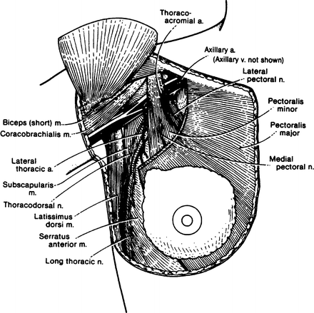
Figure 3.2.
Topography of the axilla. Anterior view (By permission of JE Skandalakis, SW Gray, and JR Rowe. Anatomical Complications in General Surgery. New York: McGraw-Hill, 1983).
Muscles and Nerves
The muscles and nerves with which the surgeon must be familiar are listed in Table 3.1.
Table 3.1
Muscles and nerves involved in mastectomy
Muscle | Origin | Insertion | Nerve supply | Comments |
|---|---|---|---|---|
Pectoralis major | Medial half of clavicle, lateral half of sternum, second to sixth costal cartilages, aponeurosis of external oblique muscle | Greater tubercle of humerus | Lateral anterior thoracic nerve | Clavicular portion of pectoralis forms upper extent of radical mastectomy; lateral border forms medial boundary of modified radical mastectomy; both nerves should be preserved in modified radical procedure |
Pectoralis minor | Second to fifth ribs | Coracoid process of scapula | Medial anterior thoracic nerve | |
Deltoid | Lateral half of clavicle, lateral border of acromion process, spine of scapula | Deltoid tuberosity of humerus | Axillary nerve | |
Serratus anterior (three parts) | 1. First and second ribs | Costal surface of scapula at superior angle | Long thoracic nerve | Injury produces “winged scapula” |
2. Second to fourth ribs | Vertebral border of scapula | |||
3. Fourth to eighth ribs | Costal surface of scapula at inferior angle | |||
Latissimus dorsi | Back, to crest of ilium | Crest of lesser tubercle and inter-tubercular groove of humerus | Thoracodorsal nerve | Anterior border forms lateral extent of radical mastectomy; injury results in weakness of rotation and abduction of arm |
Subclavius | Junction of first rib and its cartilage | Groove of lower surface of clavicle | Subclavian nerve | |
Subscapularis | Costal surface of scapula | Lesser tubercle of humerus | Subscapular nerve | Subscapular nerve should be spared |
External oblique aponeurosis | External oblique muscle | Rectus sheath and linea alba, crest of ilium | Remember the interdigitation with serratus anterior and pectoralis muscles | |
Rectus abdominis | Ventral surface of fifth to seventh costal cartilages and xiphoid process | Crest and superior ramus of pubis | Branches of 7th to 12th thoracic nerves | Rectus sheath is lower limit of radical mastectomy |
Morphology of the Breast
Each breast is composed of between 15 and 20 lobes, some larger than others, within the superficial fascia, which is loosely connected with the deep fascia. Between the superficial and deep fasciae is the retromammary (submammary) space, which is rich in lymphatics (Fig. 3.3).
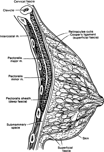
Figure 3.3.


Diagrammatic sagittal section through the nonlactating female breast and anterior thoracic wall (By permission of JE Skandalakis, SW Gray, and JR Rowe. Anatomical Complications in General Surgery. New York: McGraw-Hill, 1983).
Stay updated, free articles. Join our Telegram channel
Full access? Get Clinical Tree
