44
Anterolateral Leg
The anterolateral part of the leg is between the knee and the ankle (Fig. 44.1). The two bones of the leg are
- the tibia (the shin bone), which is a large, weight-bearing bone that contributes to the medial part of the ankle
- the fibula, a smaller bone, which is primarily for muscle attachment but also contributes to the lateral part of the ankle
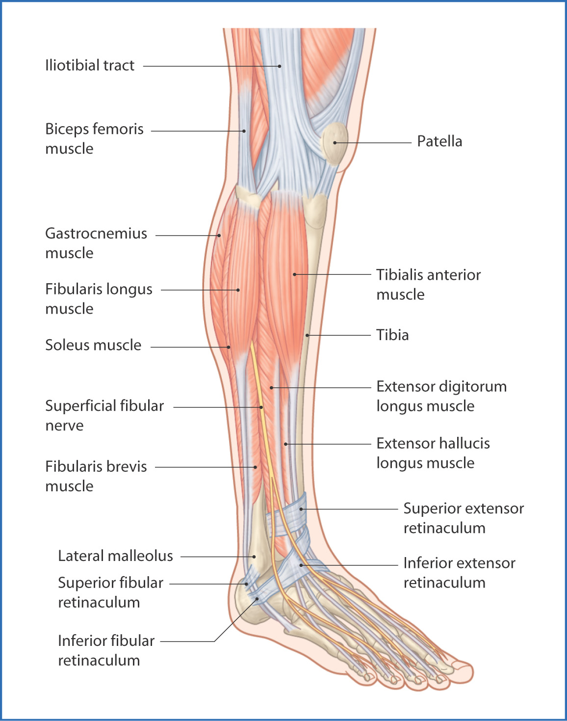
FIGURE 44.1 Muscles of the anterolateral leg.
The tibia is flattened superiorly, with medial and lateral superior articular surfaces (commonly called “plateaus”) separated by the intercondylar eminence. The roughened areas below the medial and lateral facets are the medial and lateral condyles, respectively. The tibial tuberosity is a bony prominence on the anterior aspect of the proximal end of the tibia where the patellar ligament attaches. The tibia descends as a triangular shaft toward the ankle and becomes the medial malleolus.
The fibula is on the lateral aspect of the leg and is a slender bone, with a proximal head that articulates with the lateral condyle of the tibia. The distal lateral malleolus of the fibula makes up the lateral aspect of the ankle. Together, the tibia and fibula transfer the weight of the body onto the talus bone of the foot.
Muscles
The muscles of the anterolateral part of the leg are divided into an anterior group of four muscles and a lateral group of two muscles (Table 44.1, Fig. 44.2). These muscle groups are separated by the thick intermuscular crural fascia: the anterior intermuscular septum of the leg separates the anterior and lateral muscle groups, and the posterior intermuscular septum of the leg separates the lateral group of muscles from the posterior leg muscles (see Chapter 45).
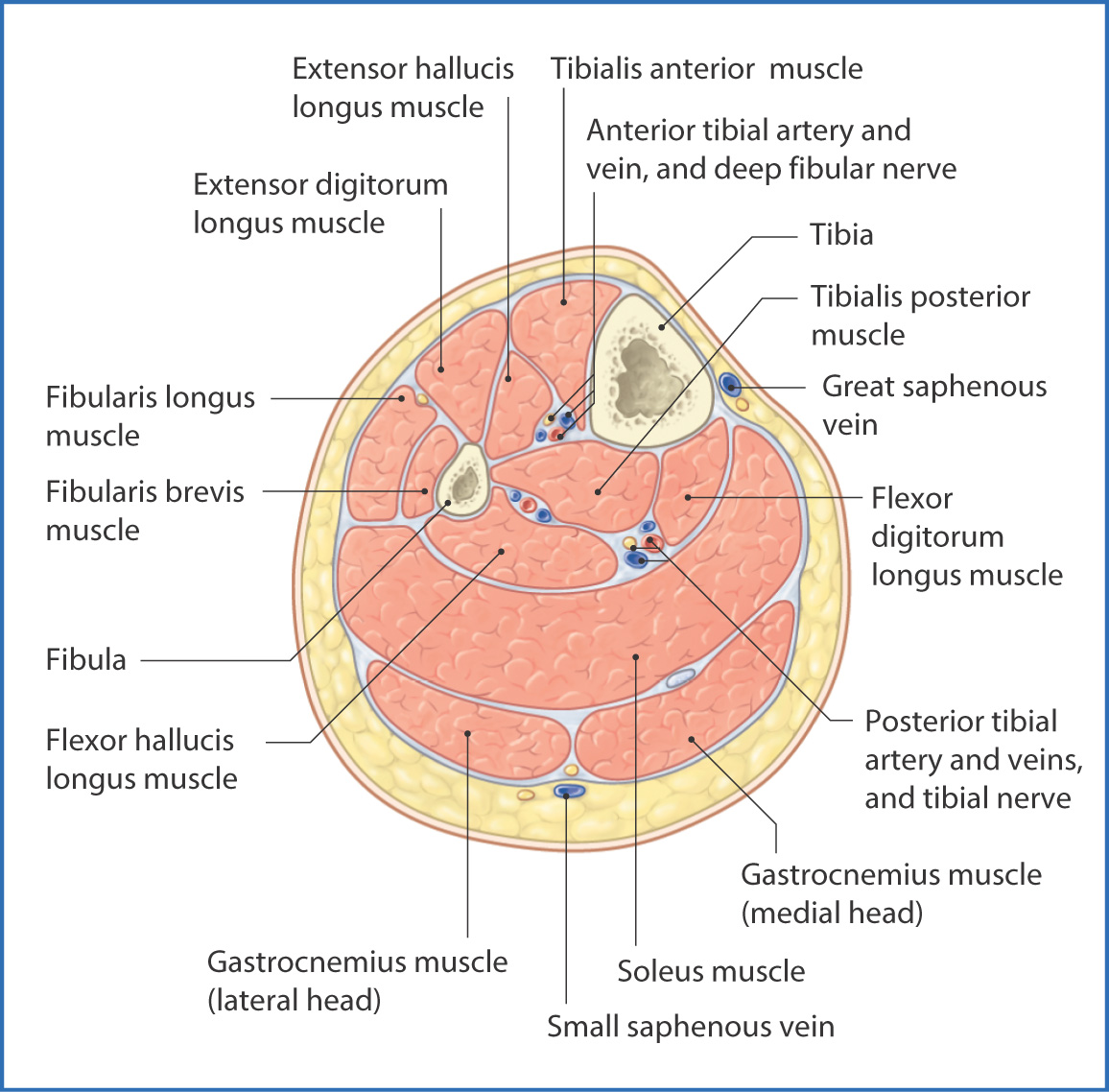
FIGURE 44.2 Cross section of the right leg viewed from distal to proximal.
TABLE 44.1 Anterolateral Leg Muscles
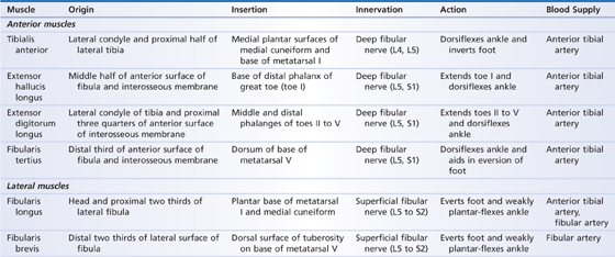
The tibialis anterior, extensor hallucis longus, extensor digitorum longus, and fibularis tertius muscles are the anterior leg muscles. They
- originate from the tibia and fibula and insert onto the dorsal metatarsals and distal phalanges of the foot
- dorsiflex the ankle and extend the toes
- originate from the lateral surface of the tibia and insert on the bases of metatarsals I and V
- evert the foot and weakly plantar-flex the ankle
Nerves
The anterolateral portion of the leg is innervated by the common fibular nerve (L4 to L5, S1 to S2 nerve roots), which is the lateral branch of the sciatic nerve. Just after the common fibular nerve separates from the tibial nerve in the popliteal fossa, it gives off a small cutaneous branch—the lateral sural cutaneous nerve. This nerve, in turn, gives off a fibular communicating branch that joins the medial sural cutaneous nerve (from the tibial nerve) to form the sural nerve. The sural nerve conveys sensation from the skin of the posterolateral part of the leg and foot.
The common fibular nerve descends through the popliteal fossa and enters the leg by wrapping around the neck of the fibula. At this point, it divides into the superficial and deep fibular nerves. The superficial fibular nerve descends in the lateral compartment of the leg to innervate the fibularis longus and fibularis brevis muscles and to convey sensation from the skin of the lateral part of the leg and foot.
The deep fibular nerve descends down the leg and innervates the anterior muscles of the leg. It enters the foot between the extensor digitorum longus and extensor hallucis longus muscles and conveys sensation from the small patch of skin of the dorsal web space between toes I and II.
Arteries
Blood is supplied to the anterior compartment of the leg by the anterior tibial artery, which is a branch of the popliteal artery.
Stay updated, free articles. Join our Telegram channel

Full access? Get Clinical Tree


