• Pools of extravasated bile may be present in wall of gallbladder
• Multinucleated giant cells common
• Over time, organization occurs, and fibrosis may predominate, further mimicking malignancy
Thickened Wall and Mucosal Ulceration This low-power view shows thickening of the wall, mucosal ulceration, and a proliferation of foamy macrophages. Note the cholesterol clefts as well, which are abundant within the foamy macrophage infiltrate.
Nodular Macrophage Infiltrate This nodular collection of macrophages in the wall of the gallbladder contains bile and ceroid-laden histiocytes.
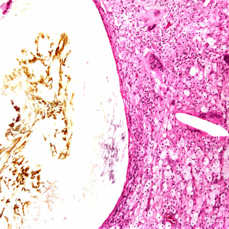
Foamy Histiocytes Xanthogranulomatous cholecystitis is often characterized by a dense infiltrate of foamy histiocytes, which may or may not contain ceroid pigment. Multinucleated giant cells are also common .
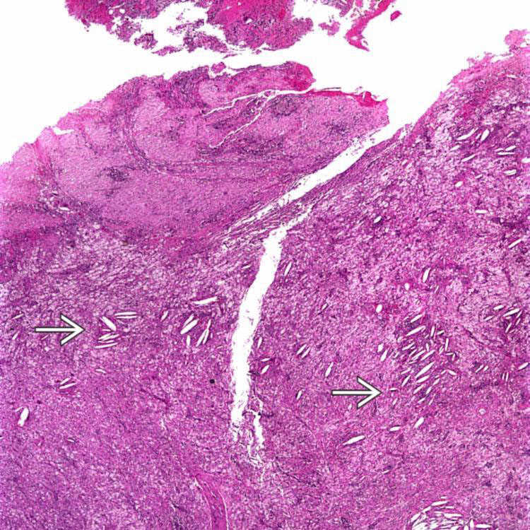
Rokitansky-Aschoff Sinus The granulomatous inflammation is often centered on a Rokitansky-Aschoff sinus, which may have previously ruptured. The one seen here is dilated and filled with bile. Note the foamy histiocytes, cholesterol clefts, and giant cells to the right.
TERMINOLOGY
Synonyms
• Cholegranulomas/cholecystic granulomas
• Ceroid granulomas
Definitions
• Variant of chronic cholecystitis featuring florid proliferation of foamy macrophages and granulomatous reaction to bile
Named due to histologic similarity to xanthogranulomatous pyelonephritis
Only gold members can continue reading. Log In or Register to continue


 as well, which are abundant within the foamy macrophage infiltrate.
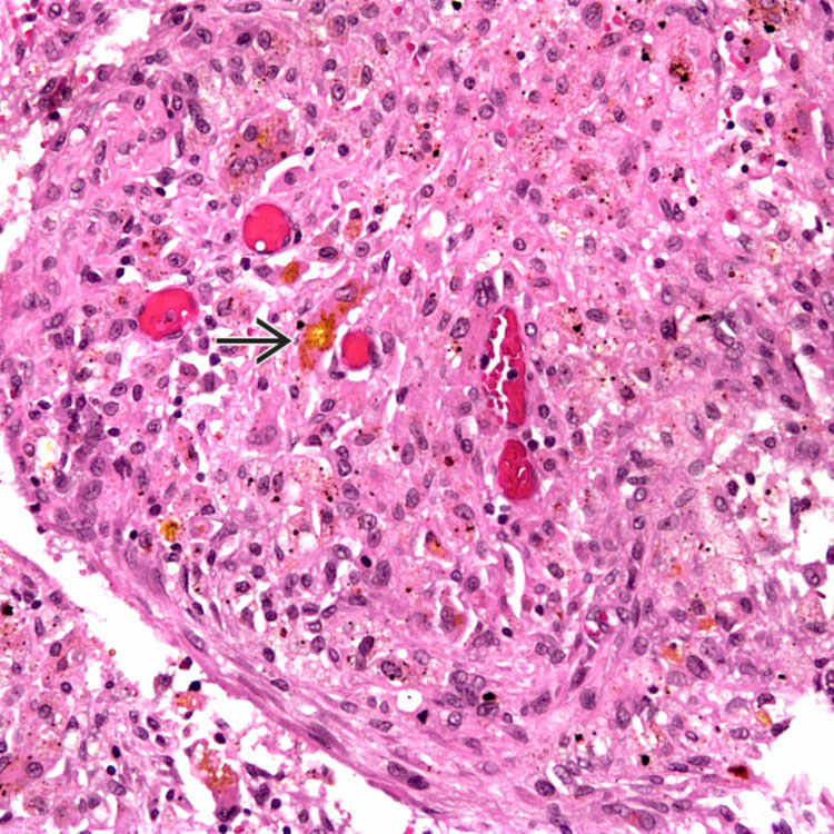
 and ceroid-laden histiocytes.
 .
.