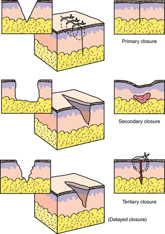
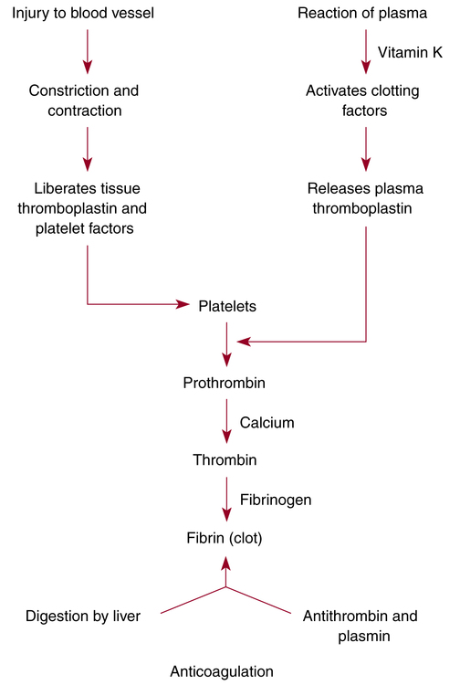
Wound healing and hemostasis

Chapter 29


Wound healing and hemostasis


Key terms and definitions


Adhesion 


Band of scar tissue that holds together or unites surfaces or structures that are normally separated.


Cicatrix 


Firm avascular scar tissue.


Contracture 


Formation of extensive scar tissue over a joint.


Dead space 


Space caused by separation of wound edges or by air trapped between layers of tissue.


Debridement 


Removal of damaged tissue and cellular or other debris from a wound to promote healing and to prevent infection.


Dehiscence 


Partial or total splitting open or separation of the layers of a wound.


Devitalized 


Tissue that is nonviable, necrotic.


Edema 


Abnormal accumulation of fluid in interstitial spaces of tissues.


Evisceration 


Protrusion of viscera through an abdominal incision.


Exsanguination 


Tissue is drained of blood.


Extravasation 


Passage of blood, serum, or lymph into tissues.


Exudate 


Fluid, cells, or other substances that have been discharged from vessels or tissues. It contains white blood cells, lymphokines, and growth factors that stimulate healing.7


Granulation tissue 


Formation of fibrous collagen to fill the gap between the edges of a wound healing with contraction (i.e., second intention).


Granuloma 


Inflammatory lesion that forms around a foreign substance, such as glove powder or a suture knot.


Hematoma 


Collection of extravasated blood in tissue.


Hemostasis 


Arrest of blood flow or hemorrhage; the mechanism is via coagulation (formation of a blood clot).


Hypertrophic scar 


Excessive raised dense scarring that does not exceed the border of the wound.


Iatrogenic 


Condition caused by treatment or procedure performed by medical personnel.


Incision 


Intentional cut through intact tissue (synonym: surgical incision).


Ischemia 


Decrease of blood supply to tissues.


Keloid 


Overgrowth of firm rounded scar that extends beyond the border of the wound. Can be painful and may bleed if injured.


Necrosis 


Death of tissue cells; devitalized tissue.


Scar 


Deposition of fibrous connective tissue to bridge separated wound edges and to restore continuity of tissues.


Seroma 


Collection of extravasated serum from interstitial tissue or a resolving hematoma in tissue.


Tensile strength 


Ability of tissues to resist rupture.


Tissue reaction 


Immune response of the body to tissue injury or foreign substances.


Wound disruption 


Separation of wound edges.


Mechanism of wound healing


Interruption of tissue integrity, either intentional or unintentional, requires understanding of the mechanism and factors that cause wounding and influence wound healing.3,7,8 When tissue is cut, the body’s inherent defense mechanisms respond immediately to begin repair. Three mechanisms of wound healing are recognized: first intention/primary union, second intention, and third intention/delayed primary closure (Fig. 29-1).3 Each mechanism has practical applications in the making and closing of incisions or traumatic wounds. The degree of contamination and the amount of viable tissues are factors in the determination of which method of healing is used.


First intention/primary union


Healing by first intention is desired after primary union of an incised, aseptic, accurately approximated wound. Well-approximated wounds form a fibrin bridge that aids healing. Key elements of first-intention closure include the following:


The rates and patterns of wound healing differ in various tissues. In general, first-intention wound healing consists of three distinct phases:


1. Lag phase of acute inflammatory response: Tissue fluids containing plasma, proteins, blood cells, fibrin, and antibodies exude from the tissues into the wound, depositing fibrin, which weakly holds the wound edges together for the first 5 days.


2. Healing or proliferative phase of fibroplasia: After the fifth postoperative day, fibroblasts multiply rapidly, bridging wound edges and restoring the continuity of body structures. Collagen, a protein substance that is the chief constituent of connective tissue, is secreted from the fibroblasts and formed into fibers.


3. Maturation or differentiation phase: From the 14th postoperative day until the wound is fully healed, scar formation occurs by deposition of fibrous connective tissue. The collagen content remains constant, but the fiber pattern reforms and crosslinks to increase the tensile strength. Wound contraction occurs over a period of weeks up to 6 months. As collagen density increases, vascularity decreases and the scar grows pale. The scar tissue is only 80% as strong as the original tissue.


Second intention


The mechanism of second-intention healing is by granulation, eventual reepithelialization, and wound contraction rather than with suturing closed by first intention. The wound heals from the bottom up; it heals spontaneously if the dermal base is preserved. The following are considerations with this type of healing:


1. Infection, excessive trauma, loss of tissue, and poorly approximated tissue are common. Inflammatory response is exaggerated.


2. The wound is left open and allowed to heal from the inner toward the outer surface. Devitalized tissue is debrided.


3. Healing is delayed. The wound may need grafting.


4. Healing may produce a weak union, which may be conducive to incisional herniation later.


5. The risk of secondary infection is proportional to the amount of necrotic tissue present in the wound and to compromised immune response in the patient. Repeated debridement may be necessary.


6. Scar formation is excessive.


7. Contracture of skin is pronounced. After healing is complete, the scar may need revision or release.


Third intention/delayed primary closure


Approximation and suturing is delayed or secondary for the purpose of walling off an area of gross infection or an area where extensive tissue was removed (e.g., in a debridement or from a traumatic injury). The edges are closed 4 to 6 days postoperatively after meticulous debridement. The following are considerations in healing by third intention:


Types of wounds


A wound is an injury, either intentional or unintentional, that disrupts the continuity of body tissues with or without tissue loss. Wounds may be surgical (intentional), traumatic, incidental, or chronic.10


Intentional wounds


Surgical-site incision or excision


An incision is a cut or an opening into intact tissue. An excision is removal of tissue. A sterile sharp scalpel, scissors, curette, or other cutting instrument may be used to separate skin and underlying tissues. Thermal instruments that both cut or vaporize tissue and coagulate surrounding blood vessels are used for incision and excision. The location, length, and depth of an incision must be planned.


The surgeon spreads the skin taut between the thumb and index finger in preparation for the skin incision. With one stroke of evenly applied perpendicular pressure on the scalpel, a clean incision is made through the epidermis and dermis into the subcutaneous layers. A number of factors influence the ease with which a primary skin incision is made:


A clean stroke with a sterile surgical scalpel, followed by attention to all of the principles of sterile technique and tissue handling, is the best insurance for healing by first intention. The line of direction of the incision in relation to the natural lines of direction of the skin may be a factor in wound healing. Excess tension on the healing wound can delay wound healing. Wounds heal side to side, not end to end.


Traumatic wounds


After traumatic injury, preservation of life is the first critical concern. The patient’s general condition is of prime consideration, and the plan of care is individualized to meet the patient’s needs. Injuries are evaluated, and those that pose the greatest hazards to life or to return to normal function are cared for first.


The primary objective after life support is wound closure with minimal deformity and functional loss. Minor injuries are cared for in the emergency department. Patients with major injuries undergo treatment in the emergency department before going to the OR as quickly as the condition warrants it. Traumatic wounds can be considered closed or open, simple or complicated, clean or contaminated. Wound closure is predicated on the type, location, severity, and extent of injury.


Contaminated wounds


When dirty objects penetrate skin, microorganisms multiply rapidly. Within 6 hours, contamination can become infection. Debridement is performed to remove devitalized tissue, and the wound is irrigated. Devitalized tissue is removed because it acts as a culture medium. The wound may be left open to heal by second or third intention. Closure may be delayed for several days.


The patient’s history should be assessed for tetanus bacillus immunization. Tetanus is most likely to occur in deep wounds contaminated by soil or animal feces. Adsorbed tetanus toxoid (0.5 mL) may be given as an initial immunizing dose or as a booster if the patient has been immunized within the previous 5 years. Tetanus immune globulin (human, 250 to 500 units) also should be given to any patient who has a severe wound or who has had the wound for more than 24 hours and has not been immunized within the previous 10 years.


Incidental and chronic wounds


Pressure sores and decubitus ulcers may result from compromised circulation over bony prominences or other pressure points for extended periods. Positioning and padding considerations in the plan of care can help prevent incidental pressure-related injuries in the perioperative environment.


Factors influencing wound healing


Each patient has internal and external forces that influence healing. Most wounds progress to healing unless the closure is poor, an infection ensues, or the tissue is devitalized by other forces. Hemostatic and inflammatory responses must be intact for healing to take place.


The degree of wound contamination is evaluated, and the potential risk for postoperative wound infection is considered. At the conclusion of the surgical procedure, the wound is assigned a classification.


Surgical wound classification


The surgical site may be clean or contaminated when the surgeon makes the initial incision. A clean site may become contaminated depending on the type of wound, the pathologic findings or circumstances that create the need for the surgical procedure, the anatomic location, and the techniques of the OR team. After completion of wound closure, the circulating nurse should verify the wound classification with the surgeon.


The wound class should not be assigned until the dressing is applied. This is documented in the patient’s intraoperative records. Surgical sites are classified by the degree of microbial contamination or exposure that may predispose a patient to a postoperative wound infection.


According to the Centers for Disease Control and Prevention (CDC), risk of infection increases in proportion to contamination of the incision and surrounding tissues exposed during the course of the surgical procedure. The true extent of risk cannot be evaluated until the procedure is completed. The wound is classified at the end of the surgical procedure as one of four types (Box 29-1):


Generalized health condition of the patient


Chronic diseases alter normal physiology. Diseases such as diabetes, uremia, fibrocystic disease, cirrhosis, active alcoholism, and leukemia can delay wound healing.


Circulatory status


Cardiovascular and respiratory insufficiency inhibits tissue perfusion. Oxygenation is essential to wound healing and to inhibition of growth of anaerobic microorganisms. If oxygen does not circulate, the wound does not heal.


Smoking.

Vasoconstriction caused by smoking decreases blood supply and oxygenation to the wound.11 Carbon monoxide in smoke binds with hemoglobin (forming carboxyhemoglobin) and further diminishes oxygenation. Smoking contributes to respiratory complications. This can cause forceful coughing that can raise intraabdominal pressure and create increased strain on an abdominal wound and impair healing.


Nutritional status


Wound healing is impaired by deficiencies in proteins, carbohydrates, zinc, and vitamins A, B, C, and K. Protein provides essential amino acids for new tissue construction. Carbohydrates are necessary energy sources for cells and prevent excessive metabolism of amino acids to meet caloric requirements. Vitamin B complex is necessary for carbohydrate, protein, and fat metabolism. Vitamin C permits collagen formation. Although vitamin A and zinc are known to be important in collagen synthesis, their mechanism in wound healing is not well understood. Vitamin K is involved in the synthesis of prothrombin and other clotting factors. Copper and iron assist in collagen synthesis. Calcium and magnesium are important in protein synthesis. Manganese serves as an enzyme activator.


Malnutrition, whether primary or secondary to disease, can be a major factor in wound healing and infection. Impairment of physiologic functions associated with a body weight loss that is greater than 10% and protein energy malnutrition increase the risk of postoperative complications. Liver function, skeletal and respiratory muscle function, overall physical and mental activity, and inflammatory response to wound healing are altered in the patient who is malnourished.


Protein and fat deficiency is especially significant in patients with extensive burns or multiple injuries who have greatly increased caloric requirements. Malnutrition caused by anorexia or cachexia has a deleterious effect on wound healing. Hyperalimentation with vitamin, trace element, and mineral supplements preoperatively and postoperatively usually is indicated for patients who are malnourished.


In obese patients, the bulk and weight of adipose tissue cause difficulty in confining excess fat and securing good wound closure. To minimize dead space, the surgeon may place drains and sutures in subcutaneous fat; both may actually potentiate infection. Of all tissues, fat is the most vulnerable to trauma and infection because of its poor vascularity. Many patients who are morbidly obese, more than 100 lbs (45.4 kg) over ideal body weight, have cardiac decompensation and respiratory insufficiency.


Fluid and electrolyte balance


The body’s system for balancing fluids and electrolytes is extremely complex. As a result of illness, injury, or infection, the patient may not be able to maintain normal fluid and electrolyte balance. Fever associated with infection, for example, can raise fluid requirements as much as 15% for each 1.5° F (or 1° C) rise in body temperature.


Body fluid is intracellular (ICF [within cells]) and extracellular (ECF [outside cells as intravascular plasma and interstitial fluid between cells]). The electrolyte content differs. ECF contains more sodium than does ICF; ICF has more potassium than does ECF. Changes in this balance can affect kidney function, cellular metabolism, oxygen concentration in the circulation, and hormonal function. Adequate ECF volume is necessary for circulation of blood to tissues.


Inflammatory and immune responses


The body repairs tissues at the cellular level in response to injury or exposure to foreign substances. It triggers an inflammatory response to mobilize cellular components associated with healing. Some foreign materials cause more inflammatory reaction than do others.


Extremes of inflammation may result in response to allergy, infection, or chronic irritation and may delay wound healing. Inflammation should not be confused with infection, which has a pathogenic microbe that causes the complications. Patients with an impaired immune response have an altered inflammatory response and do not heal appropriately.


Surgical technique


Devitalized tissue caused by laser or electrosurgery cannot regenerate. Interruption of blood supply and innervation decreases circulation and prevents epithelialization. Excess tension on the suture line that inhibits tissue perfusion prolongs healing time.


Tissue approximation


Tissue edges are brought together with precision, avoiding strangulation and eliminating dead space, to promote wound healing. A closure that is too tight or closure under tension causes ischemia, a decrease of blood supply to tissues. Approximation is critical to the healing of flaps and grafts. Constricted or interrupted blood supply can cause flap failure or loss of a vascularized graft.13


Dead space is caused by separation of wound edges that have not been closely approximated or by air trapped between layers of tissue. Serum or blood may collect in a dead space and prevent healing by keeping cut edges separated. Wound edges not in close contact cannot heal. A drain may be inserted to aid in removal of fluid or air from the surgical site postoperatively, or a pressure dressing may be applied over a closed wound to help obliterate dead space.


The choice of wound-closure materials and the techniques of the surgeon are prime factors in the restoration of tensile strength to the wound during the healing process.


Wound security


The quality of approximated tissue and the type of closure material are two factors that determine the strength of the wound. Tensile strength of the tissues themselves varies; some tissues are more friable than others. Drains or catheters may be placed in the wound to evacuate serum or fluid and prevent it from accumulating in the dead space postoperatively. Drainage tubes may cause a weak spot in the incision, and underlying tissue may protrude. Also, drains may provide an inlet for microorganisms, as well as an outlet for drainage. When possible, drains are placed through a stab wound in the skin rather than through the surgical incision.


When sutures are used, the suture material provides all of the strength of the wound immediately after closure. Closely spaced sutures give a stronger suture line. The strength of a suture should not be greater than the strength of the tissue in which it is placed. To minimize tissue reaction to sutures, the fewest and the smallest sutures consistent with the holding power of the tissues should be used. Inert surgical staples are used to approximate some tissues.


Immediately after closure, tissue along the incision is at about 40% of its original strength. It reaches its greatest strength in 7 to 15 days. The wound is about one third healed on the sixth postoperative day and two thirds healed on the eighth day. The condition of the patient, the type of surgical procedure, and many other factors may cause variance from the average patient response. As tensile strength of the wound increases, reliance on other support for wound security gradually lessens.


Physical activity


Early ambulation postoperatively is one of the most important factors in recovery for the surgical patient. Ambulation may be started immediately after recovery from anesthesia if the patient’s condition does not contraindicate it. Some surgeons exempt only the patient whose blood pressure is not stable, the patient who has a cardiac problem, or the patient whose general condition is poor. If the patient’s physical condition does not safely permit ambulation, the surgeon orders otherwise.


Ambulation is started gradually, with the patient first turning onto one side. The patient sits up with the feet over the side of the bed and then stands on the floor for a minute before returning to bed. After repeating this several times, the patient takes a few steps and finally increases the distance walked. Sitting in a chair for prolonged periods is discouraged because this contributes to stasis of blood in dependent parts. The patient must understand the value of early ambulation, which includes the following:


• Early ambulation improves circulation, which aids in the healing process and eliminates stasis of blood, which may result in thrombus and embolus formation.


• The patient is better able to cooperate in deep-breathing exercises to raise bronchial secretions; thus, pulmonary complications are reduced.


• Early ambulation decreases gas pain, distention, and the tendency toward nausea and vomiting. It helps prevent constipation. Bodily functions return to normal more readily.


• Increased exercise aids digestion. Thus, the patient’s oral intake progresses sooner after the surgical procedure, so that less supplementary intravenous fluid is necessary for hydration and nutrition.


• Early ambulation eliminates the general muscle weakness that follows bed rest.


• Fewer pain-relieving drugs are necessary.


• Early ambulation boosts patients’ morale with the knowledge that they will be out of bed early after the surgical procedure, able to care for themselves, and soon ready to go home. This helps the mental outlook and, through it, the physical recovery.


• Early ambulation shortens hospitalization.


Hemostasis


Hemostasis, the arrest of a flow of blood or hemorrhage, is essential to successful wound management. The mechanism is coagulation, or the formation of a blood clot. The clotting of blood takes place in several stages by enzyme reaction.


Hemostatic process


When severed by incision or traumatic injury, a blood vessel constricts and the ends contract somewhat. Platelets rapidly clump and adhere to connective tissue at the cut end of a constricted vessel. Interaction with collagen fibers causes platelets to liberate adenosine diphosphate (ADP), epinephrine, and serotonin from their secretory granules. In turn, ADP causes other platelets to clump to the initial layer and to each other, forming a platelet plug. This may be sufficient in small vessels to provide primary hemostasis.


The reaction of plasma from vessels with connective tissue cells at the site of injury activates clotting factors and causes a series of other reactions. Prothrombin, normally present in blood, reacts with thromboplastin, which is released when tissues are injured. Prothrombin and thromboplastin, along with calcium ions in the blood, form thrombin. This requires several minutes. Thrombin unites with fibrinogen, a blood protein, to form fibrin, which is the basic structural material of blood clots. This last reaction is very rapid.


The fibrin strands reinforce the platelet plug to form a resilient hemostatic plug capable of withstanding arterial pressure when the constricted vessel relaxes. Massive thrombosis within the vessels would occur once coagulation was initiated, if it continued. However, fibrin is digested during the process. The products of this digestion, and antithrombins normally present in blood, act as anticoagulants. The coagulation mechanism rapidly and efficiently inhibits excessive blood loss so that excessive coagulation does not occur (Fig. 29-2).


Bleeding during a surgical procedure


Two types of bleeding occur during surgical procedures: pulsating arterial bleeding and venous oozing from denuded or cut surfaces. Although the need to control gross bleeding is obvious, insidious but continuous loss of blood from small veins and capillaries can become significant if oozing is uncontrolled. Complete hemostasis, gentle tissue handling, elimination of dead space, precise wound closure, and a protective wound dressing are essential to minimize trauma to tissue and to enhance healing.


Incomplete hemostasis may cause the formation of a hematoma. A hematoma is a collection of extravasated blood in a body cavity, space, or tissue caused by uncontrolled bleeding or oozing. It may be painful and firm to the touch. Some hematomas require evacuation to prevent infection; others reabsorb with time.


Methods of hemostasis


Numerous agents, devices, and sophisticated pieces of equipment are used to achieve hemostasis and wound closure. These various methods can be classified as chemical, mechanical, or thermal.


Chemical methods of hemostasis


Chemical forms of topical hemostasis interact with blood to form a clot. Some materials reabsorb during the healing process. Hemostatic materials should not be packed into closed spaces, as in the spinal canal, where they might swell and cause pressure on nerves or other tissues. The material can be applied in closed areas until hemostasis is achieved but removed before closure.


Absorbable gelatin.

Available in either powder or compressed pad form, gelatin (Gelfoam) is an absorbable hemostatic agent made from purified porcine gelatin solution that has been beaten to a foamy consistency, dried, and sterilized by dry heat. As a pad, it is available in an assortment of sizes that can be cut as desired without crumbling. When it is placed on an area of capillary bleeding, fibrin is deposited in the interstices and the sponge swells, forming a substantial clot.


The gelatin sponge is not soluble; it absorbs 40 times its own weight in blood. It is denatured to retard absorption, which takes place in 20 to 40 days. It is frequently soaked in thrombin or epinephrine solution, although it may be used dry after compression.


Before a gelatin sponge is handed to the surgeon, it is dipped into warm saline solution, if used without thrombin or epinephrine, and pressed between the fingers or against the sides of the basin to remove air from it. The same procedure is used with thrombin (human or bovine) or epinephrine solution, but then the sponge is dropped back into the solution and allowed to absorb solution back to its original size.


In powder form, gelatin is mixed with sterile saline solution to make a paste (slurry) for application to cancellous bone to control bleeding or to denuded areas of skin or muscles to stimulate growth of granulation tissue. Gelatin film can be used in neurologic, ophthalmic, and otorhinologic procedures. Gelatin hemostatic products should not be used in infected areas.


Absorbable collagen.

Hemostatic sponges (Collastat, Superstat, Helistat) or felt (Lyostypt) of bovine collagen origin are applied dry to oozing or bleeding sites. The collagen activates the coagulation mechanism, especially the aggregation of platelets, to accelerate clot formation. The material dissolves as hemostasis occurs.


Any residual absorbs in the wound. Because of an affinity for wet surfaces, the material must be kept dry and should be applied with dry gloves or instruments. Absorbable collagen is contraindicated in the presence of infection or in areas where blood or other fluids have pooled. It is applied directly to the bleeding surface as supplied from the sterile package. Collagen hemostatics absorb within 8 to 10 weeks. Do not let this material accumulate in the skin incision because it creates a mechanical barrier to healing and causes scars.


Microfibrillar collagen.

Available in compacted nonwoven web form or in loose fibrous form, microfibrillar collagen (Avitene, Instat, Surgiflo) is an absorbable topical hemostatic agent. It is produced from a hydrochloric acid salt of purified bovine corium collagen. It is applied dry. When it is placed in contact with a bleeding surface, hemostasis is achieved by adhesion of platelets and prompt fibrin deposition within the interstices of the collagen. The collagen can swell to 20% of its volume within 10 minutes. Bovine products should not be used in patients with known allergy to bovine proteins.


Tissue cohesion is an inherent property of the collagen itself. It functions as a hemostatic agent only when it is applied directly to the source of bleeding from raw oozing surfaces, including bone and friable tissues, or directly to active bleeding from irregular contours, from crevices, and around suture lines. Firm pressure is applied quickly with a dry gauze sponge, which is held either by the fingers in accessible areas or by using forceps in less accessible areas.


The material must be compressed firmly against the bleeding surface before excessive wetting with blood can occur. Effective application is evidenced by a firm adherent coagulum with no breakthrough bleeding from either the surface or edges. Excess material should be removed from around the site without recreating bleeding. The remaining coagulum absorbs during wound healing.


Oxidized cellulose.

Absorbable oxidation products of cotton cellulose or oxidized regenerated cellulose (rayon) are available in the form of a pad or in a knitted fabric strip that is of low density (Surgicel) or high density (Surgicel Nu-Knit). These products are applied dry and may be sutured to, wrapped around, or held firmly against a bleeding site or laid dry on an oozing surface until hemostasis is obtained.


When oxidized cellulose comes into contact with whole blood, a clot forms rapidly. As it reacts with blood, it increases in size to form a gel and stops bleeding in areas in which bleeding is difficult to control by other means of hemostasis.


Except in situations in which packing is required as a lifesaving measure, only the minimal amount necessary to control capillary or venous bleeding is used. If left on oozing surfaces, it absorbs 10 times its own weight with minimal tissue reaction. It is not recommended for use on bone unless it is removed after hemostasis because it may interfere with bone regeneration. Oxidized regenerated cellulose has some bactericidal properties, but it is not a substitute for antimicrobial agents and is absorbed in 1 to 2 weeks. Oxidized cellulose is inactivated in the presence of thrombin and has an unpredictable absorption rate.


Zeolite beads.

Trauma patients may arrive in the OR with wounds packed with zeolite beads (QuikClot) used for emergency hemostasis by emergency squads in the field. The intact sterile gauze bag is radiopaque and can be placed directly into the wound. Pressure is applied. It is packaged in 3.5-ounce gauze bags wrapped in foil. The large volume is necessary to have enough to fill a traumatic wound.


The beads are derived from a form of volcanic pumice that has an exothermic reaction in the presence of moisture. The beads absorb the water from blood and reach temperatures around 140° F to 155° F. Later generations of zeolite hemostatic material have been impregnated into dressing material and have minimal exothermic action. The zeolite dressings are available with silver granules embedded for antimicrobial properties.


The OR team removes the bead pack or emergency zeolite dressing as part of the trauma surgery because the beads are not biodegradable and could cause a foreign body reaction.


Kaolin-based product.

Kaolin (QuikClot has been incorporated into a new product for hemostasis that is used in the same manner as the dressing materials impregnated with zeolite beads. No exothermic reaction occurs with kaolin. Kaolin is a natural mineral form of hydrated aluminum silicate that is insoluble in water. Used in treatment of hemorrhage, the kaolin dressings provide excellent hemostasis by activating the coagulation cascade. In its powder form, it can be used as a sclerosing agent for pleurodesis in pneumothorax. Care is taken to avoid inhaling the powder because it can cause respiratory complications.


QuikClot is approved by the U.S. Food and Drug Administration (FDA) for uncontrolled emergency bleeding and is used for eviscerating wounds. The foil-wrapped packet can be stored in warm or cool temperatures and has a shelf life of 3 years. Prolonged exposure to the air diminishes the effectiveness of the product once opened. American troops have been deployed for combat with this product since 2005.


Oxytocin.

Oxytocin is a hormone produced by the pituitary gland. It can be prepared synthetically for therapeutic injection. During cesarean section, oxytocin (10 units) may be directly injected into the uterine muscle to cause contraction after delivery of the baby and placenta. It is a systemic agent used to control hemorrhage from the uterus, rather than a local hemostatic agent per se. Oxytocin is sometimes used to induce labor. It also causes contraction of the uterus after delivery of the placenta.


Ergonovine, another oxytoxic drug commonly referred to as ergotrate maleate, can be used to treat uterine bleeding after childbirth or abortion after the delivery of the placenta. It causes sustained uterine contractions over a period of 3 hours. The drug is derived from ergot, a form of rye. Ergotrate should be stored in a cool, dry area and protected from light.


Stay updated, free articles. Join our Telegram channel

Apr 6, 2017 | Posted by in GENERAL SURGERY | Comments Off on Wound healing and hemostasis

Full access? Get Clinical Tree

Get Clinical Tree app for offline access