Water, Electrolytes, and Acid-Base Metabolism1


Water, Electrolytes, and Acid-Base Metabolism1


James L. Bailey

Jeff M. Sands

Harold A. Franch


WATER

Humans can survive only a few days without a source of water. This essential nutrient plays an integral role in the maintenance and regulation of normal cellular and metabolic processes. Drinking liquids accounts for most of our water intake, but humans also consume significant amounts of water from fruits and vegetables. Water is also formed in the metabolism of many foods, although the amount made is less than daily losses. Urine losses account for most excretion, but sweat, respiration, and stool losses are important contributors to daily excretion.


Water Content and Distribution

Water constitutes approximately 54% of body weight in hospitalized adults without fluid and electrolyte disorders (1). The fraction of body weight that is water is highest in infants and children and progressively decreases with aging; it also varies depending on body fat content. Women and obese persons, who have higher body fat content, tend to have less water for any given weight. As a consequence, age and body fat content, as well as other factors, must be taken into account when total body water is calculated.

Water is present in both the intracellular and extracellular fluid compartments of the body as an aqueous solution containing electrolytes. Each cell has its own separate environment, but it also communicates with other cells through the extracellular space. Because cell membranes are permeable to water, this arrangement allows the concentration of ions per liter of solution (i.e., osmolality) to be the same throughout both compartments (2). To maintain normal metabolic functions, optimal ionic strength is critically important, particularly in the intracellular fluid, because most metabolic activities occur there.


TABLE 6.1 VOLUMES OF BODY FLUID COMPARTMENTSa


Intracellular volume: 24.0 L (60%)


Extracellular volume: 16.0 L (40%)


Interstitial volume: 11.2 L (28%)


Plasma volume: 3.2 L (8%)


Transcellular volume: 1.6 L (4%)


aA physiologically normal man weighing 73 kg (160 lb) with 40 L of total body water is used as a model.


Reprinted with permission from Oh MS, Uribarri J. Electrolytes, water, and acid-base balance. In: Shils ME, Shike M, Ross AC et al, eds. Modern Nutrition in Health and Disease. 10th ed. Baltimore: Lippincott Williams & Wilkins, 2006:149-93.


The quantity of sodium (Na+) determines the volume of the extracellular compartment. Total body water varies from 30% to 53%, depending on whether chloride (Cl), inulin, or sulfate is used in the determination (3). It is greater in older subjects, in women, and when Cl is used as a marker (1, 4). Generally, a value of 40% of total body water is considered to represent the extracellular volume. The extracellular volume can be further divided into three fractions: interstitial (space between cells) volume, plasma volume, and transcellular (sequestered) water volume, which constitute 28%, 8%, and 4%, respectively, of the total body water (5). Thus, most extracellular fluid is partitioned between the extravascular and intravascular compartments, which are in equilibrium with each other (Table 6.1). Transcellular water represents fluids that are sequestered out of osmotic equilibrium, including luminal fluid of the gastrointestinal (GI) tract, the fluids of the central nervous system, and fluid in the eye, as well as the lubricating fluids at serous surfaces (3, 6).


Composition of the Body Fluid

Clinically, we measure electrolyte concentrations in the extracellular compartment only: plasma Na+ is 140 mEq/L, potassium (K+) is 4 mEq/L, Cl is 104 mEq/L, and bicarbonate (HCO3) is 24 mEq/L. Although Na+, Cl, and HCO3 are the main solutes in the extracellular fluid, K+, magnesium (Mg2+), phosphate (PO4), and proteins (with negative charges) are the dominant solutes in the cell (Table 6.2). The concentrations of individual electrolytes inside the cell cannot be measured, but in most circumstances, the osmolality is exactly the same inside and outside the cell (7, 8, 9).


Difference between Serum Sodium Concentration and Total Body Sodium

Because we measure extracellular fluid in which Na+ is the dominant cation, we use the serum Na+ concentration as the main determinant of the body fluid osmolality (10). Dietary intake of more or less Na+ does not usually change the blood Na+ concentration. An increase in dietary Na+ is accompanied by thirst, thus leading to a nearly proportional increase in water content as the body maintains the serum osmolality, and serum Na+ concentration remains unchanged. If dietary Na+ decreases, the kidney ensures that a proportional amount of water is also lost, and serum osmolality is again maintained. The total body Na+ content is reflected in the extracellular volume, which is the main determinant of the vascular volume. When the total body Na+ content rises, an increase in the vascular volume is expected, whereas a drop in the total body Na+ content predicts a decrease in the vascular volume. Thus, the serum Na+ concentration
is not a good marker of total body Na+. Blood pressure and physical signs of volume status such as the presence or absence of edema are much better markers of total body Na+.


TABLE 6.2 ELECTROLYTE CONCENTRATIONS IN EXTRACELLULAR AND INTRACELLULAR FLUIDS


PLASMA


INTERSTITIAL FLUID


PLASMA WATER


CELL WATER (MUSCLE)


(mEq/L)


(mmol/L)


(mEq/L)


(mmol/L)


(mEq/L)


(mmol/L)


(mEq/L)


(mmol/L)


Na+


140


140


145.3


145.3


149.8


149.8


13


13


K+


4.5


4.5


4.7


4.7


4.8


4.8


140


140


Ca2+


5.0


2.5


2.8


2.8


5.3


5.3


1 × 10⁻⁷


1 × 10⁻⁷


Mg2+


1.7


0.85


1.0


0.5


1.8


0.9


7.0


3.5


Cl


104


104


114.7


114.7


111.4


111.4


3


3


HCO3


24


24


26.5


26.5


25.7


25.7


10


10


SO4


1


0.5


1.2


0.6


1.1


0.44


PO4


2.1


1.2a


2.3


1.3


2.2


1.2a


107


57b


Protein


15


1


8


0.5


16


1


40


2.5c


Organic anions


5


5d


5.6


5.6


5.3


5.3d


Ca2+, calcium; Cl, chloride; HCO3, bicarbonate; K+, potassium; Mg2+, magnesium; Na+, sodium; PO4, phosphate; SO4, sulfate.


a The calculation is based on the assumption that the pH of the extracellular fluid is 7.4 and the dissociation constant (pKa) of dihydrogen phosphate (H2PO4) is 6.8.


b The intracellular molal concentration of phosphate is calculated from the assumption the pKa of organic phosphate is 6.1 and the intracellular pH 7.0.


c The calculation is based on the assumption that each millimole of intracellular protein has on the average 15 mEq, but the nature of cell proteins is not clearly known.


d The assumption has been that all the organic anions are all univalent.


Reprinted with permission from Oh MS, Uribarri J. Electrolytes, water, and acid-base balance. In: Shils ME, Shike M, Ross AC et al, eds. Modern Nutrition in Health and Disease. 10th ed. Baltimore: Lippincott Williams & Wilkins, 2006:149-93.


Dietary Salt Intake, Edema, and Blood Pressure

As the kidney retains Na+, total body Na+ content increases. This results in increases in vascular volume, in cardiac output, and in arterial blood pressure. At some point, pressure natriuresis occurs and causes the kidney to lose the excess Na+ (11). In individuals with hypertension, a long-term increase in cardiac output results in arteriolar constriction, an autoregulatory mechanism that prevents transmission of systemic blood pressures to the capillary beds. With chronic arteriolar constriction, cardiac output gradually returns to baseline; however, peripheral vascular resistance remains elevated (12). Hypertension ensues. The role of primary renal Na+ retention as a cause of hypertension is documented in various renal diseases, primary hyperaldosteronism, and many congenital disorders that are characterized by increased renal Na+ reabsorption. In most cases of congestive heart failure, an increase in vascular volume is not accompanied by a rise in cardiac output and arterial blood pressure. The lack of forward flow results in edema formation. Edema can also occur in conditions such as liver disease or nephrotic syndrome without an increase in vascular volume. Nevertheless, the total body Na+ content is elevated in all these conditions.

Although renal Na+ retention is well known to be the primary cause of secondary hypertension, the exact role of Na+ intake in causing essential hypertension is unknown, and the degree to which dietary Na+ should be restricted is highly debated. The National Heart, Lung, and Blood Institute of the National Institutes of Health supports the position of the National High Blood Pressure Education Program and recommends that US residents should consume no more than 2400 mg/day Na+ (6 g salt) (13). This amount is reduced to 1500 mg/ day in individuals with hypertension or with renal disease and increased in certain individuals with high Na+ losses in sweat.

A reduction in salt intake is not accompanied initially by a reduction in renal Na+ excretion, and so salt excretion temporarily exceeds salt intake. This imbalance leads to a reduction in extracellular volume and the effective vascular volume. Eventually, the kidney reduces salt excretion in response to a reduction in the extracellular volume, and a new balance between Na+ intake and output is achieved. Until then, salt excretion exceeds salt intake. A reduction in the total body Na+ content is accompanied by a reduction in the extracellular volume and a decrease in blood pressure. Those persons who have substantial salt loss before a new balance is achieved are likely to have a greater decrease in blood pressure than are those who lose little salt before a new salt balance is achieved (14, 15).


Chloride

Whereas we consider that Na+ largely determines extracellular volume, total body Cl is usually regulated in exactly the same proportion as Na+, so it is difficult to measure the effect of Cl on extracellular fluid volume. Thus, except in acid-base disorders, one could use the Na+ or the Cl concentration to calculate changes in osmolality, and when total body Na+ rises, total body Cl rises as well. The Cl concentration does vary in certain acid-base disorders, however, so for practical reasons the clinical standard is to use the serum or plasma Na+ concentration for osmolality. Evidence indicates that Cl has effects independent of Na+: for example, a dose of sodium chloride (NaCl) raises blood pressure to a much greater extent than does an equal dose of sodium bicarbonate (NaHCO3) (16). Moreover, administration of Cl, but not Na+, relieves metabolic alkalosis (see later).


Sodium and Chloride Content of Food

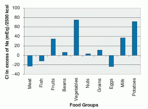
Despite possible differences, Na+ and Cl are consumed together in most foods. Although Na+ and Cl are major extracellular solutes, the amount of Na+ contained in food is quite small because the interstitial fluid represents a small fraction of the total fluid content of foods. Moreover, although the intracellular content of Cl is somewhat higher than Na+, intracellular content of both ions is still quite low (17). For these reasons, the salt content of food is low before preparation. The high intake of Na+ and Cl results from salt added to food in its preparation or during cooking. On average, the Na+ and Cl content in foods before processing tends to be equal; many plant-derived foods such as nuts, vegetables, fruits, and cereals contain more Cl than Na+ (17), whereas meat, fish, and eggs contain more Na+ than Cl (Fig. 6.1).


Fig. 6.1. Sodium chloride difference of major food groups. Most sodium (Na) and chloride (Cl) in food is now in the form of added salt (1:1 sodium-to-chloride ratio). In natural foods without added salt, chloride content is greater than sodium, with the exception of meat, fish, and eggs, which contain more sodium than chloride. (Reprinted with permission from Oh MS, Uribarri J. Electrolytes, water, and acid-base balance. In: Shils ME, Shike M, Ross AC et al, eds. Modern Nutrition in Health and Disease. 10th ed. Baltimore: Lippincott Williams & Wilkins, 2006:149-93.)


PATHOPHYSIOLOGY OF WATER AND OSMOLALITY


Osmolar Relations and Regulations

Measurement of Plasma Osmolality The plasma osmolality can be measured with an osmometer or estimated as the sum of the concentration of all the solutes in the plasma. NaCl, glucose, and urea are the major constituents of the plasma that contribute to plasma osmolality. The plasma osmolality is estimated from the following formula:

Plasma osmolality = Plasma Na+ (mEq/L) × 2 + Glucose (mg/dL)/18 + Urea (mg/dL)/2.8

Na+ always partners with its anion, Cl, to preserve electroneutrality, whereas the contribution of glucose and urea to the osmolality depends on fractional molecular weight. The molecular weight of glucose is 180 daltons, and that of urea is 28 daltons. Unlike NaCl or glucose, which largely remain in the plasma, urea can cross cell membranes and is not restricted to the extracellular fluid. As such, it is considered an ineffective osmol. Although urea can attain substantial concentrations in the plasma, its normal concentration is only 5 mOsm/L. Because of urea’s small contribution to the total osmolality, the total osmolality is nearly equal to the effective osmolality in normal plasma.


Dangers of Changes in Osmolality

Solutes that are restricted to the extracellular fluid and that contribute to the osmolality are called effective osmols, whereas solutes that can enter the cell freely are called ineffective osmols. Examples of effective osmols include glucose and Na+; examples of ineffective osmols are urea and alcohol. When the concentration of effective osmols increases, osmotic equilibrium is reestablished by water shifting from the cell to the extracellular fluid. Intracellular osmolality then increases to the same level as the extracellular osmolality (18, 19, 20). If ineffective osmols are added to the extracellular fluid, the osmotic equilibrium is reestablished by entry of those solutes into the cell. Because most of the solutes normally present in the extracellular fluid are effective osmols, loss of extracellular water, which can occur through insensible losses, will result in an increase in the effective osmolality and will cause a shift of water from the cells into the extracellular fluid. If extracellular osmolality is reduced, either by loss of normal extracellular solutes or by retention of water, a shift of water into the cells will occur to maintain osmolality. When the effective osmolality changes, cellular metabolism is affected, and cell swelling or shrinkage occurs as intracellular volume changes. Some of the most serious manifestations of altered osmolality are related to changes in brain cell volume because the brain is confined to a fixed space. Brain cells have the capacity to regulate their volume with time, and this explains why rapidity of alteration in osmolality is an important determinant of severity of symptoms (21).

Most of the signs and symptoms of a reduced concentration of Na+ (hyponatremia, representing a low osmolality) are caused by brain swelling and increased intracranial pressure and include nausea or vomiting, headache, papilledema, and mental confusion (22). With increasing severity, lethargy, weakness, hyperreflexia and hyporeflexia, delirium, coma, psychosis, focal weakness, ataxia, aphasia, generalized rigidity, and seizures occur and are caused by an increase in cell volume and reduced electrolyte concentration of the brain cells. GI manifestations include abdominal cramps, a temporary loss of sense of taste and flavor, decreased appetite, nausea, vomiting, salivation, and paralytic ileus. Cardiovascular effects of hypoosmolality are usually manifested as hypotension and other signs of low effective vascular volume. Hyponatremia can also be accompanied by muscle cramps, twitching, and rigidity (23, 24).

Increased effective osmolality need not be accompanied by a high serum Na+ concentration (hypernatremia), but hypernatremia is always accompanied by hyperosmolality. As in the hypoosmolal states, the signs and symptoms of hyperosmolality depend on the rapidity of development as well as the severity of hyperosmolality. In both human subjects and in animals, acute hyperosmolality from hypernatremia leads to subdural, cortical, and subarachnoid hemorrhages; it causes sudden shrinkage of brain cells and it creates negative pressure in the brain (25). Depression of mental status ranges from lethargy to coma. If the condition is severe, generalized seizure may also be observed but less commonly than in hypoosmolality. Muscular symptoms of hyperosmolality include muscular rigidity, tremor, myoclonus, hyperreflexia, spasticity, and rhabdomyolysis. In children with chronic hyperosmolality, spasticity, chronic seizure disorder, and mental retardation may occur (25).


Regulation of Thirst and Antidiuretic Hormone Release

If the effective osmolality rises, the hypothalamic osmoreceptor cells will shrink; this process then stimulates the thirst center in the cerebral cortex and stimulates antidiuretic hormone (ADH) production in the supraoptic and paraventricular nuclei (26, 27). If the effective osmolality declines, the osmoreceptor cells will swell; ADH production is inhibited. ADH produced in the hypothalamus is carried through long axons and is secreted from the posterior pituitary (28, 29, 30). Stimulation and inhibition of osmoreceptor cells affect production by the hypothalamus and ADH secretion by the posterior pituitary.

ADH secretion is extremely sensitive to changes in effective osmolality. A rise in the effective osmolality by just 2% to 3% stimulates ADH secretion sufficiently to result in maximally concentrated urine (25), whereas a decline in plasma osmolality of only 2% to 3% produces maximally dilute urine (<100 mOsm/L). ADH release is also regulated by nonosmotic factors such as nausea, pain,
and volume (22). A low effective vascular volume (˜10% decrease) provokes thirst and ADH release (31, 32, 33). These effects are mediated through baroreceptors and some humoral factors released in response to reduced blood flow. This response explains the severe thirst despite hyponatremia seen in heart or liver failure. Other factors, including β-catecholamines, angiotensin II, and physical and emotional stress, enhance ADH output. Ethanol and catecholamines inhibit the output of ADH. Lithium, certain tetracycline antibiotics (demeclocycline), foscarnet, methoxyflurane, amphotericin B, and V-2 receptor antagonists (Vaptans) inhibit the effect of ADH on the kidney (22).

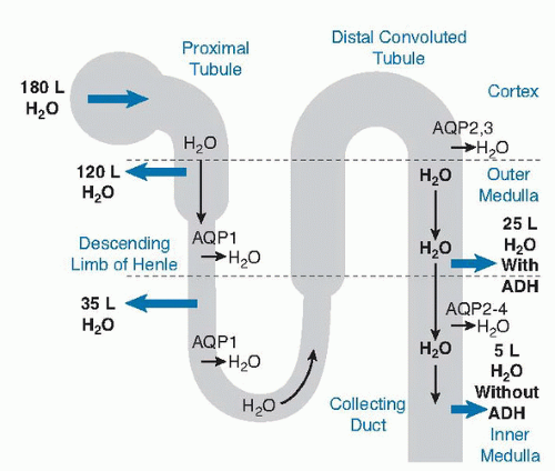
To understand the effect of ADH, consider that 180 L of water are filtered through the kidney daily; 120 L are reabsorbed in the proximal tubule, and 35 L are reabsorbed in the descending limb of Henle. All this water absorption is accompanied by salt absorption (in the case of the loop of Henle in the ascending limb), however, so no net change in osmolality occurs (34). The distal convoluted and collecting tubules reabsorb salt without water, so approximately 25 L of dilute urine are delivered to the collecting duct. When ADH is totally absent, approximately 5 L of water are reabsorbed in the inner medullary collecting duct, and 20 L are excreted as the final urine. In the presence of maximal ADH, urine volume can be as low as 0.5 L/day as the urine is concentrated to as high as 1200 mOsm/L and water is reabsorbed in the cortical and medullary collecting duct (Fig. 6.2). The reabsorption of water in the collecting duct is regulated by ADH. Water is conserved as the urine is concentrated. The net effect is osmotically concentrated urine (35, 36, 37, 38, 39, 40, 41, 42, 43, 44).


Fig. 6.2. Of the 180 L of water filtered through the kidney daily, 120 L are reabsorbed in the proximal tubule and 35 L are reabsorbed in the descending limb of Henle. Most of the remaining 25 L are reabsorbed in the collecting duct in the presence of antidiuretic hormone (ADH). When ADH is totally absent, approximately 5 L are reabsorbed in the collecting duct, and the remaining 20 L are excreted as the final urine. AQP, aquaporin.


Nonrenal Control of Water and Electrolyte Balance

Besides urinary losses of water, water is also lost from the skin and through normal respiration. Water is lost from the skin primarily as a means of eliminating heat, and the amount of water lost depends on the amount of heat generated in the body. In the absence of sweat or febrile illness, water loss from the skin is called insensible perspiration. Water loss from the skin depends mainly on the amount of heat generated in the body. Water loss from the skin is 30 mL/100 cal or approximately (˜300 to 1000 mL/24 hours). Besides water, sweat contains Na+ and K+ at a concentration of approximately 50 mEq/L and 5 mEq/L, respectively, and is approximately equivalent to 0.45% normal saline. The Na+ content of sweat varies, depending on the conditioning of the individual. An unconditioned individual placed in a hot environment (e.g., a new recruit in basic training) may have sweat that contains up to 100 mEq/L Na+, whereas after training the Na+ sweat content may be as low as 30 mEq/L. This difference explains why a higher dietary Na+ intake is required for unconditioned individuals (45).

Both fats and carbohydrates serve as the major energy sources for the body. In turn, these are broken down into carbon dioxide (CO2) and water. Both can be excreted through ventilation. Water is lost during normal ventilation because the water content of inspired air is less than that of the expired air. Ventilation is determined by the amount of CO2 production, which is determined by the caloric expenditure. The amount of water lost during ventilation also depends on caloric expenditure:

Respiratory water loss = 13 mL/100 kcal at normal partial pressure of CO2 (PCO2)

By burning calories, water is produced that is largely lost during normal respiration. In calculating water balance, respiratory water loss may be ignored in the measurement of insensible water loss, provided metabolic water gain is also ignored. In cases of hyperventilation or fever, respiratory water loss increases disproportionately to metabolic water production (1).

The net activity of the GI tract to the level of the jejunum is secretion of water and electrolytes. The net activity from jejunum to colon is reabsorption of water and electrolytes. Most of the fluid entering the small intestine is absorbed there, and the remainder is absorbed in the colon, leaving only approximately 100 mL of water to be excreted daily in the feces. The contents of the GI tract are isotonic with plasma, and any fluid that enters the GI tract becomes isotonic. Thus, if water is ingested and vomited, solute is lost from the body.


Dehydration and Volume Depletion

In any discussion of salt and water losses, the terms dehydration and volume depletion occur. Dehydration is characterized by water loss alone or an excess loss of water to
salt. Volume depletion describes the equal loss of salt and water. Salt in this case refers to NaCl, which is the main solute in the vascular space. Depending on the quantity of NaCl losses in relation to water losses, mixed forms of volume depletion and dehydration are encountered. In hypotonic dehydration, NaCl losses exceed water losses.


Volume Depletion

NaCl may be lost isotonically (i.e., at the same concentration as in the plasma) through the GI tract or directly from aspiration of the extracellular fluid from pleural effusions or ascites. With GI fluid loss, NaCl is lost with an equal or larger amount of water loss, and the osmolality of the body fluids is subsequently adjusted to isotonicity by changes in oral intake or urinary excretion of water. Isotonic fluid loss reduces only the extracellular fluid volume and can be treated with isotonic salt solution (0.9% normal saline).


Dehydration

The primary aberration in dehydration is water loss, and hypernatremia results from an increase in the concentration of Na+ in the extracellular space. This disproportionate excess of NaCl to water in the extracellular space can occur if water intake is inadequate or water loss is excessive. Dehydration resulting from excessive water loss usually develops more rapidly than dehydration caused by reduced water intake. A lack of water intake is always caused by one of two mechanisms: (a) a defect in the thirst sensing mechanism or impaired consciousness (46, 47) or (b) a lack of available water or an inability to drink water.


Hypotonic Dehydration.

Hypotonic dehydration (volume depletion with more Na+ than water loss) occurs when the patient loses NaCl and replaces the salt with water or with water containing less NaCl than the fluid that has been lost (see the later discussion of hyponatremia). In the presence of normal renal function, NaCl loss in excess of water loss is difficult to achieve because the kidney readily excretes the excess water through the suppression of ADH. This response is blunted or absent in patients with hypotonic dehydration (48).


PRINCIPLES OF FLUID THERAPY


Goals of Salt and Water Replacement

The goal of therapy is to restore the patient to normal. Deficits in volume and water must be identified and repleted; basal requirements for electrolytes and water must be supplied daily, and ongoing salt and water losses must be quantified and provided for in the treatment plan (49, 50).


Basal Requirements

The basal requirement for water depends on sensible ( urinary) and insensible losses of water (51). Fever increases both respiratory and skin water losses as a result of an increase in the basal metabolic rate. To some degree, urinary loss of water declines to compensate for these losses; however, urinary water losses do depend in part on the total amount of solute excreted and the degree to which the kidney can concentrate the urine. Solute excretion depends mainly on salt ingestion and protein intake, but severe glycosuria causes osmotic diuresis and increases urinary water losses.


Daily Water Requirements

In the absence of fever or exercise, water loss through the skin is relatively fixed, but urinary water losses vary greatly and depend on the total amount of solute to be excreted and urinary concentrating ability. For example, for a total solute excretion of 600 mOsm/day, the urine volume will be 500 mL if urine is concentrated to 1200 mOsm/L and 6 L if urine osmolality is 100 mOsm/L. For the former individual who can maximally concentrate the urine, the minimum water requirement would be 1100 mL (500 mL for urinary water loss plus 600 mL for skin water loss at 2000 cal/day). For the latter individual who is unable to concentrate the urine, the maximal allowable water intake would be 6.6 L. In the absence of an abnormality in urinary concentrating or diluting ability, large ranges of water intake are well tolerated as the kidney adjusts and maintains fluid homeostasis (2, 52). Nevertheless, in hospitalized patients, it is best not to overestimate water requirements to avoid water intoxication. Impairment in urinary dilution, as occurs in the syndrome of inappropriate ADH secretion (SIADH), is more common than impairment in urine concentration. In a conscious patient, thirst is an effective defense mechanism, whereas patients with severe hyponatremia often lapse into coma without warning (1).


POLYURIA

Polyuria, which is arbitrarily defined as an unintentional urine volume in excess of 2.5 L/day, can be caused by either osmotic diuresis or water diuresis (1). In osmotic diuresis, urine output increases as a result of an excessive rate of solute excretion. Certain solutes such as glucose, urea, mannitol, radiopaque media, and NaCl can cause osmotic diuresis in which the solute excretion rate exceeds 60 mOsm/hour or 1440 mOsm/day in the adult (1). In water diuresis, urinary osmolality is lower than plasma osmolality because the kidney excretes dilute urine and water is not reabsorbed in the collecting duct. Major reasons for reduced water reabsorption in the collecting duct can be attributed to drinking large amounts of water, lack of ADH (53, 54, 55, 56, 57, 58, 59, 60, 61, 62), or unresponsiveness to ADH (nephrogenic diabetes insipidus [DI]).

Nephrogenic DI can be either congenital or acquired. The lack of ADH can stem from a primary deficiency of ADH (central DI) or from physiologic suppression of ADH by a low serum osmolality. The latter results from the consumption or infusion of large amounts of water and is common among institutionalized patients with psychosis, particularly among those with schizophrenia (53, 54, 55, 63, 64). Various gradations of ADH deficiency can
be seen. In the setting of a partial ADH deficiency, the urine osmolality is close to normal. ADH deficiency can be congenital or acquired (56, 57, 58, 59, 60, 61). During pregnancy, ADH deficiency may be caused by excessive production of vasopressinase (gestational DI) (65, 66). Causes of polyuria are listed in Table 6.3. Note that urine outputs of more than 2.5 L may be considered desirable in patients with kidney stone formation.

Primary polydipsia is an increase in water intake in the absence of a physiologic stimulus such as hyperosmolality or volume depletion (53, 54, 55, 63, 64). It is usually psychogenic in origin, hence the term psychogenic polydipsia. An increase in urine output is caused by physiologic suppression of ADH secretion, and the serum Na+ is usually at the low range of normal. Occasionally, the serum Na+ may be low and indicates that the capacity of the GI tract to absorb water exceeds the normal capacity of the kidney to excrete water. In contrast, secondary polydipsia results from thirst stimulation in response to hyperosmolality. This condition is seen in patients with DI or patients with diabetes with severe glycosuria; the serum Na+ is usually in the high normal range.


TABLE 6.3 CAUSES OF POLYURIA


Water diuresis


A. Lack of ADH


1. Central diabetes insipidus


Congenital


Acquired (destruction of posterior pituitary)


Tumors


Granulomas


Pituitary surgery


Trauma


Infarction


Infection of the pituitary or hypothalamus


2. Primary (psychogenic) polydipsia


B. Failure of the kidney to respond to ADH


1. Congenital nephrogenic diabetes insipidus


Defect in the ADH receptor


Defect in aquaporin expression


2. Chronic renal failure


3. Acquired nephrogenic diabetes insipidus


Drugs


Lithium


Demeclocycline


Methoxyflurane


Heavy metals


Interstitial kidney disease


Amyloidosis


Sickle cell anemia or trait


Sarcoidosis


Electrolyte imbalance


Hypercalcemia


Hypokalemia


Obstructive uropathy


Solute diuresis


A. Saline loading


B. Postobstructive diuresis


C. Hyperglycemia


D. High-protein tube feedings


E. Salt-wasting nephropathy


ADH, antidiuretic hormone.


DISORDERS OF SODIUM METABOLISM


Hyponatremia

Hyponatremia occurs when the plasma Na+ concentration falls to less than 135 mEq/L. It is the most common electrolyte disorder and generally causes clinical concern when the concentration is less than 130 mEq/L. Pseudohyponatremia is a spurious reduction in serum
Na+ concentration resulting from a systematic error in measurement. Changes in methodology have largely reduced this problem in most clinical centers (75); however, the presence of a nonosmotic substance (in vitro hemolysis, hyperlipidemia, hyperproteinemia, and mannitol), may still cause pseudohyponatremia (76, 77, 78, 79).


Causes and Pathogenesis

The mechanisms responsible for a reduction in extracellular Na+ concentration (hyponatremia) are as follows:


  • Water can shift from the cell in response to an accumulation of extracellular solutes other than Na+ salts (78, 79, 80, 81, 82). Hyperglycemia causes hyponatremia by this mechanism, and the serum Na+ decreases by 1.6 mEq/L for every 100 mg/dL rise in the serum glucose. Serum osmolality is unchanged.


  • The body can retain excess water.


  • The body can fail to retain Na+ (83).


  • Na+ shifts into the cells.

In examples 2, 3, and 4, hypotonicity results, and the appropriate physiologic response is suppression of ADH release, which leads to rapid excretion of excess water and correction of hyponatremia. Therefore, persistence of hyponatremia indicates the failure of this compensatory mechanism. In most instances, hyponatremia is maintained because the kidney fails to produce a water diuresis, but sometimes ingestion of water in excess of the limits of normal renal compensation is responsible. The reasons for the inability of the kidney to excrete water include renal failure, reduced delivery of glomerular filtrate to the distal nephron, and the presence of ADH. After ruling out causes of pseudohyponatremia, an assessment of extracellular fluid volume provides a useful working classification of hyponatremia (84, 85, 86).

In most cases of hyponatremia, the main reason for the fall of serum Na+ is abnormal retention of water, which is either ingested or administered in the form of hypotonic fluids (83). Water retention can still occur despite the administration of isotonic fluid. This is seen in the setting of an increased amount of ADH, which leads to the excretion of hypertonic urine. The response is considered appropriate when ADH is released in response to hypertonicity of the body fluid or when the effective vascular volume is reduced. Hyponatremia in clinical states such as congestive heart failure and cirrhosis of the liver is associated with reduced effective vascular volume and is caused by increased secretion of ADH. Similarly, decreased perfusion may cause ADH secretion despite hyponatremia in hypothyroidism (87) and glucocorticoid deficiency states. The cerebral salt-wasting syndrome, which is defined as renal loss of salt caused by humoral substances released in response to cerebral disorders, such as acute subarachnoid hemorrhage, causes volume depletion that results in hyponatremia (88, 89, 90).

The term syndrome of inappropriate ADH (SIADH) is therefore reserved for ADH secretion that occurs despite hyponatremia and a normal or increased effective vascular volume. Causes of SIADH include tumors, pulmonary diseases such as tuberculosis and pneumonia, central nervous system diseases, and drugs, among others (Table 6.4) (21, 22, 91, 92, 93, 94, 95, 96, 97, 98, 99, 100, 101). Finally, mild hyponatremia may be caused by resetting of the osmostat to an osmolality lower than the usual level. In such cases, urine dilution occurs normally when the plasma osmolality is brought down to less than the reset level. Patients with chronic debilitating diseases such as pulmonary tuberculosis often manifest this phenomenon (102).


TABLE 6.4 CAUSES OF HYPONATREMIA


Disorders in which renal water excretion is impaired


A. Effective circulating volume depletion


1. Gastrointestinal losses


Vomiting


Diarrhea


Tube drainage


Intestinal obstruction


2. Renal losses


Diuretics


Hypoaldosteronism


Sodium-wasting nephropathy


3. Skin losses


Ultramarathon runners


Burns


Cystic fibrosis


4. Edematous states


Heart failure


Hepatic cirrhosis


Nephrotic syndrome


5. Potassium depletion


B. Thiazide diuretics


C. Renal failure


D. Nonhypovolemic states of ADH excess


1. Syndrome of inappropriate ADH secretion


2. Cortisol deficiency


3. Hypothyroidism


E. Decreased solute intake


Disorders in which renal water excretion is normal


A. Primary polydipsia


B. Reset osmostat


1. Pregnancy


2. Psychosis


3. Quadriplegia


4. Severe malnutrition


ADH, antidiuretic hormone.


Hypernatremia

Hypernatremia is defined as an increased Na+ concentration in plasma water. Whereas hyponatremia may not be accompanied by hypoosmolality, hypernatremia is always associated with an increase in the effective plasma osmolality and a reduced cell volume. An increase in the plasma osmolality should stimulate thirst. Hence, hypernatremia can occur only if the thirst mechanism is blocked, such as in the setting of altered mental status, or when an immobile patient has no access to water. Although the extracellular volume in hypernatremia may be normal, decreased, or increased, hypernatremia almost always occurs in the setting of volume depletion.


Causes and Pathogenesis

Theoretically, hypernatremia is caused by loss of water, reduced water intake, gain of Na+, or a combination of these (Table 6.5). In gain of Na+ in a person who has normal perception of thirst and the ability to drink water, however, the availability of water does not result in hypernatremia because a proportional amount of water is retained to maintain normal body fluid osmolality. The physiologic defense against hyponatremia is increased renal water excretion, whereas the physiologic defense against hypernatremia is an increase in water intake in response to thirst. Because thirst is such an effective and sensitive defensive mechanism against hypernatremia, it is virtually impossible to increase serum Na+ by more than a few milliequivalents (mEq) per liter if the mechanism for drinking water is intact. Therefore, a patient with hypernatremia always has reasons for reduced water intake. Reduced water intake occurs most commonly in comatose patients, in those
patients with a defective thirst mechanism, in those patients with continuous vomiting or who lack access to water, or in those patients with mechanical obstruction resulting from a condition such as an esophageal tumor.


TABLE 6.5 CAUSES OF HYPERNATREMIA


Water loss


A. Insensible loss


1. Increased sweating: fever, exercise


2. Burns


3. Respiratory infections


B. Renal loss


1. Central diabetes insipidus


2. Nephrogenic diabetes insipidus


3. Osmotic diuresis


Glucose


Mannitol


C. Gastrointestinal loss


1. Osmotic diarrhea


Lactulose


Malabsorption


Infectious enteritides


D. Hypothalamic disorders


1. Primary hypodipsia


2. Reset osmostat from volume expansion in primary mineralocorticoid excess


E. Water loss into cells


1. Seizures


2. Severe exercise


3. Rhabdomyolysis


Reduced water intake


A. Defective thirst


1. Altered mental status


2. Thirst center defect


B. Inability to drink water


C. Lack of access to water


Sodium retention


A. Administration of hypertonic sodium chloride or sodium bicarbonate


B. Ingestion of sodium


The excess gain of Na+ leading to hypernatremia is usually iatrogenic. This occurs in the setting of hypertonic saline infusion, accidental entry into the maternal circulation during abortion with hypertonic saline solution, or administration of hypertonic NaHCO3 during cardiopulmonary resuscitation or treatment of lactic acidosis. Reduced renal Na+ excretion leading to Na+ gain and hypernatremia usually occurs in response to dehydration caused by a primary water deficit. Water depletion resulting from DI, osmotic diuresis, or insufficient water intake leads to secondary Na+ retention in patients who continue to ingest Na+ or who are given Na+ (124).

Only gold members can continue reading. Log In or Register to continue

Stay updated, free articles. Join our Telegram channel

Jul 27, 2016 | Posted by in PUBLIC HEALTH AND EPIDEMIOLOGY | Comments Off on Water, Electrolytes, and Acid-Base Metabolism1

Full access? Get Clinical Tree

Get Clinical Tree app for offline access