Cobalamin (Vitamin B12)1,2


Cobalamin (Vitamin B12)1,2


Ralph Carmel


HISTORICAL BACKGROUND

The history of cobalamin is inextricably bound to the disease that provides the most common setting for its clinical deficiency, even though cobalamin deficiency can arise from many other causes. The reader is referred to excellent reviews of the dramatic scientific and clinical story (1, 2). In 1849, Addison reported several patients with a “remarkable form of anemia” that was accompanied by languor and restlessness, among other signs and symptoms. Although Addison mistakenly attributed the anemia to adrenal disease, his report is considered to be the first of the disease whose often fatal course later led Biermer to name it “pernicious anemia” (PA). That name is less apt now because the easily treated disease is no longer pernicious and because the disease is defined by its underlying gastric defect and not its anemic manifestation, which is sometimes minimal or even absent. Indeed, the strikingly megaloblastic anemia, although characteristic, is not specific to PA or even to cobalamin deficiency.

The classical experiments by Minot and Murphy (3) transformed the lethal course of PA by feeding affected patients large amounts of liver and documenting their hematologic improvement. For this work, they shared a Nobel Prize. The second important contribution was Castle’s discovery that patients with PA responded effectively to an “extrinsic factor” in the ingested liver or meat
when it was combined with an “intrinsic factor” (IF) in gastric juice (4). This demonstration sealed the longsuspected connection of PA with achylia gastrica. The third critical achievement was the identification of cobalamin as the extrinsic factor. The synthesis of cobalamin (5, 6) was accompanied by elucidation of its structure by Hodgkin (7), who was also awarded a Nobel Prize for her crystallographic work.

As biosynthetic fermentation made cyanocobalamin readily available, PA became easy to treat. The vitamin also became one of the most frequently given injections in the United States and acquired the dubious status of a frequently misused placebo and “energizer.” Having lost its grim implications, cobalamin deficiency began to be viewed with complacency by some health professionals, at times to their patients’ disadvantage.

The past several decades have extended methodologic advances in accurate and sensitive metabolic assays that allow identification of cobalamin deficiency at even earlier stages of development. As a result, asymptomatic subclinical cobalamin deficiency (SCCD) (8, 9) is now understood to be far more prevalent than the relatively rare state of clinical deficiency (10). This subclinical expansion has had major epidemiologic ramifications. Molecular understanding of cobalamin transport and metabolism and of their varied disorders has also advanced, with exploration of genetic influences and interactions with the environment and with acquired disorders.


BIOCHEMISTRY

Cobalamin contains a planar tetrapyrrole (corrin), at whose center sits a cobalt atom, and has critically attached moieties (Fig. 27.1). The cobalt fluctuates among monovalent, divalent, and trivalent states, with the reduced, monovalent cob(I)alamin being the active form. Linked to cobalt in the α position below the corrin plane is the 5,6-dimethylbenzimidazole nucleotide. Also linked to the cobalt atom but extending above the plane (β position) is any one of several interchangeable prosthetic moieties that lend their names to the cobalamin. The most important cobalamins are methylcobalamin, in which methyl is the prosthetic moiety, and deoxyadenosylcobalamin, in which 5′-deoxyadenosine is the β-linked moiety. Methylcobalamin predominates in cytoplasm and serves as a cofactor with 5-methyltetrahydrofolic acid (methylTHF) in the methylation of homocysteine to methionine (Fig. 27.2). Deoxyadenosylcobalamin predominates in mitochondria, where it serves as a cofactor in the intramolecular rearrangement of L-methylmalonylcoenzyme A (CoA) to succinyl-CoA in propionate metabolism (Fig. 27.3). These two are the only known roles for cobalamin in humans.

Other cobalamins include hydroxocobalamin, which is very stable and occurs widely; aquocobalamin; and sulfitocobalamin. Cyanocobalamin is a stable biosynthetic pharmaceutical that requires conversion to other cobalamins to become metabolically active; the term vitamin B12 refers specifically to cyanocobalamin (5), but it often serves as a catch-all name for cobalamins as a whole. Altered corrinoids with structural deletions are nonfunctional in humans but can find their way into tissues (11), even though cobalamin carriers, other than transcobalamin (TC) I, bind them poorly compared with functional cobalamins (12, 13, 14).


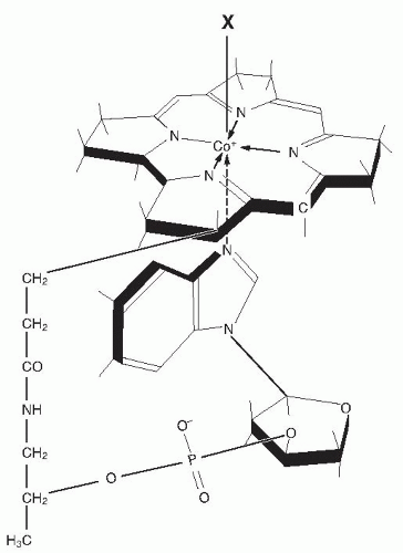
Fig. 27.1. The structure of cobalamin. Attached to the central cobalt atom of the corrin tetrapyrrole and to one of the pyrrole rings is the α-ligand, the 5,6-dimethylbenzimidazole nucleotide, extending below the corrin plane. The β-ligand (marked as X in the figure) above the plane can be any of several moieties such as methyl, 5′-deoxyadenosyl, hydroxyl, or cyanide. (Reprinted with permission from Carmel R. Megaloblastic anemias: disorders of impaired DNA synthesis. In: Greer JP, Foerster J, Lukens JL et al, eds. Wintrobe’s Clinical Hematology. 11th ed. Philadelphia: Lippincott Williams & Wilkins, 2004.)

Many confusing terminologies have been applied to the cobalamin-binding proteins. This chapter uses TC I and TC II for the two plasma carriers, terms with the longest usage and in conformity with the genetic nomenclature of TCN1 and TCN2, respectively. Others introduced the names haptocorrin and transcobalamin, respectively. An older, common name for TC I in the literature was R binder.


ANALYTIC METHODS

Analytic methods to diagnose cobalamin deficiency fall into two categories: measures of cobalamin amount, such
as cobalamin and holotranscobalamin (holo-TC) II assays; and measures of functional metabolic status, such as the metabolite biomarkers, methylmalonic acid (MMA) and homocysteine, or complex cellular metabolic indicators such as the deoxyuridine suppression test. When clinical signs of deficiency are evident, one test usually suffices as confirmation (15), but in research and epidemiologic surveys, the frequent absence of clinical identifiers usually requires the application of more than one diagnostic biomarker (16). Unfortunately, no diagnostic gold standard exists.


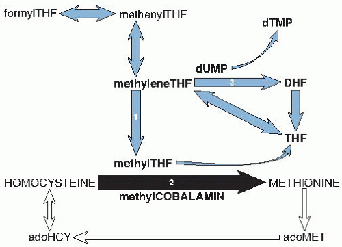
Fig. 27.2. Schematic diagram of the intersection of cobalamin (black arrow) with folate metabolism (blue arrows) and the methionine cycle (white arrows). The direct role of folate in thymidylate synthesis (reaction 3) is also shown. Reaction 1: Reduction of 5,10-methylene tetrahydrofolic acid (THF) to 5-methylTHF by methyleneTHF reductase, which requires riboflavin. Reaction 2: Remethylation of homocysteine to methionine by methionine synthase, with methylTHF and methylcobalamin as cofactors; the THF that is produced is reused in the folate metabolic cycle. Reaction 3: Conversion of deoxyuridylate (dUMP) to deoxythymidylate (dTMP) by thymidine synthase, in which 5,10-methyleneTHF is converted to dihydrofolic acid (DHF). (See the chapter on folic acid for fuller details of folate metabolism.) adoHCY, S-adenosylhomocysteine; adoMET, S-adenosylmethionine.


Serum Cobalamin

Cobalamin exists as methylcobalamin and other forms in serum (17). Serum cobalamin is stable on long-term storage (although specific forms may be converted upon exposure to light) and can be assayed by various techniques. The earliest methods employed microorganisms, such as Euglena gracilis and Lactobacillus leichmannii, whose growth is proportional to the unknown sample’s cobalamin content (17). The assays, now automated, are still considered the standard by some reference laboratories. Radioisotopic methods rely on competitive binding of the sample’s cobalamin by purified IF as the added cobalamin-binding protein; IF must not be contaminated with TC I, which also binds nonfunctional corrinoids and causes falsely high cobalamin results in samples with high levels of such corrinoids. Immunoenzymatic chemiluminescence techniques using anti-IF antibody to capture IF-complexed cobalamin now predominate in diagnostic use. These highly proprietary, automated assays have been less well defined and monitored than previous methods. They also appear susceptible to falsely normal results in some cobalamin-deficient sera (18, 19), probably by failing to inactivate PA samples’ endogenous anti-IF antibody (19). This selective error does not seem to affect normal sera, which complicates detection of the error; it may explain puzzling reports of normal cobalamin levels despite severe cobalamin deficiency (16, 20).


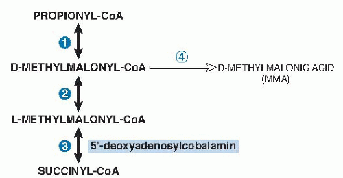
Fig. 27.3. The sole function of 5′-deoxyadenosylcobalamin in humans. The mitochondrial conversion of propionyl-coenzyme A (CoA), which derives from diverse sources to succinyl-CoA, which enters the tricarboxylic acid cycle, passes through three reversible reactions. Reaction 1: Carboxylation of propionyl-CoA by propionyl-CoA carboxylase, requiring adenosine triphosphate (ATP), biotin, and magnesium. Reaction 2: Racemization of D-methylmalonyl-CoA by methylmalonyl-CoA racemase. Reaction 3: Intramolecular rearrangement of L-methylmalonyl-CoA to succinyl-CoA by L-methylmalonyl-CoA mutase, which requires 5′-deoxyadenosylcobalamin. In addition, an irreversible side reaction converting D-methylmalonyl-CoA to methylmalonic acid, mediated by D-methylmalonyl-CoA hydrolase, produces methylmalonic acid (reaction 4). The metabolic fate of methylmalonic acid is largely unknown, but a fraction is excreted by the kidneys.

The cutoff point between subnormal and normal serum cobalamin values varies from method to method and laboratory to laboratory (16). Most laboratories use traditional decision limits (cut-points) of 200 to 250 ng/L (148 to 185 pmol/L) to define normality. The sensitivity of low cobalamin levels for cobalamin deficiency has been questioned but much depends on what kind of deficiency is considered (15, 16). Sensitivity exceeded 95% in patients who had obvious clinical manifestations of deficiency, such as megaloblastic anemia or neurologic abnormalities (16, 21, 22, 23). The lower the cobalamin level, the greater is the likelihood of clinically severe deficiency (17, 24, 25), but exceptions exist (26, 27). As with all biomarkers, diagnostic sensitivity decreases in subclinical conditions, and it is 38% to 39% in SCCD (16). Low-normal cobalamin levels with associated metabolic abnormalities are not unusual in population surveys (28, 29, 30), but metabolic abnormalities can be spurious sometimes (16).

The cobalamin insensitivity in such surveys led some investigators to raise the cut-point for deficiency from the traditional 200 or 250 ng/L to 350 ng/L (258 pmol/L) or higher to ensure that no case of deficiency was
missed (28). This change, adopted by many laboratories, instantly increased the frequency of diagnosed deficiency. The overdiagnosis it creates is substantial. For example, the 5.3% to 24.8% frequency of “abnormal” cobalamin values in four surveys became 40.5% to 71.7% in the unexceptional elderly populations (15). More relevantly, reanalysis showed that only one third of the persons thus recategorized had MMA or homocysteine abnormalities, thus, making two thirds of the new diagnoses metabolically suspect; moreover, very few of the one third had clinical deficiency (10, 16). Furthermore, the advantages of overdiagnosis remain unrealized because the health risks of SCCD and the benefits of intervention in such cases remain unproven.

As mentioned, low cobalamin levels’ specificity may be more limited than their sensitivity. Table 27.1 lists the conditions associated with low serum cobalamin. The most notable causes of falsely low levels include pregnancy and folate deficiency (17, 31), although true deficiency may accompany occasional cases. Substantive genetic influences on cobalamin levels are also becoming evident. TCN1 mutations often cause TC I deficiency (32, 33), and this deficiency may explain 15% of low cobalamin levels (27) and thus may make it a more frequent cause of low serum cobalamin than PA.

Genetic influences can increase cobalamin levels, too, such as the unexplained moderately higher levels in homozygotes for a common polymorphism of the α-1,2 fucosyltransferase gene (34, 35). Cobalamin levels are higher in Africans than in Asians or whites (36), perhaps for genetic reasons. Spuriously, high cobalamin levels in medical settings most often result from renal failure or are idiopathic (37). Autoantibodies to TCs can be therapy-induced (38) or spontaneous (39, 40), and they may explain 8% of elevated cobalamin levels (41). Less frequent but often dramatic elevations accompany extremely high plasma TC I levels in chronic myelogenous leukemia and some cancers (42).


TABLE 27.1 CONDITIONS FREQUENTLY ASSOCIATED WITH LOW SERUM COBALAMIN LEVELS


Cobalamin deficiency


Clinically expressed deficiencya


Subclinical deficiencyb


Normal cobalamin status


Pregnancy


Transcobalamin I deficiency (severe or mild)c


Human immunodeficiency virus infection; acquired immunodeficiency syndromed


Folate deficiency


Multiple myelomad


Aplastic anemia; myelodysplastic syndrome


Multiple sclerosis


Drugs (e.g., metformin, omeprazole)e


a Deficiency can be severe or very mild. The disorders usually arise from severe intrinsic factor-related malabsorption, such as pernicious anemia.

b Deficiency is expressed only biochemically. The causes of the deficiency are usually unknown; some arise from cobalamin malabsorption limited to food-bound cobalamin.

c Severe deficiency is rare but mild deficiency may be common.

d Most patients have no evidence of cobalamin deficiency or malabsorption, but some may have coexisting deficiency or malabsorption (described in some patients with acquired immunodeficiency syndrome) or coexisting pernicious anemia (described in some patients with multiple myeloma).

e Drugs taken briefly or irregularly rarely affect cobalamin status. Metformin decreases cobalamin levels by an unknown mechanism, and evidence of deficiency is weak. Omeprazole reduces cobalamin absorption, but low cobalamin levels have been few (it may require many years of constant use to induce deficiency).


Despite its limitations, cobalamin assay remains the first test of choice for now in patients suspected of cobalamin deficiency (16, 43). Whatever the circumstances, including laboratory errors, incongruous laboratory results that conflict with a patient’s clinical picture must always be pursued further (15). Additional tests of value when the diagnosis remains uncertain are discussed next. Cobalamin rise after therapy lacks specificity and, therefore, diagnostic value.


Methylmalonic Acid

MMA accumulates in serum, and some MMA is excreted in the urine, when deoxyadenosylcobalamin-dependent methylmalonyl-CoA mutase activity is reduced (see Fig. 27.3). MMA can be measured reliably by gas chromatography-mass spectrometry. Most laboratories define serum values higher than 280 nmol/L as abnormal, although cut-points have varied between 210 and 480 nmol/L (16, 21, 44, 45). MMA is elevated in 98% of patients with PA who have clinically expressed cobalamin deficiency, often to levels exceeding 1000 nmol/L (21, 22, 23, 46). MMA elevation reverses soon after cobalamin therapy (22, 23).

MMA elevation is usually milder in SCCD because cobalamin stores are not severely depleted, and the sensitivity of high MMA, although not established because a gold standard comparator is lacking, is probably lower than in clinical deficiency (16, 29, 30, 47). Specificity is clearly superior to that of homocysteine because folate status does not affect MMA (22, 23, 46). However, major known influences on MMA include, in order of frequency, glomerular filtration (even a minimal decrease raises serum MMA), cobalamin status, age, and perhaps sex (45, 48), and they explain only 16% of the MMA variation (45). Asymptomatic babies can have moderate MMA elevation that remits spontaneously after the first year of life (49). The cause is unknown, but the association with mild homocysteine and cobalamin changes (neither of which usually reach abnormality) and their improvement after cobalamin treatment raise the possibility of relative cobalamin deficiency (50).

Many experts consider MMA the best metabolic test available to confirm cobalamin deficiency, and normal MMA values provide strong evidence against deficiency. However, MMA cannot be the diagnostic gold standard because its specificity is undefined (16). It is particularly
unclear what mild MMA elevations without any other abnormalities mean (16, 51, 52). The improvement of many isolated MMA elevations after cobalamin is given, which suggests that these elevations represented mild SCCD (23, 30, 51). However, normal MMA levels often decline after cobalamin therapy, too—a finding suggesting regression to the mean or supersaturation of methylmalonyl-CoA mutase by the cobalamin as alternative explanations. A noteworthy longitudinal study of 432 patients left untreated over 1 to 3.9 years observed that 44% of isolated mild elevations of MMA improved spontaneously and only 16% progressed (53). Antibiotics sometimes reduce cobalamin-unresponsive MMA elevation (23, 54)—a finding suggesting that increased propionate metabolism by some intestinal bacteria may directly elevate MMA without cobalamin deficiency.


Homocysteine

Elevation of total homocysteine because of impaired methionine synthase activity is nearly as sensitive as MMA elevation for cobalamin deficiency. Sensitivity is 95% when cobalamin deficiency is clinically overt, and the homocysteine elevation is often striking (23, 46). However, hyperhomocysteinemia has many causes (55, 56), including preanalytic influences such as delayed blood sample processing or using serum instead of plasma, both of which promote artifactual release of red blood cell homocysteine. Renal status and folate status affect homocysteine more than does cobalamin status (28, 55, 56), as observed best in countries that have not fortified the diet with folic acid (48). The relative impact of cobalamin status on homocysteine rises in the aged, who have high rates of cobalamin deficiency. Other important influences on homocysteine include sex, genetic polymorphisms (especially methylene THF reductase), drugs, alcohol abuse, lifestyle factors, and disorders of homocysteine transsulfuration (55, 56). Cut-points for homocysteine results have varied widely, and this affects case definition. Plasma levels lower than 10 µmol/L are considered optimal, but many laboratories use cut-points of 12 to 14 µmol/L in adult men and 10 to 12 µmol/L in premenopausal women.

Homocysteine is more reliable than cobalamin and is as reliable as MMA in monitoring therapeutic response in cobalamin deficiency; both homocysteine and MMA elevation respond to cobalamin but not to folic acid (22, 23).


Holotranscobalamin II

Lindemans et al (57) first suggested that measuring only the serum cobalamin attached to TC II, the carrier that promotes cellular uptake of cobalamin, may enhance diagnostic specificity and sensitivity of cobalamin testing. Holo-TC II, the TC II with attached cobalamin, originates in the ileal cell but may also have renal origins. Because holo-TC II is taken up by cells rapidly, less than 20% to 30% of plasma cobalamin is in holo-TC II at any moment; the remainder is carried by TC I, which does not promote specific cellular uptake (42).

Holo-TC II assays are now accurate, and a fully automated method is commercially available (58). As with other cobalamin-related biomarkers, holo-TC II cut-points vary widely between 19 and 50 pmol/L (16). Direct comparisons show a minor advantage for holo-TC II over (total) cobalamin; areas under the curve by receiver operator curve analyses varied between 0.75 and 0.90 for holo-TC II versus between 0.72 and 0.85 for cobalamin (16). The claim that holo-TC II does not decline spuriously like cobalamin in pregnancy and therefore permits better characterization of cobalamin status in pregnant women (59) may be premature: an unnoticed holo-TC II rise actually followed delivery (prenatal levels were not determined)—a finding suggesting that holo-TC II had probably indeed declined during pregnancy (16). Most holo-TC II studies have involved large amorphous groups defined almost solely by their MMA levels, whose specificity is itself uncertain. Clinical or absorptive status was rarely assessed, and disagreements between holo-TC II and cobalamin results were rarely explored (16). One of the few clinical studies of patients with mild clinically expressed deficiency did not find holo-TC II to be superior to cobalamin in predicting response to therapy (60).

Controversy persists about holo-TC II because very little is known about other influences on it. Renal failure can produce striking holo-TC II elevation (37), but preliminary reports of many cobalamin-unrelated influences on holo-TC II such as alcohol abuse, folate deficiency, myelodysplasia, and Gaucher disease still await clarification (16, 61). Moreover, holo-TC II levels lower than control values in cobalamin-repleted patients with PA (62) suggest that cobalamin absorption affects holo-TC II independently of metabolic status. Such dual influences could introduce diagnostic nonspecificity; for example, it seems possible that even transient dietary change or malabsorption (e.g., drug induced) lasting only several days or weeks and unaccompanied by and unlikely to evolve into cobalamin deficiency might cause low holo-TC II levels. These and other influences may explain isolated holo-TC II elevations that have heretofore been attributed to an unusual sensitivity of holo-TC II for SCCD.


NUTRITION AND BIOAVAILABILITY

Certain bacteria and archaea synthesize cobalamin (63); some also synthesize corrinoids that are nonfunctional in humans. Animals that ingest the microorganisms incorporate the cobalamin (17, 64, 65). Animals and their products contain diverse amounts of cobalamin: from 139 µg/100 g in dark muscle of skipjack to 83 µg in cooked beef liver, 10 µg in shellfish, and 3 to 8.9 µg in salmon and other fish, to 0.9 to 1.4 µg in eggs and 0.3 µg
in milk (65). Plants are negligible sources of cobalamin, although dried green and purple algae contain cobalamin that may be bioavailable (65). Better methods are needed to quantitate food content and to differentiate cobalamins that are usable by humans from corrinoids that are not (65). As important as content are features such as bioavailability, which can vary 10-fold among different foods and stability after cooking or processing. Milk and cobalamin-fortified cereals are particularly efficient sources in the US diet and fish in the Norwegian diet, and all exceed meat in their cobalamin bioavailability (65, 66, 67).

The uniqueness of active cobalamin absorption resides in an IF-mediated system of limited capacity that maximizes the bioavailability of ingested cobalamin, whether free or food-bound, while simultaneously preventing excess absorption, perhaps especially to exclude nonfunctional or even harmful corrinoid analogs. Great disparities in efficiency exist between active and passive absorption, both of which begin with cobalamin release from food. More than 50% of the cobalamin in a typical meal will be absorbed actively if the IF system, which includes IF and its uptake system, is intact. However, IF cannot accommodate much more than 2 µg cobalamin at a time (Table 27.2). Larger doses, such as those found in many supplements, exceed the capacity of the IF system. The excess cobalamin then becomes dependent on passive, nonspecific absorption, which is much less efficient (1% to 2% of the dose is absorbed) even though it is nonsaturable and linearly related to the amount of cobalamin presented. The absorption characteristics of free cobalamin in supplements are often assumed to remain unchanged when taken together with food, but the assumption appears unjustified in persons with PA (68) or food-bound cobalamin malabsorption (FBCM) (69).


ABSORPTION

IF-mediated absorption of cobalamin predominates in the ileum, where IF receptors are most abundant (17, 31). This efficient process, designed to secure and concentrate cobalamin maximally, is illustrated in Figure 27.4.

The food-bound cobalamin is released by gastric pepsin, whose activity becomes optimal at the acid pH of the normal stomach and degrades the food proteins that bind cobalamin (70) (see Fig. 27.4, panel 1). The parietal cells that provide the acid also synthesize and secrete IF, a 48-kDa glycoprotein that binds cobalamin specifically. However, the released cobalamin is preferentially bound at the low gastric pH by TC I (also called haptocorrin or R binder), a glycoprotein secreted by salivary gland epithelial cells.

Pancreatic proteases degrade TC I/haptocorrin as it leaves the stomach and is exposed to pancreatic alkalinization, which potentiates trypsin activity (71). The rereleased cobalamin is then bound by IF in the intestine, as presumably is biliary cobalamin that is exposed to proteases there (13) (see Fig. 27.4, panel 2). IF, unlike TC I/haptocorrin, binds inactive corrinoid analogs poorly (12).

The IF-cobalamin complex travels to the ileum (see Fig. 27.4, panel 3), where it is taken up by the IF receptor, cubilin, a nontransmembrane, multiligand receptor on gut epithelial cells (72); the membrane localization and function of the 460-kDa cubilin are supported by amnionless, the 45-kDa protein that provides the transmembrane and cell signaling function in a complex called cubam (73, 74). After classical internalization and endocytosis, the cubilin-IF-cobalamin complex is split in the ileal cell endosomes. The cobalamin eventually reaches the abluminal surface of the ileal cell, where it exits into the bloodstream bound to TC II (75) approximately 4 hours after oral ingestion (17).


TABLE 27.2 COBALAMIN BIOAVAILABILITY FROM A SINGLE ORAL DOSE GIVEN TO PERSONS WITH (A) NORMAL ABSORPTION AND (B) ABNORMAL ABSORPTION WITHOUT INTRINSIC FACTOR (PERNICIOUS ANEMIA)a


ORAL DOSE


(A) AMOUNT (AND %) ABSORBED BY BOTH INTRINSIC FACTOR-MEDIATED AND PASSIVE PROCESSESb


(B) AMOUNT (AND %) ABSORBED BY PASSIVE PROCESS ONLYc


0.25 µg


0.19 µg


(75%)


1 µg


0.56 µg


(56%)


≈0.02 µg


(≈2%)


2 µg


0.92 µg


(46%)


3 µg


≈0.08 µg


(≈3%)


5 µg


1.4 µg


(28%)


10 µg


1.6 µg


(16%)


≈0.2 µg


(≈2%)


50 µg


1.5 µg


(3%)


≈0.5 µg


(≈1%)


100 µg


≈1.8 µg


(≈1.8%)


500 µg


≈6 µg


(≈1.2%)


aData are not available for the partially compromised absorption in patients with food-bound cobalamin malabsorption, whose absorption efficiency is unknown but is presumed to be intermediate between (A) normal and (B) severely impaired absorption.

b In normal subjects, as evident from comparing columns A and B, intrinsic factor-mediated absorption in normal subjects exceeds passive diffusion more than tenfold until intake exceeds 10 µg.

c Intrinsic factor activity is lost in patients with pernicious anemia, so absorption is by passive diffusion only. The numbers in this column were obtained in a different study from those of normal subjects in the left-hand column. Comparability may therefore be limited, but the striking patterns illustrate the essential points.


Data combined from Chanarin I. The Megaloblastic Anaemias. 2nd ed. Oxford: Blackwell Scientific, 1979:94; and Berlin H, Berlin R, Brante G. Oral treatment of pernicious anemia with high doses of vitamin B12 without intrinsic factor. Acta Med Scand 1968;184:247-58.


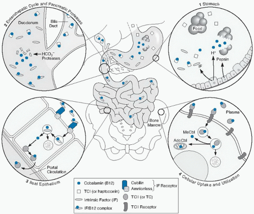
Fig. 27.4. Absorption and cellular uptake cycle of cobalamin in humans: 1, secretion of gastric intrinsic factor (IF), acid, and pepsin and release of cobalamin from food and binding to TC I (R binder or haptocorrin); 2, biliary and pancreatic secretion and degradation of TC I by pancreatic enzymes; 3, ileal cell uptake of IF-cobalamin by cubam (the cubilin-amnionless receptor), lysosomal processing, and transfer of transcobalamin (TC) II-cobalamin to the portal circulation; 4, cellular uptake (e.g., in bone marrow) of plasma TC II-cobalamin, lysosomal processing, and release of cobalamin for mitochondrial or cytoplasmic attachment to enzymes. AdoCbl, adenosylcobalamin; MeCbl, methylcobalamin. (Modified from Carmel R, Rosenblatt, DS. Disorders of cobalamin and folate metabolism. In: Handin RI, Lux SE, Stossel TP, eds. Blood: Principles and Practice of Hematology. 2nd ed. Philadelphia: Lippincott Williams & Wilkins, 2003. Originally adapted with permission from Carmel R. Cobalamin deficiency. In: Carmel R, Jacobsen DW, eds. Homocysteine in Health and Disease. Cambridge: Cambridge University Press, 2001.)

IF-mediated absorption of ingested cobalamin and, presumably, also reabsorption of much of biliary cobalamin are efficient but saturable. The only alternative pathway is nonspecific, passive diffusion. This inefficient process, discussed in the previous section, assumes importance only when the IF mechanism is damaged, as in PA, or overwhelmed by cobalamin doses larger than a few micrograms. Diffusion is not limited to the ileum and also occurs at nongastrointestinal surfaces, such as the sublingual or nasal epithelium. The quantitative efficiencies of active and passive intestinal absorption are compared in Table 27.2.


TRANSPORT, METABOLISM, AND EXCRETION

Cobalamin crosses membranes poorly and depends on several binding proteins for its transport throughout the body. The IF-mediated absorption process is limited to the gastrointestinal tract, although cubilin is abundant in renal tubular cell brush borders (72) and inexplicable IF fragments have been found in urine (76). Once absorbed
cobalamin reaches the bloodstream, its transport and uptake depend on TC II. TC I also binds cobalamin in the blood, but its role appears to involve withholding cobalamin and, possibly more importantly, nonfunctional corrinoid analogs from cells. Hepatic clearance of cobalamin through bile approximates 1.4 µg daily; approximately 70% is normally reabsorbed, presumably by IF, whereas the remainder is lost in feces (13) along with most corrinoid analogs.


Transcobalamin II (also called Transcobalamin)

The TCN2 gene shares considerable homology with the GIF gene for IF, although it is located on a different chromosome (77). A common genetic variant substitutes proline for arginine in TCN2 codon 259; its effects are uncertain, but it may impair TC II function slightly (78). TC II has a critical role in the blood, but small amounts also exist in milk, cerebrospinal fluid, semen, and elsewhere (42). Holo-TC II rapidly delivers its cobalamin to all tissues (see Fig. 27.4, panel 4), and its plasma half-life is only 90 minutes. A specific, calcium-dependent, 282-amino acid cell membrane receptor, whose gene has been identified (79), is ubiquitous and seems to be regulated in synchrony with cell cycles (80). The holo-TC II-receptor complex is internalized by endocytosis. However, megalin, a separate calcium-dependent, 600-kDa, multiligand receptor for holo-TC II also exists in enterocytes, yolk sac, and other tissues (72). Megalin has been studied best in the renal tubule (81), where it may help conserve cobalamin by reabsorbing large amounts of filtered holo-TC II. The need for two receptor systems for TC II awaits explanation.


Cellular Metabolism

After uptake, cobalamin is released within endosomes and enters the cytoplasm, where it exists primarily as methylcobalamin or is taken up by mitochondria (see Fig. 27.4, panel 4). The cytoplasmic methylcobalamin participates in homocysteine remethylation (see Fig. 27.2), and the mitochondrial 5′-deoxyadenosylcobalamin takes part in propionyl-CoA metabolism (see Fig. 27.3).


Transcobalamin I (also called Haptocorrin or R binder)

Plasma TC I originates in the specific granules of granulocyte precursors (82). It is structurally and immunologically identical to TC I in secretions, which derives from exocrine gland epithelial cells, but glycosylation varies considerably throughout (42). TC I appears to have no specific cellular receptors, thus leaving the plasma TC I-cobalamin complex (holo-TC I) to circulate with a half-life of 9 to 10 days; therefore, holo-TC I usually carries 70% or more of plasma cobalamin (83). Cross-species animal experiments suggest that plasma holo-TC I eventually becomes desialylated and is cleared by the nonspecific asialoglycoprotein receptors in the liver (84). This process may initiate much of the enterohepatic recycling of cobalamin. Plasma TC I also carries 100 to 380 pmol/L of corrinoid analog (14) and may divert unusable or even potentially harmful corrinoids from cells and also promote their fecal excretion through bile.

TC I exists in saliva, breast milk, tears, and other secretions, often in very high concentrations. The TC I presence in both secretions and granulocytes and its ability to withhold cobalamin and analogs from both tissues and microorganisms suggest that it may also serve an antibacterial role (42).

Jul 27, 2016 | Posted by in PUBLIC HEALTH AND EPIDEMIOLOGY | Comments Off on Cobalamin (Vitamin B12)1,2

Full access? Get Clinical Tree

Get Clinical Tree app for offline access