 Vessels of the Thigh
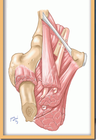
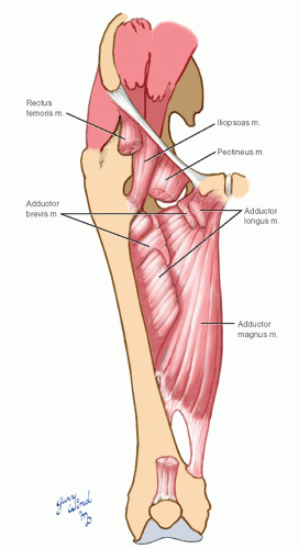 Fig. 16-1 The adductor muscles of the thigh fan out to attach along the linea aspera of the femur. |
Surgical Anatomy of the Thigh
Muscles
Between the bifurcation of the common femoral artery in the femoral triangle and the beginning of the popliteal artery at the adductor hiatus, the deep and superficial divisions of the femoral artery traverse the thigh anteromedial to the femoral shaft, in intimate contact with the adductor muscles. The adductor muscles (Fig. 16-1) originate from the inferior ramus of the pubis and ischium and fan out to attach to the linea aspera along the posterior side of the femur. The deepest of these muscles, the adductor magnus, attaches to the full length of the linea aspera beginning below the lesser trochanter and ending at the adductor tubercle. It is interrupted by four small apertures through which perforating branches of the deep femoral artery reach the posterior compartment, and by the large adductor hiatus at the lower third of the femur through which the superficial femoral artery passes. The lower part of the adductor brevis muscle is located between the magnus and the adductor longus muscles. The pectineus muscle, from the superior pubic ramus, covers the superior part of the adductor brevis muscle.
The posterior view of the adductor magnus muscle (Fig. 16-2) shows the more horizontal direction of the pubic fibers and the predominantly longitudinal ischial fibers. The tendinous openings can be seen along the linea aspera.
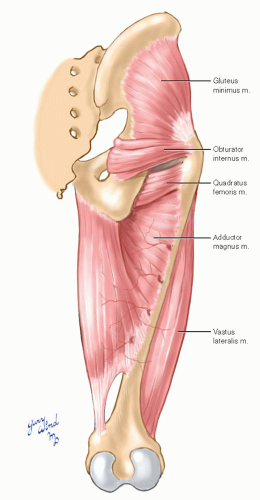 Fig. 16-2 In this posterior view, perforating branches of the deep femoral artery can be seen passing through openings in the tendinous portion of the adductor magnus muscle. |
The anterior compartment of the thigh consists of the quadratus femoris muscle, which is made up of four heads: rectus femoris, vastus medialis, vastus lateralis, and vastus intermedius (Fig. 16-3). These muscles enlarge from tapered origins proximally to a bulky teardrop form distally. The deep and superficial femoral vessels lie in the cleft between the vastus medialis and adductor muscles.
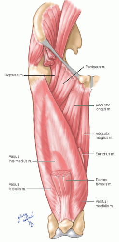 Fig. 16-3 The anterior compartment of the thigh consists of the bulky quadratus femoris muscle. |
Posteriorly, the long hamstrings, the biceps femoris, semimembranosus, and semitendinosus muscles run the length of the thigh from the ischial tuberosity to the tibia and fibula. They lie across the lower portion of the posterior surface of adductor magnus muscle. The upper portion of the adductor magnus muscle is covered by the insertion of the gluteus maximus muscle into the upper part of the linea aspera (Fig. 16-4).
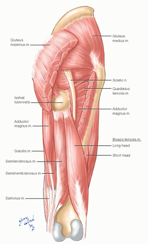 Fig. 16-4 The posterior thigh musculature is shown. |
The medial adductor compartment forms a superiorly based pyramid between the quadriceps muscle and the hamstrings (Fig. 16-5). In cross section (Fig. 16-6), the bulky adductor magnus muscle has a roughly triangular profile, with a narrow linear medial attachment along the medial lip of the linea aspera.
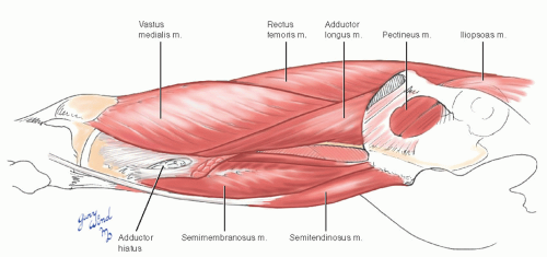 Fig. 16-5 The medial adductor compartment is interposed between the quadratus femoris and hamstring muscles. The body of the adductor magnus has been resected in this view. |
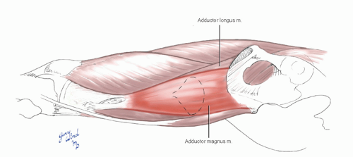 Fig. 16-6 The adductor magnus muscle tapers medially to form a narrow linear attachment along the linea aspera of the femur. |
Vessels
The common femoral artery enters the femoral triangle beneath and slightly medial to the midpoint of the inguinal ligament (Fig. 16-7). Within the femoral triangle it divides into deep and superficial branches. The superficial branch crosses the adductor longus muscle to lie beneath the sartorius muscle. The deep (profunda) branch passes between the pectineus and adductor longus muscles to lie beneath the latter muscle.
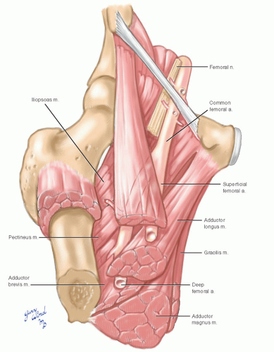 Fig. 16-7 The relationship between the branches of the femoral artery and thigh musculature is shown. |
The superficial femoral artery supplies the adjacent adductor muscles and the quadriceps muscle (Fig. 16-8). The deep femoral branch supplies the adjacent adductor muscles and sends three perforating branches and its termination through the tendon of the adductor magnus muscle to supply the hamstrings in the posterior compartment.
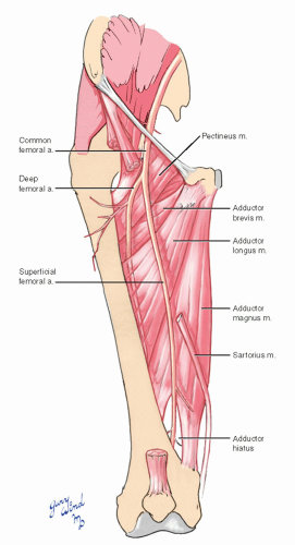 Fig. 16-8 The deep and superficial femoral branches are separated by the adductor longus muscle. |
At the apex of the femoral triangle, the superficial femoral artery enters a triangular fascialined cleft, the adductor (Hunter’s) canal, between the vastus medialis, the sartorius, and the adductor longus (upper portion) and adductor magnus (lower portion) muscles. The canal takes a 90° twist as it descends toward the knee (Fig. 16-9). The roof of the adductor canal is a sling of tough fascia crossing from the vastus medialis to adductor muscles lying just deep to the sartorius muscle. The superficial femoral artery lies superficial to the robust accompanying superficial femoral vein. Two branches of the femoral nerve, the sensory saphenous nerve and the motor nerve to the vastus medialis muscle, accompany the superficial femoral vessels in the canal.
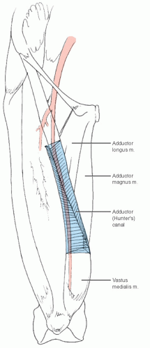 Fig. 16-9 Hunter’s canal twists 90° as it descends toward the knee. |
The deep femoral vessels (Fig. 16-10) lie close to the femur beneath the adductor longus muscle. They first lie on the adductor brevis muscle, then directly on the adductor magnus muscle. Its upper perforating branch or two transverse both deeper adductor muscles, whereas the lower branches penetrate only the tendon of the magnus to reach the posterior compartment.
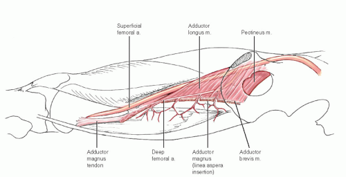 Fig. 16-10 The relationship of the deep femoral vessels to the adductor muscles is shown. |
Cross sections of the thigh show the relationships of the vessels to the muscular compartments (Fig. 16-11). The lateral intermuscular septum of the thigh between the vastus lateralis and biceps femoris muscles is dense and well developed. There is firm adhesion between the surface of the adductor longus muscle and the adjacent vastus medialis muscle that requires sharp dissection to separate. The remaining interfaces between muscle groups are less well defined.
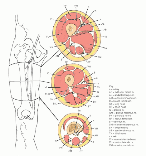 Fig. 16-11 Cross-sectional (caudal) views of the thigh demonstrate the relationship of the femoral vessels to the surrounding musculature. |
Stay updated, free articles. Join our Telegram channel

Full access? Get Clinical Tree


