Most commonly on palate, gingiva, or alveolar ridge mucosa
Usually single lesion, but on rare occasions, there may be multiple
Well-circumscribed, yellow, red, or gray plaque
Rough or granular surface
• In skin
Most commonly occur in anogenital area
Usually single lesion but may be multiple
Well-circumscribed, pink or grayish nodule or plaque
Rough or bosselated surface similar to warts
• No clear disease association
Microscopic
• Verrucous epithelial/epidermal hyperplasia
• Epidermal ridges extend downward but all extend to same depth
• Epidermal ridges are wide and bulbous
• Often keratinocyte necrosis
• Neutrophilic infiltrate is common
• Dermal pegs between elongated epidermal ridges are cellular
Vacuolated or foamy macrophages
Top Differential Diagnoses
• Condyloma acuminatum
• Verrucous carcinoma
• Granular cell tumor
• Verruca vulgaris
• Seborrheic keratosis
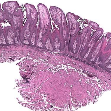
Verrucous Mucosa Note the verrucous and papillomatous nature of the overlying mucosa. The lamina propria is abnormally pale , indicative of the histiocytic infiltrate in those areas.
Pale Lamina Propria At a slightly higher power, the pale nature of the superficial lamina propria is better appreciated when compared to the eosinophilic deeper soft tissue.
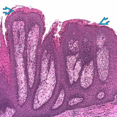
Higher Magnification of Mucosa Although the mucosa is verrucous and papillomatous, no keratinocytic atypia is present. Overlying parakeratosis with intracorneal neutrophils are seen. A mild lymphocytic infiltrate is seen in the lamina propria with some extension into the mucosa.
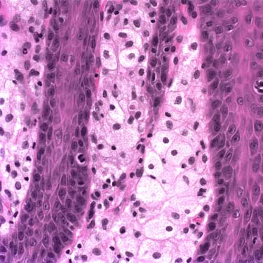
High Magnification Showing Foamy Histiocytes High power clearly shows abundant foamy histiocytes in the superficial lamina propria. No atypia is present. Also, the cytoplasm is not granular as seen in granular cell tumors of the oropharynx.
TERMINOLOGY
Definitions
• Verrucous epithelium/epidermis with foamy macrophages in lamina propria/dermis
CLINICAL ISSUES
Presentation
• Usually occurs in oral cavity, but may occur in skin and other mucosal sites
Anogenital region, ear, nose, digits
• In oral cavity
Most commonly on palate, gingiva, or alveolar ridge mucosa
Usually single lesion, but on rare occasions, there may be multiple
Well-circumscribed, yellow, red, or gray plaque
Rough or granular surface
May be raised or depressed
Only gold members can continue reading. Log In or Register to continue


 , indicative of the histiocytic infiltrate in those areas.
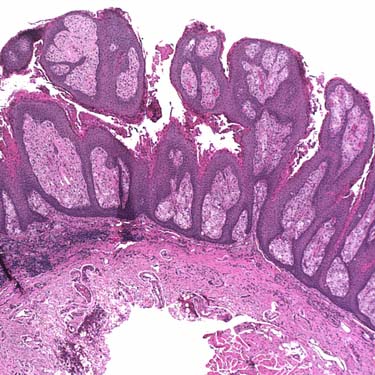
 with intracorneal neutrophils are seen. A mild lymphocytic infiltrate is seen in the lamina propria with some extension into the mucosa.