Fig. 7.1
(a–d) Examples of Grade 0—no rejection. Note the normal epidermis and minimal, superficial perivascular, lymphocyte-predominant inflammatory infiltrate
7.1.2 Grade I Rejection: Mild
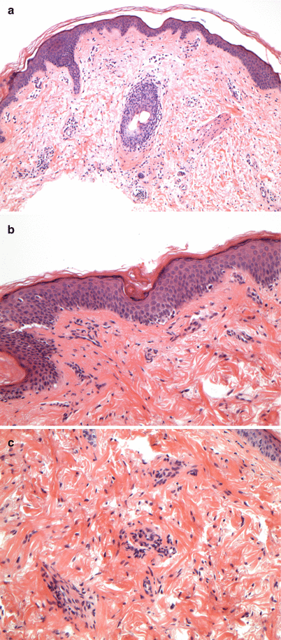
The presence of mild inflammation characterizes mild, acute cellular rejection (ACR). There should be no involvement of the overlying epidermis (Figs. 7.2).


Fig. 7.2
(a–c) Examples of Grade I—mild rejection. Mild, superficial perivascular, lymphocyte-predominant inflammatory infiltrate. No epidermal involvement
7.1.2.1 Differential Diagnosis
Viral exanthem
Drug eruption
7.1.3 Grade II Rejection: Moderate
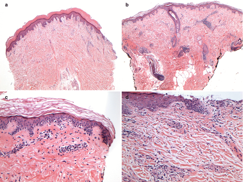
The presence of a moderate to severe degree of perivascular inflammation, with or without mild epidermal and/or adnexal involvement (limited to spongiosis and exocytosis) without epidermal dyskeratosis/apoptosis characterizes moderate ACR (Fig. 7.3).


Fig. 7.3
(a–d) Examples of Grade II—moderate rejection. Moderate to severe perivascular inflammation with or without mild epidermal and/or adnexal involvement. Note the lymphocyte exocytosis into the epidermis without epidermal dyskeratosis/apoptosis (c). Note the epidermal spongiosis and lymphocyte exocytosis without epidermal dyskeratosis/apoptosis (d)
7.1.3.1 Differential Diagnosis
Drug eruption
Eczematous process (allergic/irritant contact dermatitis)
Id reaction
Infectious process (fungal, bacterial, viral)
7.1.4 Grade III Rejection: Severe
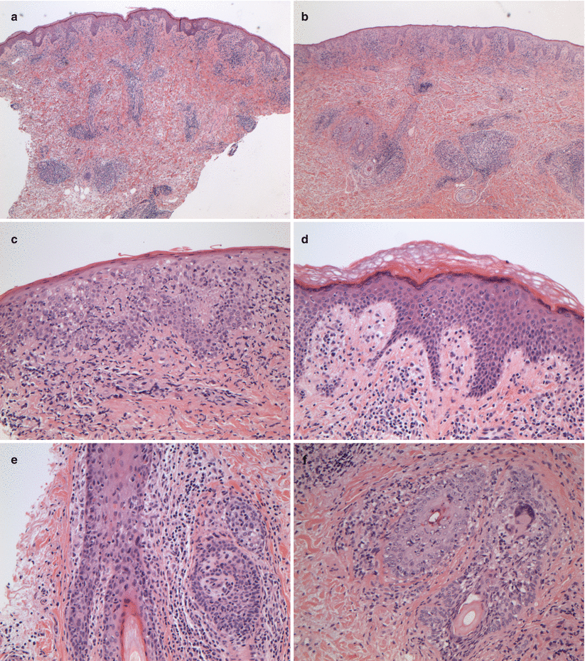
The presence of dense inflammation and epidermal involvement with epithelial apoptosis, dyskeratosis, and/or keratinolysis characterizes severe ACR (Fig. 7.4).


Fig. 7.4
(a–f) Examples of Grade III—severe rejection. Dense inflammation and epidermal involvement with epithelial apoptosis and dyskeratosis. Note the epithelial dyskeratosis/apoptosis involving the epidermis and the follicular epithelium (c–f)
7.1.4.1 Differential Diagnosis
Drug eruption
Eczematous process (allergic/irritant contact dermatitis)
Stay updated, free articles. Join our Telegram channel

Full access? Get Clinical Tree


