Figure 82-1. Schematic diagram of the fibrous cardiac skeleton.
Two papillary muscles arise directly from the ventricular wall: the anterolateral and posteromedial papillary muscles. Importantly, the anterolateral papillary muscle usually is supplied by two coronary arteries, the left anterior descending artery and branches of the circumflex artery. On the other hand, the posteromedial papillary muscle is usually supplied by a single coronary artery, either from the right coronary or the circumflex artery, which makes it twice as likely to rupture from ischemia and infarction as the anterolateral papillary muscle. The papillary muscles play an important role in the proper function of the MV. MV closure and appropriate leaflet coaptation are permitted by end-diastolic and early systolic lengthening of the papillary muscles.3
Chordae tendineae attach the leaflets to the papillary muscles or directly to the ventricular wall and can be categorized based on the attachments. Primary chordae attach to the leaflets at the free edge to ensure proper coaptation without prolapse or flail. The secondary chordae attach along the line of coaptation and are more prominent on the anterior leaflet. Tertiary chordae arise directly from the ventricle or trabeculae carneae and are only present on the posterior leaflet. Finally, commissural chordae attach to both leaflets and arise from either papillary muscle.
The structures in close proximity to the MV and, therefore, susceptible to injury during MV surgery include the AV, the atrioventricular node, and the circumflex coronary artery (Fig. 82-3).

Figure 82-2. Schematic diagram of the relationship of the aortic valve to the underlying structures.
AORTIC STENOSIS
Prevalence and Etiology
1 Aortic stenosis (AS) is the most prevalent valvular heart disease in developed countries. The most common cause of AS is degenerative calcific disease, followed by congenital AS due to bicuspid valve anatomy. Rheumatic AS is becoming exceedingly uncommon in developed countries due to efficient prevention of rheumatic heart disease.

Figure 82-3. Schematic diagram of the relationship of the mitral valve to the underlying structures.
Degenerative Calcific Aortic Stenosis
The most frequent cause of AS is degenerative calcification of the AV. The prevalence of degenerative AS in persons older than 65 years, which is the most commonly affected age group, is 2%.4 The degenerative process that leads to stiffening of the aortic leaflets is the result of proliferative and inflammatory changes with lipid accumulation and infiltration of macrophages and T lymphocytes.4–9 Fibrosis and calcification initially affect the base of the leaflets, but ultimately progress to immobilization of the leaflets due to large calcific deposits that can extend deep into the annulus. These deposits may also extend onto the ventricular surface of the anterior leaflet of the MV, as well as into the wall of the ascending aorta. The risk factors for the development of calcific AS are similar to those for atherosclerosis and include elevated serum levels of low-density lipoprotein (LDL) cholesterol, diabetes, smoking, and hypertension.10,11
Bicuspid Aortic Stenosis
Calcification of bicuspid AVs, which are present in approximately 2% of the general population, represents the most common form of congenital AS. In patients with bicuspid AVs, the left and right coronary cusps are usually fused, while the noncoronary cusp is freestanding. Gradual calcification of the bicuspid valve results in AS with typical onset of symptoms in the fifth or sixth decade of life, in contrast to degenerative AS, which causes symptoms in elderly individuals. Bicuspid AS is frequently associated with degenerative changes in the wall of the ascending aorta with resultant dilation or aneurysm formation.
Rheumatic Aortic Stenosis
Introduction of effective antibiotic therapy has resulted in a decline in the prevalence of rheumatic fever and rheumatic valve disease. Rheumatic AS is caused by inflammation and thickening of the AV leaflets, producing a mixture of AS and regurgitation. Rheumatic AS is rarely an isolated disease, and usually occurs in conjunction with MV stenosis.12
Pathophysiology
Regardless of the etiology of AS, the pathophysiologic consequences are similar. Narrowing of the AV to one-quarter of its normal area of 3 to 4 cm2 produces a significant pressure gradient between the left ventricle and aorta. There is a resultant increase in LV workload and compensatory LV hypertrophy.
Even though this hypertrophy is an appropriate response to the increased afterload, there are numerous harmful effects. First, the increased wall thickness makes the ventricle stiff and less compliant. This leads to diastolic dysfunction and increased wall tension. In addition to diastolic dysfunction, systolic dysfunction, typically occurring later in the course of the disease, can develop from chronic ischemia. All of the following contribute to increased myocardial oxygen demand: increased LV muscle mass, increased wall tension, increased systolic ventricular pressure, and increased systolic ejection time. There is also decreased coronary artery perfusion, which occurs during diastole, due to increased wall tension, increased diastolic ventricular pressure, and decreased diastolic aortic pressure.13,14 The subsequent ischemia of the subendocardium due to increased oxygen demand and decreased perfusion leads to cell death and fibrosis. Chronically, this ischemia results in systolic dysfunction.
Diagnosis
Symptoms
The most common symptoms of AS are angina pectoris, syncope, and heart failure.15 Angina pectoris occurs in 30% to 50% of patients with severe AS. It is a reflection of myocardial ischemia caused by increased metabolic demands and decreased coronary perfusion. Coronary artery disease, which affects more than 70% of elderly patients with degenerative AV disease, causes further deterioration of myocardial perfusion and lowers the threshold for angina.
Syncope is most commonly due to reduced cerebral perfusion that occurs during exertion. Reduced cerebral perfusion is a result of decreased mean arterial pressure from peripheral vasodilation in the presence of a fixed cardiac output. Approximately 15% of patients present with syncope and only 50% of these survive for 3 years.
Congestive heart failure in patients with severe AS is typically a sign of advanced and longstanding disease. It is marked by shortness of breath and dyspnea with exertion, and results from ongoing LV outflow obstruction. Heart failure is a consequence of the aforementioned diastolic and systolic dysfunction from decreased compliance and ischemia, respectively. In addition, as the left ventricle becomes less compliant, atrial systole becomes more important for maintaining cardiac output and the onset of atrial fibrillation may result in worsening of congestive heart failure.
Some patients with severe AS may develop serious gastrointestinal bleeding secondary to angiodysplasias, occurring predominantly in the right colon, and also in the small bowel and stomach. These result from shear stress–induced platelet aggregation with reduction in high–molecular-weight multimers of von Willebrand factor.
Signs
Signs of AS include a loud systolic ejection murmur that radiates to the neck and is often accompanied by a thrill. “Pulsus parvus et tardus” describes a weak and prolonged arterial pulse characteristic of advanced AS. The weak pulse is a reflection of a narrowed pulse pressure, while the slow rise in pulse reflects a prolonged ejection of blood volume through a stenotic valve.16
Electrocardiogram and Imaging
2 The electrocardiogram typically shows signs of LV hypertrophy, which is found in the majority of patients with AS. Echocardiography represents the “gold standard” modality for the diagnosis of AS. Two-dimensional (2D) echocardiography with Doppler allows precise real-time analysis of ventricular and valvular anatomy and function. The most important objective of echocardiography is correct assessment of the severity of AS using Doppler echocardiography to calculate jet velocity, mean transvalvular pressure gradient, and valve orifice area (Table 82-1). It is also used to assess valve thickening and calcification, as well as reduced leaflet motion. Distinction between bicuspid and TVs is often possible, particularly when the amount of calcification is small.

Two-dimensional echocardiography is also invaluable in detecting associated MV disease and in assessing LV hypertrophy, systolic function, and diastolic performance. Ejection fraction is used to measure LV systolic function. However, a severe decrease in ejection fraction can falsely lower estimates of severity of AS due to low-pressure gradients. Stress echocardiography with dobutamine administration may be required to properly assess the severity of valvular disease and to distinguish it from primary contractile dysfunction with lack of contractile reserve.17
3 Cardiac catheterization with direct measurement of the pressure gradients across the AV to calculate the severity of stenosis has been replaced by less invasive echocardiography. The current indication for cardiac catheterization is limited to preoperative evaluation of coronary artery disease.
Natural History
The natural history of AS is marked by a prolonged latent period with few symptoms and minimal morbidity. Even patients with moderately severe AS have a slow decrease in AV area, generally by approximately 0.1 cm2 per year.18,19 The natural history of severe AS correlates well with the onset and severity of symptoms. Life expectancy of patients with severe, untreated AS and angina is approximately 5 years. Patients presenting with syncope have life expectancies of 3 years. Presence of congestive heart failure in patients with severe, untreated AS is associated with a worse prognosis, with the time of death occurring less than 2 years from the onset of symptoms (Fig. 82-4).20

Figure 82-4. Natural history of aortic stenosis without operative treatment. Onset of symptoms identifies patients at high risk of death over the next 2 to 5 years.
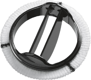
Figure 82-5. St. Jude Medical Regent Valve.
Treatment
4 There is no effective medical therapy for patients with severe AS. Diuretics and digitalis may improve the symptoms of congestive heart failure. Mechanical relief of AS is accomplished by surgical AV replacement (AVR), percutaneous AVR, or percutaneous balloon aortic valvotomy (PBAV).
Surgical Aortic Valve Replacement
The primary indication for surgery is the presence of symptoms in patients with severe AS. AVR is also indicated in patients with severe AS and reduced LV function and in patients with moderate to severe AS who also require coronary, other valve, or aortic surgery.17 Recent studies suggest that AVR may also be beneficial in patients with asymptomatic severe AS and severe LV hypertrophy.21
Choice of valve prosthesis for AVR is primarily influenced by the patient’s age. Mechanical prostheses are made of carbon, require lifelong anticoagulation, and are very durable (Fig. 82-5). Mechanical prostheses are therefore indicated in patients younger than 65 years old. Stented biologic prostheses are most commonly made of bovine pericardium or porcine valve leaflets, do not require anticoagulation, and have a limited durability of approximately 15 years (Fig. 82-6). Biologic prostheses are used in elderly patients (older than 65 years) and in younger patients in whom long-term anticoagulation with warfarin is contraindicated (bleeding diathesis, peptic ulcer disease, etc.).
Percutaneous and Transapical Aortic Valve Replacement
Percutaneous AVR is an emerging therapy for patients previously deemed inoperable due to prohibitive operative risk. This is a novel treatment utilizing a bioprosthesis sutured to a balloon or self-expandable stainless steel or nitinol stent (Fig. 82-7). The prosthesis is introduced through the femoral artery retrogradely into the aorta and placed at the midpart of the native stenotic AV. The radial forces of the stent push the native AV aside to increase the valve orifice area. The prospective trial (Placement of Aortic Transcatheter valves: PARTNER) randomized “high-risk” patients (operative mortality >15%) to either transfemoral AVR (TF-AVR) or conventional AVR.22 Thirty-day mortality was doubled in the conventional group, but at 1 year, survival was similar. In the “inoperable” subset, TF-AVR was shown to be significantly superior to optimal medical therapy with improvement in NYHA scores to 1–2 and a 20% improvement in mortality at 5-year follow-up. Hemodynamic benefits and valve integrity also persisted at 5 years.23 In patients with severe peripheral vascular disease, the retrograde arterial approach cannot be used. In these patients, similar prostheses can be inserted directly into the beating heart through the LV apex (transapical approach). This approach has been shown to have a lower rate of vascular complications, postoperative heart block necessitating permanent pacemaker implantation and paravalvular regurgitation as compared to the TF-AVR with similar 1-year survival.24–26 In conclusion, appropriately selected high-risk or inoperable patients can benefit significantly from TF-AVR in both survival and functional status, and in those where femoral access is not an option, the transapical technique is warranted.

Percutaneous Balloon Aortic Valvotomy
PBAV is a procedure in which a balloon is placed across a stenotic AV and inflated in order to decrease the degree of valve narrowing. This procedure is a valuable tool for treatment of AS in children and young adults. Rapid development of restenosis and clinical deterioration limits its application in the treatment of adults to those with severe AS who are not candidates for conventional surgical or transcatheter AVR.
AORTIC REGURGITATION
Prevalence and Etiology
5 AR results from the improper coaptation of the AV leaflets due to either intrinsic leaflet abnormalities or aortic root distortion. The most common causes of AR include bicuspid valve disease, rheumatic fever, and endocarditis. Regurgitant bicuspid AVs are often partly calcified, which results in limited opening and closing of the valve and a mixture of AR and AS. Rheumatic fever causes inflammation and fibrosis of the leaflets, while endocarditis causes destruction of leaflets. In addition, AR can occur as a secondary phenomenon of aortic root enlargement due to Marfan syndrome, anuloaortic ectasia, or aortic dissection. Most of these causes produce chronic AR with a slow increase in LV size. Some lesions, in particular acute aortic dissection and endocarditis, cause acute AR with a sudden decrease in cardiac output.
Pathophysiology
In patients with AR, inappropriate coaptation of the aortic leaflets causes diastolic reflux of blood from the aorta into the LV with a consequent increase in LV end-diastolic volume and pressure. In chronic AR, compensatory mechanisms of the LV result in dilation and hypertrophy. LV hypertrophy and dilation ultimately result in a decrease in LV function and heart failure.
Diagnosis
Symptoms
Chronic AR is usually well tolerated and patients with even severe AR often remain asymptomatic for years. Initial symptoms of severe AR include fatigue, shortness of breath, and dyspnea on exertion. Advanced AR is marked by the onset of congestive heart failure, syncope, and angina. Acute onset of severe AR, such as in cases of aortic dissection or endocarditis-induced valve destruction, causes a sudden increase in LV end-diastolic volume and pressure with subsequent cardiogenic shock and pulmonary edema.
Signs
Lateral displacement of the point of maximum impulse is seen in chronic AR due to LV dilation and hypertrophy. The classic auscultatory findings of AR are best heard at the right sternal border of the second intercostal space and include a high-pitched, blowing holodiastolic decrescendo murmur; an S3 heart sound; and a systolic ejection murmur.

Figure 82-7. Schematic diagram of percutaneous aortic valve replacement. A: Balloon valvuloplasty. B: Balloon catheter with valve in the native diseased valve. C: Balloon inflation to secure the valve. D: Valve in place.

Additionally, the widened pulse pressure leads to a number of interesting findings, such as bounding pulses (water-hammer pulse), Quincke pulse (pulse in fingernail bed), De Musset sign (rhythmic bobbing of the head), Duroziez sign (systolic and diastolic murmurs heard over the femoral artery when it is gradually compressed), and Müller sign (pulsations of the uvula).16
Imaging
Echocardiography represents the diagnostic “gold standard” for patients with AR. It allows assessment of the valvular and aortic anatomy, the severity of regurgitation, and the size and function of the left ventricle. Assessment of the severity of AR is determined by color Doppler jet width, regurgitant volume, regurgitant fraction, and regurgitant orifice area (Table 82-2). The role of coronary angiography is reduced to the preoperative diagnosis of coronary artery disease.
Natural History
The natural history of AR is strongly dependent on the presence of symptoms and LV dysfunction at the time of presentation. Asymptomatic patients with normal LV function develop symptoms and/or LV dysfunction at 6% per year. Asymptomatic patients with LV dysfunction at presentation develop cardiac symptoms at 25% per year. Finally, the mortality rate of symptomatic patients is 10% per year.17
Treatment
Management of patients with AR depends on the severity of symptoms and the size and function of the left ventricle. Asymptomatic patients with chronic severe AR and preserved LV size and function are treated with vasodilator medical therapy, which reduces afterload and improves forward flow.
Surgery is indicated in symptomatic patients with chronic severe AR and in asymptomatic patients with severe AR and evidence of LV dilation (end-systolic LV diameter >55 mm) or dysfunction (ejection fraction <50%). Additionally, acute severe AR with consequent pulmonary edema and shock caused by endocarditis or aortic dissection is a surgical emergency.
AVR should be performed before irreversible changes in LV function occur. Mechanical valves are indicated for patients younger than 65 years of age. Stented bioprostheses are used for AVR in patients older than 65 and in younger patients with contraindications to long-term anticoagulation. AV repair can be performed in patients with severe AR caused by bicuspid valve disease or connective tissue disorders. Traditionally, patients with proximal aortic root pathology, including dissection and aneurysms with secondary AR underwent aortic root replacement with a mechanical valve. This was done regardless whether or not their native valve was diseased. The valve sparing root replacement (David procedure) has been shown to have similar morbidity and mortality with minimal risk of thromboembolism or endocarditis. Additionally, it precludes the need for anticoagulation and has an acceptably low reoperation rate. This makes it ideal for young patients, especially those with connective tissue disorders.27–30
MITRAL STENOSIS
Prevalence and Etiology
6 Mitral stenosis (MS) is predominantly caused by rheumatic fever. The steady rate of decline of rheumatic fever in developed countries has resulted in a similar decline in the prevalence of MS. Other far less common causes of MS include severe annular and leaflet calcification, congenital malformations, malignant carcinoid, left atrial myxoma, left atrial thrombus, and endocarditis. A definitive history of rheumatic fever can only be obtained in 50% to 60% of cases and women are affected more often than men by a 2:1 ratio.31,32 Rheumatic fever commonly occurs in childhood or adolescence and can lead to a postinfective pancarditis, affecting to various degrees the valves, endocardium, myocardium, and pericardium. In MS due to rheumatic fever, there is leaflet thickening and calcification, chordal shortening and fusion, and commissural fusion, which all lead to a smaller, funnel-shaped mitral orifice. This deformation can also prevent complete closure of the valve, which is evidenced by concomitant regurgitation in about half of patients with MS.
Pathophysiology
The cross-sectional area of the normal MV is 4 to 5 cm2. Normally, there is a trivial diastolic pressure gradient present to move blood across the MV from the left atrium into the left ventricle. An increasing gradient is required as the MV becomes more narrowed, and a significant transvalvular gradient first develops when there is reduction of the mitral orifice to less than 2.5 cm2, which represents mild MS. The increased atrial pressure leads to left atrial enlargement and the pressure is subsequently transmitted retrograde into the pulmonary veins, capillaries, and arteries. The determinants of the transvalvular gradient, and therefore the determinants of the left atrial pressure, are atrial contractility, cardiac output, and heart rate. Consequently, the gradient and atrial pressure are increased if atrial kick is lost (decreased atrial contractility), flow rate across the valve increases (increased cardiac output), or transit time across the valve is shortened (increased heart rate).
Diagnosis
Symptoms
Symptoms of left-sided heart failure, such as dyspnea, orthopnea, and paroxysmal nocturnal dyspnea, are the primary indicators of MS and are typically triggered by exertion, stress, infection, pregnancy, or the abrupt onset of atrial fibrillation. The increased heart rate and cardiac output that can occur under these circumstances, and the mechanical obstruction inherent to MS, lead to an increased transvalvular gradient and left atrial pressure. The ensuing pulmonary congestion results in dyspnea. Likewise, the loss of atrial kick with atrial fibrillation increases the gradient and atrial pressure. Patients with atrial fibrillation may also present with palpitations or systemic embolization.
Patients infrequently present with hoarseness, dysphagia, hemoptysis, and symptoms of right-sided heart failure. Hoarseness and dysphagia may result if left atrial enlargement is sufficient to compress surrounding structures. Hemoptysis may occur from significant pulmonary venous hypertension. Symptoms of right-sided heart failure arise when right ventricular function is impaired due to an increased afterload from the stenotic MV and secondary pulmonary hypertension. Still, other patients may present without symptoms but have an abnormal physical examination.
Signs
The left ventricle is typically not enlarged; thus, the point of maximum impulse is not displaced. The typical auscultatory findings of MS are best heard at the apex and include a low-pitched, rumbling middiastolic murmur; an accentuated first heart sound; and an opening snap. These findings may be absent with a heavily calcified immobile valve, severe pulmonary hypertension, or low cardiac output. Physical findings of pulmonary hypertension, such as a loud pulmonic component of the second heart sound (P2), a right ventricular heave, distended neck veins, hepatomegaly, ascites, and peripheral edema, can also be observed with MS.16
Imaging
Echocardiography is the principal tool used in the diagnosis of MS. It is used to assess the morphologic characteristics of the valve apparatus, which include leaflet mobility, flexibility, and thickness; presence of calcifications and subvalvular fusion; and appearance of the commissures. Additionally, Doppler echocardiography is utilized to determine the hemodynamic severity by measurement of the mean transvalvular pressure gradient, pulmonary artery systolic pressure, and valve area (Table 82-3). This morphology and severity information is fundamental in determining the timing and type of intervention to be used.17
The enlarged left atrium gives rise to characteristic findings on the chest radiograph, which includes displacement of the left main-stem bronchus superiorly and displacement of the esophagus posteriorly. Additionally, calcification of the mitral leaflets and enlarged pulmonary arteries with cephalization of pulmonary blood flow can be seen.
Similar to the diagnosis of other valvular heart disease, cardiac catheterization with direct pressure measurement has largely been replaced by echocardiography.
DIAGNOSIS
Table 82-3 Classification of Mitral Stenosis Severity

Stay updated, free articles. Join our Telegram channel

Full access? Get Clinical Tree


