OBJECTIVES
Prostate
1 Define the anatomy and physiology of the prostate gland.
2 Compare the clinical presentation, workup, and management of a patient with acute prostatitis, a patient with chronic prostatitis, and a patient with nonbacterial prostatitis.
3 Define the clinical presentation, workup, and management of a patient with benign prostatic hypertrophy.
4 Describe how clinical evaluation and diagnostic studies can help a practitioner to distinguish between benign prostatic hypertrophy and prostate cancer.
5 Discuss the use of prostate-specific antigen determinations in evaluating patients with carcinoma of the prostate and benign prostatic hypertrophy.
6 Outline the staging and management of prostate cancer, both localized and advanced.
Kidneys
1 Discuss the types of renal trauma, the mechanisms involved, and the appropriate management of each.
2 Discuss two congenital urinary tract anomalies that require intervention.
3 Discuss the etiology and management of inflammatory renal disease, and outline the treatment of pyelonephritis.
4 Describe the workup for a renal mass lesion. Discuss the characteristic findings in benign and malignant renal masses.
5 Describe the workup and treatment options in the management of patients with calculous disease of the urinary system.
Ureters
1 Describe the etiology, clinical presentation, sequelae (if untreated), and management of ureteral obstruction.
2 Outline the management of iatrogenic ureteral injuries.
Bladder
1 Describe the different tests used in a urodynamic evaluation.
2 Describe the pathophysiology of vesicoureteral reflux, its evaluation, and modes of treatment.
3 Discuss the evaluation and treatment of bladder trauma.
4 Describe the pathophysiology, diagnosis, and treatment of bacterial cystitis and interstitial cystitis.
5 Describe the symptoms, evaluation, and treatment of bladder fistulae.
6 Discuss the physiology of normal bladder function and disorders of micturition (e.g., incontinence, neurogenic bladder).
7 Discuss the etiology, presentation, and treatment of bladder cancer.
Penis
1 Describe the etiology, clinical presentation, evaluation, and management of penile trauma.
2 Describe the etiology, clinical presentation, evaluation, and treatment of penile cancer.
3 Describe the clinical presentation and management of four acquired penile disorders.
4 Describe the clinical presentation and management of three congenital penile anomalies.
5 Discuss six sexually transmitted diseases. Demonstrate knowledge of their causative pathogens, clinical presentation, evaluation, and treatment.
6 Discuss the indications for and complications of circumcision.
Urethra
1 Describe the etiology, clinical presentation, evaluation, and management of urethral trauma.
2 Describe the natural history, evaluation, and treatment of male and female urethral cancer.
3 Discuss the etiology, presentation, evaluation, and management of urethral stricture disease.
4 Describe the clinical presentation, evaluation, and management of posterior urethral valves and hypospadias.
5 Name the common pathogens responsible for urethritis.
Testes, Male Infertility, and Impotency
1 Discuss three congenital anomalies involving the testes.
2 Discuss the evaluation and differential diagnosis of the patient with acute testicular pain.
3 Describe three scrotal infections.
4 Describe the evaluation of a patient with a scrotal mass.
5 Discuss germ cell tumors of the testicle, their staging, and their treatment.
6 Provide a concise evaluation plan for the infertile male.
7 List the four major categories of erectile impotency.
Urology is a surgical specialty that deals with disorders of the male and female urinary tracts, the male genital tract, and the adrenal glands. Diseases treated by urologists account for a substantial percentage of patient visits. For example, prostate cancer accounts for 29% of all diagnosed cancers in men, and urinary tract infections (UTIs) will affect approximately 10% to 20% of women during their lifetime.
PROSTATE
Anatomy and Physiology
The normal prostate gland is located just distal to the bladder neck and surrounds the urethra for a distance of 2 to 3 cm. The membranous urethra, which is surrounded by the external urinary sphincter, is located just distal to the prostatic urethra. The ejaculatory ducts empty into the prostatic urethra at the level of the verumontanum (pronounced vee-roo-mon-tán-um), a small midline portion of the prostate that protrudes into the prostatic urethra (Fig. 7-1). The prostate gland is anatomically divided into zones that can be distinguished both histologically and grossly. The transition zone enlarges substantially in men with benign prostatic hyperplasia (BPH). The peripheral zone is the origin of nearly 90% of prostatic cancers.

Figure 7-1 A, Schematic transverse image of the midportion of the prostate. A, peripheral zone; C, central zone; V,verumontanum; U, urethra; EJ, ejaculatory ducts; D, anterior fibromuscular stroma. B, Schematic longitudinal representation of the prostate. A, peripheral zone; B, central zone; C, transition zone; D, anterior fibromuscular stroma; U, urethra; V, verumontanum; EJ, ejaculatory ducts; VD, vas deferens.
The blood supply to the prostate originates primarily from branches of the hypogastric artery that enter the prostate posterolaterally (Fig. 7-2). A large dorsal vein complex is located on the anterior surface of the prostate. The puboprostatic ligaments anchor the prostate gland anteriorly to the pubic bone. Microscopically, the prostate gland is composed of glandular epithelium contained within a fibromuscular stroma.

Figure 7-2 Sagittal view of male pelvic anatomy showing the relation of the prostate gland to adjacent pelvic structures.
Secretions from the prostate gland account for a major portion of the volume of a normal ejaculate. The prostatic fluid provides nutrients that are necessary for normal sperm motility and function. The prostate remains relatively dormant from birth until puberty and then begins to function as an exocrine gland. Enlargement begins in the fourth decade of life and may lead to BPH.
Inflammatory Diseases: Prostatitis
Prostatitis is an inflammatory condition of the prostate that may be bacterial in origin but often has no defined etiologic agent. Prostatodynia is a symptom complex that consists of an aching perineal discomfort, urinary frequency, urgency, and dysuria, in the absence of any known infectious etiology. More classic obstructive symptoms (e.g., urinary hesitancy, dribbling, difficulty emptying the bladder) may occur. A distinction between bacterial and nonbacterial prostatitis cannot reliably be made based on symptoms alone.
Acute Prostatitis
Acute prostatitis is a relatively unusual bacterial infection of the prostate that may have protean manifestations. The disease typically occurs after the second decade of life and is characterized by fever, back pain, chills, and dysuria. The symptoms are usually of relatively rapid onset. On physical examination, the prostate gland is swollen and often described as boggy, sometimes warm to the touch because of inflammation, and often exquisitely tender. The urine sediment shows white blood cells and the serum white blood cell count may be increased. Patients may present in acute urinary retention because of prostatic edema.
The infectious agent is usually a Gram-negative bacterium, most often Escherichia coli. Treatment consists of broad-spectrum antibiotics. When acute prostatitis is associated with urinary retention, catheterization of the bladder is required. Because placing a catheter transurethrally could lead to an exacerbation of the condition, it is usually recommended to place the catheter suprapubically. In some cases, infection of the prostate can progress, leading to the formation of an abscess. A prostate abscess can be diagnosed by fluctuance on digital rectal examination, or on imaging studies such as a computed tomography (CT) scan. The treatment of an abscess, in addition to that described previously, is transurethral unroofing of the abscess with a resectoscope (most common treatment) or needle aspiration by transrectal ultrasonography (TRUS).
Chronic Prostatitis
Chronic prostatitis has a more indolent clinical course than acute prostatitis. It is uncertain whether chronic bacterial prostatitis is a consequence of recurring independent infections or of failure to adequately eliminate an initial infection. Most often, there is no antecedent history of acute prostatitis. Patients typically present with discomfort in the perineum, back, or pelvis that is associated with urinary frequency, hesitancy, and dysuria. A voided urinalysis may show white blood cells. Prostatic fluid obtained by “milking” the urethra after digital prostatic massage characteristically has more than 10 white blood cells per high-power field. Cultures of prostatic fluid should be positive for bacteria before a diagnosis of chronic bacterial prostatitis can be made. Treatment consists of oral broad-spectrum antibiotics (e.g., trimethoprim-sulfamethoxazole, quinolone), often for a 6-week course. Voiding symptoms usually improve on antibiotics, but may recur after antibiotics are discontinued.
Nonbacterial Prostatitis
Nonbacterial prostatitis is a common and often frustrating problem for patients and physicians. The subjective manifestations may be indistinguishable from those associated with chronic prostatitis. However, no bacterial or other etiologic agent is consistently identified, although Chlamydia trachomatis is isolated in some patients. Frequently, there are no objective findings and the prostatic fluid is normal on microscopic examination. Treatment options may include antibiotics (despite the absence of a documented bacterial etiology), prostatic massage, and symptomatic treatment consisting of sitz baths and nonsteroidal anti-inflammatory drugs.
Degenerative Diseases: Benign Prostatic Hyperplasia
Epidemiology
BPH is a process of hyperplasia (proliferation in the number of cells) and hypertrophy (enlargement of the prostate) associated with voiding symptoms. The pathologic process primarily occurs in the transition zone and can be demonstrated histologically as early as the third decade of life. Symptomatic manifestations of BPH are uncommon before 50 years of age. There is no apparent causal association between BPH and other pathologic conditions of the prostate (e.g., prostatitis, cancer). The natural history of BPH is poorly understood and variable. Significant spontaneous symptomatic improvement is relatively uncommon, but not all patients have progressive symptoms if left untreated. Histologic evidence of BPH is almost universal in aging men, but there is no direct correlation between prostate size and symptoms.
Clinical Presentation and Evaluation
Because of its anatomic position surrounding the urethra, enlargement of the prostate typically results in a relative bladder outflow obstruction. Classic obstructive symptoms are hesitancy in initiating voiding, a decrease in the force of the urinary stream, terminal dribbling, intermittence, or a feeling of incomplete bladder emptying. Because of bladder changes that may result from obstruction, typical irritating voiding symptoms (e.g., frequency, urgency, nocturia) are also common. The symptoms are usually of gradual onset and may progress to the point of acute urinary retention.
Digital rectal examination of the prostate often shows palpable enlargement. There may be some asymmetry of the prostate, but BPH characteristically has a smooth contour and a soft consistency. It is important to remember that marked enlargement of the prostate can occur without associated symptoms, and urologic consultation and evaluation is not indicated simply because of prostate enlargement on physical examination alone. Moreover, symptoms may occur without marked enlargement. In the absence of infection, a voided urinalysis is normal. A urine flow rate test shows diminished force of the urinary stream (usually less than 15 mL/sec in symptomatic patients). An abnormal amount of urine in the bladder after voiding, termed postvoid residual (PVR), may be demonstrable by direct catheterization or ultrasonography. Intravenous pyelography (IVP), when obtained for other symptoms or signs, may suggest thickening of the bladder detrusor muscle (trabeculation); it may also show J-hooking of the distal ureters, which is caused by cephalad displacement by the enlarged prostate. However, IVP is not routinely obtained or indicated in the evaluation of uncomplicated BPH. A decompensated bladder with poor emptying is a relatively unusual manifestation of long-standing BPH; it could result in hydronephrosis and renal failure because of the chronically increased intravesical pressure.
Treatment
Occasionally, patients may have objective indications for treatment (e.g., renal failure, poor bladder emptying resulting in recurrent infection or bladder stones). However, symptomatic relief is the best reason to pursue treatment of BPH. Patients should be counseled that symptomatic improvement can be achieved with treatment in most patients, but that a lack of treatment does not necessarily imply progressive symptoms or detrimental consequences. Symptoms may be quantified by administering the American Urological Association symptom score test. Improvement in symptom score is often used as an objective measure of treatment response. Because some patients tolerate marked symptoms with little bother, the degree of patient annoyance or bother with symptoms is most often the initiating factor for treatment.
Medical Therapy
Medical treatment for BPH can provide symptomatic benefit in many patients. With the advent of modern medical therapy for BPH, surgical treatment is much less frequently performed; surgery is usually reserved for patients who do not tolerate or are unresponsive to medical management. α1-Adrenergic blocking agents are most commonly used in medical management of BPH. The exact mechanism of action is uncertain, although α-adrenergic receptors are present in the bladder neck and prostatic urethra. Three α blockers currently are used to treat lower urinary tract voiding symptoms: terazosin (Hytrin), doxazosin (Cardura), and tamsulosin (Flomax). Each agent is administered as a single daily dose. A favorable response can occur rapidly, sometimes within 24 hours of dose administration. Each of the available drugs is usually well tolerated, although drug-related weakness may occur and postural hypotension is occasionally observed.
Finasteride (Proscar) is a 5α-reductase inhibitor used for the treatment of symptomatic BPH. The usual dose is 5 mg/day. 5α-Reductase converts testosterone to the active intracellular metabolite dihydrotestosterone. Consequently, finasteride acts by blocking androgenic activity on the prostate cells. A modest decrease in prostatic size (20%–30%) occurs, and some patients have mild symptomatic improvement or a small increase in urinary flow rates. The drug is well tolerated, with very few side effects, although a small percent of patients notice sexual dysfunction (e.g., decreased libido, impotence). Finasteride usually lowers serum prostate-specific antigen (PSA), a marker for prostate cancer. When a patient is taking finasteride, serum PSA should be doubled to calculate a useful relative value. In comparative studies, finasteride was less effective than α-adrenergic blocking drugs in relieving lower urinary tract symptoms when the overall prostate size is less than 50 g. For men with large prostates, the drugs show equal efficacy, but response may not be observed with finasteride until after several months of usage.
Surgical Therapy
The surgical removal of obstructing prostatic tissue may be performed through an open or a transurethral route. An open surgical approach is usually chosen for patients with a very large (>60 g) prostate size. The prostate is approached through a lower midline abdominal incision or, rarely, a perineal incision. The enlarged prostatic adenomatous tissue is enucleated by sharp dissection with scissors and blunt finger dissection through either the bladder (suprapubic prostatectomy) or the prostatic capsule (simple retropubic prostatectomy). During open surgery for BPH, the capsule of the prostate is not removed and there is no disruption of urethral continuity.
More often, the transurethral route is chosen for the performance of transurethral prostatectomy (TURP). An electrocautery loop is used to successively remove prostatic tissue under direct visualization. The resection is usually carried to the level of the prostatic capsule, and all obstructing tissue is removed. Alternatively, a transurethral incision of the prostate may be performed in patients with smaller glands. With either technique, hemostasis is obtained with electrocautery.
Usually, the patient is hospitalized for one or two evenings after TURP; patients should be voiding well at the time of hospital discharge. The risk of incontinence is low (1%–2%) and treatment-related impotence occurs in less than 5% of patients. The risk of future prostate cancer is not affected by surgery for benign enlargement because the tissue removed is transitional tissue (the origin of most prostate cancers is the peripheral zone). In properly selected patients, treatment results are excellent and are usually marked by a substantial increase in urinary flow rate.
Minimally Invasive Techniques
Procedures for the treatment of lower urinary tract symptoms have proliferated. These procedures have fewer side effects than those of surgery, can be conducted on an outpatient basis, and are less expensive. Transurethral microwave thermotherapy is performed on an outpatient basis and does not require anesthesia. This procedure involves delivery of microwave energy to the prostate. Long-term improvement in symptoms occurs in most patients, but objective measurements show less improvement than with surgery. Transurethral needle ablation uses radiofrequency energy transmitted through a specially designed needle inserted transurethrally into the prostate. Outpatient and even office-based treatment is feasible. Improvement in symptom score is observed in most patients.
Various methods are used for laser treatment of the prostate. Most commonly, neodymium:yttrium-aluminum-garnet (Nd:YAG) laser or Holmium:YAG energy is used to vaporize prostate tissue. Bleeding complications are reduced with laser treatment compared with electrocautery resection.
Malignant Diseases
Epidemiology
Carcinoma of the prostate is among the most common cancers in men in the United States. More than 95% of prostatic cancers are adenocarcinoma arising from the prostatic acinar structures. The incidence of prostate cancer increases with age. A familial pattern has been identified, and the disease is more common in African Americans than in Caucasians. A high-fat diet has been implicated as a contributing etiology in some studies. A pattern of autosomal dominant inheritance has been identified in some patients, especially those with an early age of onset.
Histologically, adenocarcinoma can be identified at autopsy in over 30% and 70% of men over the age of 50 and 80, respectively. Thus, there is a large discrepancy between histologic and clinically significant disease.
Clinical Presentation and Evaluation
Most men with early-stage prostate cancer have no disease-related symptoms. Prostate cancer and BPH may occur simultaneously, but there is no apparent causal relation. Obstructive voiding symptoms may be from BPH or, as the cancer enlarges, from malignant tissue. Patients with advanced disease may have weight loss, pelvic pain, ureteral obstruction, gross hematuria, or bone pain from distant metastasis.
Early Detection
Given the recent efforts to promote early detection by digital rectal examination and PSA testing, the current trend is for patients to present with an abnormality on screening. Although issues involving screening remain controversial, many feel that screening should begin at age 50 years. For those at high risk, African Americans, and those with a family history of prostate cancer, screening should begin at 40 years of age. In elderly patients and those with a life expectancy of less than 10 years, there may not be a significant benefit to prostate cancer screening.
Digital Rectal Examination. Digital rectal examination is an important method for early detection of prostate cancer. A normal prostate is smooth, symmetrical, and has a consistency similar to that of the muscles of the thenar eminence of the hand. Eighty percent of prostate cancers arise in the peripheral zone and, once they attain sufficient size, are palpable as an area of induration or nodularity within the substance of the prostate (Fig. 7-3).

Figure 7-3 Schematic representation of a palpable prostate cancer with extension into the seminal vesicle (left) and the levator ani muscle (right). Most prostate cancers arise in the peripheral zone, and once they attain sufficient size, are palpable by digital rectal examination as an area of induration or nodularity.
For digital rectal examinations, patients are directed to lie in the knee–chest position or to stand while bending forward at the waist. A gloved and lubricated index finger is inserted into the rectum. The prostate gland is palpable beneath the anterior rectal wall (normal seminal vesicles are usually not palpable). The margins of the prostate should be distinct, and any areas of induration, nodularity, or asymmetry should be noted.
Prostate-Specific Antigen Testing. PSA is a serine protease enzyme whose function is to cleave the proteins in the postejaculatory semen. Serum PSA is specific for the prostate but not prostate cancer because PSA is expressed by both benign and malignant prostatic epithelial cells. In normal conditions, PSA is secreted into the prostatic lumen. But with any conditions that destroy the normal prostatic architecture, the cellular polarity is lost and PSA is secreted both into the prostatic lumen and the blood vessels. Thus, the PSA level may be elevated in men with prostatitis, BPH, or prostate cancer.
PSA values differ somewhat, depending on the assay used. In general, a level less than 4.0 ng/mL is considered normal. However, prostate cancer is present in 20% to 25% of patients with “normal” PSA levels. A number of factors can be used to adjust PSA levels to increase specificity. The PSA level increases gradually with age, probably because of overall prostate enlargement. By age-adjusted PSA standards, a PSA level of 3.9 ng/mL is considered high for a man in the sixth decade of life, whereas a level greater than 4.0 ng/mL is within the normal range for older men.
PSA density (i.e., the amount of PSA per volume of prostate tissue) may also be measured. Men with a large prostate may have a higher PSA because of the benign component of the enlargement, even in the absence of cancer. PSA density may account for the age-adjusted variations that have been observed. A year-to-year comparison of PSA levels provides some useful information. A change of less than 0.75 ng/mL/yr is often considered acceptable, although this is both age dependent and related to the overall PSA level.
Serum PSA occurs in two forms, one conjugated to α1-antichymotrypsin and the other unconjugated, or free. The relative proportion of the two forms can be used to improve the specificity of PSA testing. A greater proportion of free PSA is seen in men with a modest increase in PSA from BPH compared to those with prostate cancer. In general, a percent-free fraction of less than 20% to 25% is more commonly associated with prostate cancer than higher levels.
Evaluation
Transrectal Ultrasonography. The best imaging test for the prostate is TRUS, which can distinguish the zonal anatomy of the prostate and accurately measure the prostate size. Prostatic cancers typically are located in the peripheral zone and have a hypoechoic pattern (Fig. 7-4). Because of its lack of sensitivity and specificity, TRUS is not used as a screening test. It is used to direct prostate biopsy in men with a palpable abnormality of the prostate or an abnormal PSA reading.
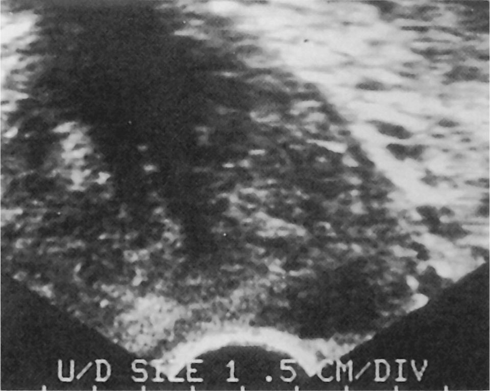
Figure 7-4 Transverse ultrasound image of the prostate showing a hypoechoic area in the peripheral zone and the left base, which is characteristic of prostate cancer. Ultrasound can help direct biopsies to specific areas of the prostate that may be suspicious for cancer.
Prostate Biopsy. Obtaining a biopsy for detection of prostate cancer is almost always performed via the transrectal route, although it can be accomplished through the perineum. Accurate needle placement is facilitated by TRUS but can also be performed with finger guidance. A spring-loaded automatic gun is used to obtain cores of tissue in a systematic fashion. Usually 10 to 12 cores are obtained.
Patients are usually given a Fleet enema on the morning of the examination. In addition, premedication with a broad-spectrum antibiotic for 1 to 2 days after the biopsy is required.
Tumor Grade. The degree of differentiation of the tumor provides important prognostic information. Most often, the Gleason grading system is used. This system assigns a number (1–5) to the dominant grade and a secondary number to arrive at a Gleason sum or score. Gleason scores of 2 to 4 are usually considered well differentiated, 5 to 7 moderately differentiated, and 8 to 10 poorly differentiated. Prognosis is strongly linked to grade. Most cancers found through early detection or screening programs are of an intermediate grade (i.e., Gleason 4 to 6).
Staging of Prostate Cancer
Staging of prostate cancer defines the local, regional, and distant extent of disease. The tumor-node-metastasis (TNM) system is currently the standard and allows categorization of nonpalpable tumors detected because of PSA or ultrasound abnormalities (Table 7-1).

The primary staging modality for local disease is digital rectal palpation. TRUS is used primarily to direct biopsy and is of limited value for staging. Serum PSA levels correlate only roughly with disease extent. However, bone metastasis is quite uncommon in patients with a PSA less than 20 ng/mL. Radionuclide bone scanning is the most sensitive method for detection of bone metastases (Fig. 7-5). Bone scan is not recommended if the PSA value is less than 10 ng/mL and the grade of the tumor is less than a Gleason sum of 7. Pelvic CT scanning is not routinely used because grossly positive nodes are detected rarely with a clinically localized tumor. Prostate cancer, when metastatic, typically affects the axial skeleton and forms osteoblastic metastases on plain radiograph (Fig. 7-6). Soft tissue metastasis may also occur but is unusual without concomitant bone metastasis.

Figure 7-5 Radionuclide bone scan showing multiple areas of abnormal uptake in the pelvis and spine, typical of metastatic prostate cancer.

Figure 7-6 Radiograph of the pelvis showing characteristic osteoblastic metastases from prostate cancer.
Lymph node staging is of critical importance in selecting patients for therapy. CT scanning may show enlarged lymph nodes in patients with high-volume or high-grade primary tumors. Laparoscopic pelvic lymphadenectomy is technically feasible and, when indicated, can provide adequate sampling of the pelvic lymph nodes. Most often, though, lymph node dissection is performed through an open incision immediately before radical prostatectomy. The anatomic limits of a staging lymph node dissection for prostate cancer are the bifurcation of the common iliac artery proximally, the circumflex iliac vein distally, the midportion of the external iliac artery laterally, and the bladder wall medially. The dissection is carried posteriorly to the obturator nerve (Fig. 7-7).

Figure 7-7 Anatomic boundaries of a staging pelvic lymph node dissection for prostate cancer.
In the past, patients who had a new diagnosis of prostate cancer usually underwent a metastatic workup that included a bone scan and lymph node dissection. However, studies have shown that when both the PSA level and Gleason grade are low the disease is almost always localized. Therefore, patients with both a PSA less than 10 and a Gleason score of 7 or less usually do not require a bone scan or lymph node dissection.
Treatment
Localized Disease
The optimal therapy for localized prostate cancer is uncertain and a point of continual controversy. For men with a life expectancy of less than 10 years, observation alone (i.e., “watchful waiting”) may be appropriate. Surgical removal of the prostate (i.e., radical prostatectomy) and radiation therapy are the most commonly used treatments. Ten-year survival rates are similar after external beam radiation or surgery, but valid comparisons are difficult and follow-up beyond 10 years is important. Brachytherapy using interstitial implantation of either iodine-125 or palladium-103 is also used, but there is insufficient long-term follow-up to assess results adequately. Cryotherapy (i.e., freezing of the prostate) is being investigated in some centers, but results published to date indicate that this method is inferior to established treatments for intracapsular tumors.
Radical prostatectomy can be performed via a perineal approach (Fig. 7-8). An inverted U-incision is made anterior to the rectum. The dissection continues in the plane between the prostate and the rectum. The posterior layer of Denonvilliers’ fascia is opened and the prostate gland, the capsule, and the seminal vesicles are dissected free entirely. The perineal route, associated with minimal postoperative pain, does not allow simultaneous lymph node dissection.

Figure 7-8 Radical perineal prostatectomy. The rectum is retracted posteriorly, and the posterior layer of Denonvilliers’ fascia has been incised to expose the prostatic capsule.
More often, radical prostatectomy is accomplished by a retropubic route. An incision is made from the umbilicus to the pubis. Usually, pelvic lymphadenectomy is performed. During radical retropubic prostatectomy, the entire prostate—including the prostatic capsule, the seminal vesicles, and the ampullary portion of the vas deferens—is removed. After the prostate is removed, a direct anastomosis is performed between the reconstructed bladder neck and the urethra (Fig. 7-9). In patients who are sexually active before therapy, potency can be retained in nearly two thirds by preservation of the neurovascular bundle that lies immediately posterolateral to the prostate and urethra. In patients with negative surgical margins, a 15-year, disease-free survival rate of nearly 50% can be anticipated. In patients who have positive surgical margins or histologically positive lymph nodes, adjuvant radiation treatment or hormonal therapy may be used.

Figure 7-9 Radical retropubic prostatectomy. The surgical specimen has been removed and the bladder neck reconstructed. A direct anastomosis is performed with the stump of the urethra.
A recent surgical advancement for prostate cancer is laparoscopy and robotic prostatectomy. These techniques remove the prostate using multiple small incisions. Currently, approximately 20% of prostatectomies are performed in this manner. It is anticipated that the percentage will markedly increase as more urologists and patients become familiar with the approach. Available data suggest that robotic and laparoscopic radical prostatectomies have the following advantages over the open surgery: (a) better cosmesis, (b) decreased risk of transfusion, (c) shorter time to recovery, and (d) earlier return to continence. Short-term results indicate that this technique is equivalent to open approach in controlling the cancer.
Serum PSA levels should decrease to an undetectable range after radical prostatectomy because all PSA-producing cells, both benign and malignant, ideally are removed. After radiation therapy, superior results are achieved in patients in whom the PSA level decreases to less than 1.0 ng/mL or, perhaps, 0.5 ng/mL. An increasing serum PSA is evidence of tumor recurrence, although other studies may not identify metastatic disease initially. There is controversy and uncertainty about when to initiate hormonal therapy in men with an increasing PSA level after treatment.
Metastatic Disease
Prostate cancer is a partially androgen-dependent disease. Therefore, the primary treatment for metastatic carcinoma of the prostate is deprivation of androgens from the cancer cell. Suppression of serum testosterone can be achieved by bilateral surgical orchiectomy or medical castration. Oral administration of estrogens effectively lowers serum testosterone, but is associated with cardiovascular side effects in up to 20% of patients. Estrogen therapy, common in the past, has been replaced by luteinizing hormone– releasing hormone (LHRH) analogs, which effectively suppress testosterone to the castrate range within 1 month of administration. LHRH analogs are associated with few serious side effects; however, they cause vasomotor hot flashes in approximately two thirds of patients. Loss of libido and impotence are consequences of orchiectomy or LHRH administration.
Less than 10% of circulating androgens in men are of adrenal origin. The contribution of these androgens to the growth of prostate cancer is uncertain. Some studies have shown that antiandrogen can prolong the duration of response when used in conjunction with LHRH analogs or orchiectomy. However, other studies have shown conflicting results. The drug is administered orally and can cause some degree of gynecomastia and gastrointestinal side effects (usually diarrhea). Hepatotoxicity is seen occasionally.
The duration of response to hormonal therapy in patients with metastatic prostate cancer is usually 18 to 24 months. After that time, disease progression occurs. Taxol-based chemotherapy has been shown to be moderately effective in hormone-refractory cancer. Radiation can be effective for isolated sites of bone metastasis. In the PSA era, the median survival of patients with hormone-refractory prostate cancer is 53 months.
THE KIDNEYS
Anatomy
The kidneys are paired retroperitoneal organs that lie on either side of the vertebral column, opposite the 12th thoracic and the first through third lumbar vertebrae. They are bordered by the diaphragm posteriorly and superiorly, and by the psoas and quadratus lumborum muscles posteriorly. The right kidney is bordered by the right lobe of the liver anteriorly and superiorly and by the right colon inferiorly (Fig. 7-10). The duodenum lies over the anteromedial portion of the right kidney. The left kidney lies adjacent to the spleen, with the left colon over its anterior lateral surface. The stomach borders the anterior surface of the upper pole, the jejunum overlies the anterior lower pole, and the tail of the pancreas overlies the hilum. Normally, the left kidney lies more cranially than the right one. The dimensions of the average kidney are approximately 11 cm in length, 6 cm in breadth, and 3 cm in anteroposterior thickness. The kidney typically weighs 150 g in the male and approximately 135 g in the female.

Figure 7-10 Anatomic relation of the pleural cavity and some intra-abdominal structures to the kidneys.
The kidney is covered by a renal capsule composed of fibrous tissue that is closely applied to the renal cortical surface. At the hilum, this layer becomes continuous with the fibrous sheaths of the renal and great vessels. The capsule is easily stripped from the parenchyma. The layer of adipose connective tissue surrounding the kidney and its vessels, the perirenal fat, is thickest at the borders of the kidney. Surrounding the perirenal fat is a layer of fibroareolar connective tissue, the renal fascia (Gerota’s). Superiorly, the fascial layers envelop the adrenal glands. Inferiorly, the layers remain separate and surround the ureters. Medially, the layers fuse and adhere to the renal vessels and kidney pelves, limiting extravasation of urine, blood, or purulence.
Physiology
A clear understanding of renal and acid–base physiology is the basis for the management of many urologic disorders. Examples include hyponatremia as a complication of fluid absorption during TURP (TURP syndrome), renal tubular acidosis, hyperchloremic metabolic acidosis after urinary diversion using intestinal segments, acute tubular necrosis after renal ischemia, and renal artery stenosis. A complete discussion of renal physiology should be reviewed in a physiology text.
Blood Supply
Twenty percent of cardiac output is directed to the kidneys. The renal vascular pedicle is anterior to the renal pelvis, entering at the hilum (Fig. 7-11). Usually, a single renal artery supplies each kidney. Later, this artery divides into branches that supply the various kidney segments. Variation in number and configuration of the renal arteries is extremely common. In one study, 65% of kidneys examined had at least one aberrant vessel. Interruption of these end arteries may result in ischemia and infarction of that portion of the kidney. The potential long-term sequela of this type of injury is hypertension. Additionally, aberrant lower pole arteries may be associated with congenital ureteropelvic junction obstruction.

Figure 7-11 Blood supply to the kidneys.
Trauma
Blunt trauma accounts for 70% to 80% of all renal injuries. It usually results from motor vehicle accidents or, less commonly, from accidental falls or contact sports. The mechanism of injury is forces of rapid deceleration or actual impact on the upper abdomen, flank, or back. Hematuria occurs in many patients. Associated injuries can include rib fractures, vertebral body and transverse process fractures, and flank contusions and abrasions. Retroperitoneal hematoma secondary to renal injury must be considered in patients who present in shock.
Evaluation
In the past, imaging was routinely performed when a renal injury was suspected based on mechanism of injury, microhematuria, or gross hematuria. It has subsequently been shown that adults with microhematuria after blunt renal trauma but no history of a major deceleration injury or hypotension do not necessarily need renal imaging. Children and patients with gross hematuria, hypotension, or a major deceleration injury require evaluation. A concern in children with rapid deceleration injury is an avulsion of the ureteropelvic junction; urinalysis can be negative in this situation.
A trauma patient with a suspected renal injury can be hemodynamically stable or unstable. The evaluation of a stable patient with a suspected renal parenchymal injury includes a CT scan. Any renal injury noted is classified as shown in Fig. 7-12. Minor contusions are the most common, and lacerations may be minor or major. In minor lacerations, the injury extends no further than the renal cortex, there is no urinary extravasation or large hematoma, and the capsule may remain intact. In major lacerations, there is a transcapsular rupture through the corticomedullary junction of the kidney and, often, associated urinary extravasation or a large perirenal hematoma.

Figure 7-12 Grades of renal trauma. Grades I and II are considered minor. Grades III, IV, and V are considered major. A, Grade I trauma showing microscopic or gross hematuria. A contusion or a contained subcapsular hematoma is present. There is no parenchymal laceration. B, Grade II trauma showing a nonexpanding, confined perirenal hematoma or cortical laceration that is less than 1 cm deep. No urinary extravasation is present. C, Grade III trauma showing laceration of the parenchyma that extends less than 1 cm into the cortex. No urinary extravasation is present. D, Grade IV trauma showing laceration of the parenchyma that extends through the corticomedullary junction and into the collecting system. Laceration of a segmental vessel may also occur. E, Grade IV trauma showing thrombosis of a segmental renal artery. There is no laceration of the parenchyma, but there is ischemia of the corresponding parenchyma. F, Grade V trauma showing thrombosis of the main artery of the kidney. An intimal tear and a distal thrombosis are seen in the inset. G, Grade V trauma showing a kidney that is shattered because of multiple lacerations. H, Grade V trauma showing avulsion of the main artery or vein of the kidney. (Modified from Tanagho EA, McAninch JW, eds. Smith’s General Urology, 13th ed. Norwalk, Conn: Appleton & Lange; 1992.)
An unstable patient should be taken to the operating room for an emergency laparotomy regardless of whether a renal injury is suspected. In this situation, renal imaging may be accomplished with a “one-shot” IVP. One protocol for this test involves administering an intravenous bolus of 2 mL/kg contrast, with a film taken 10 minutes later.
In the stable patient who sustains blunt trauma, renal exploration is required only if the CT scan shows a major renal injury. There is no general agreement as to exactly what CT findings justify exploration. The trend is toward observation. In the unstable patient who undergoes surgery, indications for renal exploration include an expanding or pulsatile retroperitoneal hematoma or an abnormality on the IVP. The technical approach during renal exploration includes incision of the posterior peritoneum medial to the inferior mesenteric vein, anterior to the aorta, with isolation of the renal vessels before reflection of the colon and exploration of the kidney. This avoids exsanguinating hemorrhage.
The patient with penetrating trauma is managed similarly to the patient with blunt trauma. However, patients who have penetrating injuries more often require exploration, and the threshold to explore the kidney tends to be lower.
Congenital Disorders
Scores of congenital anomalies are found in the urinary tract. Some are symptomatic and discovered in children, others are asymptomatic and discovered serendipitously, and yet others do not become symptomatic until adulthood. Two common disorders that require intervention are horseshoe kidney and congenital obstruction of the urinary tract.
Horseshoe kidney occurs in 1:400 to 1:1,800 live births. It is the most common type of renal fusion anomaly and usually occurs at the lower pole. Symptoms are usually associated with obstruction or infection and include hematuria and vague abdominal discomfort. Diagnosis is usually made by IVP, although ultrasonography and CT are useful as well. When intervention is necessary, the renal isthmus connecting the kidneys is divided surgically (symphysiotomy). Revision of the ureteropelvic junction may need to accompany the symphysiotomy.
Congenital obstruction of the urinary tract occurs most often at the junction between the ureter and the renal pelvis, the ureteropelvic junction. The etiologic factors in this obstruction are myriad and complex and result in varying degrees of hydronephrosis. Bilateral involvement occurs in 10% to 40% of cases. An intrinsic obstruction caused by maldevelopment of the ureteropelvic junction is the likely cause of obstruction in children. In cases presenting in adulthood or late childhood, an aberrant vessel that crosses the ureteropelvic junction may also cause obstruction. The resulting stenosis or functional obstruction leads to hydronephrosis. Presenting signs and symptoms include palpable abdominal mass, pain, hematuria, urinary infection, fever, hypertension, and renal stones. Today, many cases of ureteropelvic junction obstruction are diagnosed by antenatal ultrasonography. Diagnosis is made by IVP or ultrasonogram. Nuclear scan (Lasix renography) is preferable to IVP in children because this type of scan is superior in quantifying renal function and obstruction. Surgical repair of the obstruction is performed to prevent loss of renal function and recurrent UTIs.
Inflammatory Diseases
Pyelonephritis
Pyelonephritis is usually a clinical diagnosis. Patients present with fever, flank pain, UTI, and costovertebral angle tenderness on the side of the involved kidney. In 80% of cases, E. coli is the causative organism. Chronic pyelonephritis often leads to renal failure and is a common reason for renal transplantation. Findings on IVP, usually nonspecific, include diffuse renal enlargement with calyceal distortion. If the infection is uncomplicated, treatment is outpatient oral antibiotic therapy. When the patient has evidence of sepsis or significant compromise requiring hospitalization, intravenous antibiotics are administered. It is not uncommon for fever and flank pain to persist for several days. If symptoms persist for a longer period, it is reasonable to image the kidneys with a CT or ultrasound to exclude an abscess. If ureteral obstruction is suspected, renal imaging must be immediately obtained and any obstruction must be emergently relieved (e.g., obstructive pyelonephritis associated with an obstructing ureteral stone) with a ureteral stent or a percutaneous nephrostomy. Obstructive pyelonephritis represents a urologic emergency because the infected urine proximal to the obstruction is similar to an abscess, but worse because the abscess is under pressure. These patients can become septic, and antibiotics alone do not represent adequate therapy. Treatment with removal of the stone is contraindicated as initial treatment because stone manipulation can lead to worsening of the sepsis. When emergently treating obstructive pyelonephritic, the goal is to relieve the obstruction. Treatment of a renal abscess is usually by percutaneous drainage in addition to antibiotics.
Emphysematous Pyelonephritis
Emphysematous pyelonephritis is a life-threatening infection in which bacteria, often E. coli, form gas in the renal parenchyma. The condition is associated with poorly controlled diabetes. Patients are usually septic and can deteriorate rapidly. A CT scan is diagnostic. Patients are managed with intravenous antibiotics, supportive measures, and an emergency nephrectomy. Percutaneous drainage of the affected area was recently reported as an alternative to nephrectomy. However, nephrectomy remains the standard treatment option.
Xanthogranulomatous Pyelonephritis
Xanthogranulomatous pyelonephritis (XGP) is another inflammatory disease of the kidney. Females are affected more often (75% of cases). Patients with XGP, a disease generally involving patients in the fifth to seventh decades of life, often have a history of failure to thrive and chronic UTI. The diagnosis can be difficult because the symptoms are usually nonspecific. There is often a delay in the diagnosis of XGP, and it is important for primary care physicians to consider this disease with the described presentation. When urine culture shows UTI, E. coli and Proteus species are the most common causes. Currently, the CT scan is the imaging test of choice used to diagnose XGP. The lesions in the kidney, which may be quite large, are characterized by diffuse enlargement, central nephrolithiasis, and spherical areas surrounding the kidney in a hydronephrotic pattern. The affected kidney is usually nonfunctional. The treatment is usually a nephrectomy. The kidney can be adherent to adjacent structures, and the nephrectomy is frequently technically challenging.
Genitourinary Tuberculosis
Painless frequency, especially at night, is a common complaint in patients with genitourinary tuberculosis (TB). In a patient with sterile pyuria, TB should be suspected. A PPD skin test can help establish the diagnosis. A positive test does not necessarily indicate active disease. A definitive diagnosis is made by urine culture with isolation of Mycobacterium tuberculosis. Further testing includes chest films and spine imaging. When genitourinary TB is diagnosed, an IVP is mandatory. Findings may include calcifications at any point along the genitourinary tract, and extensive renal calcifications can be seen. Ureteral obstruction can occur secondary to stricture of the ureter. The management of genitourinary TB begins with antituberculosis drugs. Subsequent nephrectomy may be indicated in some cases when extensive renal destruction and nonfunction are present. Ureteral stricture management options may include temporary internal stenting, corticosteroids, or ureteral reimplantation in distal ureteral strictures. Treatment depends on the location and length of the stricture and the response to medical management.
Neoplasms
Renal masses are classified as benign or malignant; malignant tumors are primary or metastatic. The most commonly encountered renal mass lesion is a simple cyst (70% of cases). Renal cell carcinoma is the most common primary neoplasm of the kidney and accounts for more than 85% of all primary renal cancers in adults. This chapter discusses only the most frequently encountered tumors.
Clinical Presentation and Evaluation
Patients with renal cancer often have painless hematuria, but the cancer is being discovered with increasing frequency on CT scans and ultrasound as an incidental finding. Many texts refer to the classic triad (a complex of flank pain, abdominal mass, and hematuria) as characteristic of renal cancer. However, patients seldom have all three findings.
CT scanning plays a prominent role in the workup of solid renal masses and may be valuable in characterizing some atypical cystic masses. Cystic lesions are often identified by IVP and confirmed by ultrasound. Arteriography, once a standard preoperative study, is recommended only in selected cases where the diagnosis is in doubt or aberrant vasculature is expected. If tumor invasion of the inferior vena cava is suspected, a magnetic resonance image (MRI) or inferior venacavogram is indicated to define the extent of involvement.
Benign Neoplasms
Most simple cystic lesions are asymptomatic and benign and require no intervention. However, some are complex (e.g., septations, wall thickening, or calcifications) and require further investigation to rule out malignancy. Benign simple renal cysts are usually asymptomatic. Simple cysts are found in as many as 33% of adults. A simple cyst noted on imaging (e.g., CT, ultrasound) is managed by observation. A complex cyst is usually considered cancerous until proven otherwise, and partial or radical nephrectomy may be indicated. A needle biopsy or cyst aspiration is usually of little value in most cases because a negative result may be a false negative and does not rule out malignancy. Benign solid tumors of the kidney are encountered occasionally. An angiomyolipoma is usually diagnosed by the characteristic appearance of fat within the lesion on CT scan. Fat is black on CT, and when fat is seen within a renal mass, angiomyolipoma is almost always the diagnosis.
Malignant Neoplasms
Renal Cell Carcinoma
Renal cell carcinoma, a tumor that usually arises from the proximal convoluted tubules, is by far the most common primary solid tumor affecting the kidney. Although etiologic factors are not well documented in human beings, nitrosamines are implicated in animal studies. Carcinogens found in cigarette smoke have also been implicated, but no specific carcinogen has been identified. Renal cell carcinoma has a 2:1 male-to-female preponderance. Typically unilateral, this lesion is spherical with a pseudocapsule of parenchyma and fibrosis. Five percent of renal carcinoma may be bilateral.
Hematuria is the single most common sign, occurring in 29% to 60% of reported cases. Flank pain and palpable flank mass occur next most often, but the classic triad of hematuria, flank pain, and a palpable abdominal mass are reported in only 4% to 17% of cases. Other common signs and symptoms are fever, anemia, and elevated sedimentation rate. Although serum lactate dehydrogenase and alkaline phosphatase may be elevated, there are no reliable tumor markers for renal cell carcinoma. Renal cell cancers can cause only nonspecific symptoms such as weight loss, fever, or weakness.
In later stages, this tumor invades the renal vein and vena cava and may even extend into the right atrium. A staging system such as the TNM system (Table 7-2) is used to determine the extent of the primary lesion, the involvement of contiguous structures, the extent of vascular involvement, and whether the tumor has metastasized. Renal cell carcinoma metastasizes most often to lungs, bone, and brain, in that order. Metastatic lesions may appear in both the ipsilateral and contralateral kidney, and late metastasis may occur to the liver. Five-year survival rates for stages I and II are 80% to 90%, and for stages III and IV are 40% to 60%. Treatment for renal cell carcinoma is radical nephrectomy (Fig. 7-13
Stay updated, free articles. Join our Telegram channel

Full access? Get Clinical Tree


