Upper Limb
Cards 6-1 to 6-66
Bones and Joints
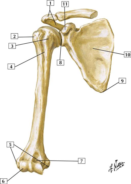
6-1 Humerus and Scapula: Anterior View
Comment:
The clavicle and scapula form the pectoral girdle, or shoulder, which connects the upper extremity to the trunk. The clavicle serves as a strut, keeping the upper limb away from the trunk and free for movement. It is vulnerable to fracture.
The scapula, or shoulder blade, articulates with the clavicle and the head of the humerus (glenohumeral joint). Sixteen different muscles attach to the scapula. Fractures of the scapula are uncommon.
The humerus is a long bone. Its proximal end forms part of the shoulder joint, and its distal end contributes to the elbow joint. The surgical neck of the humerus (the region just below the lesser tubercle) is a common fracture site. Fractures at this site may injure the axillary nerve of the brachial plexus.
Atlas Plate 405
See also Plate 183
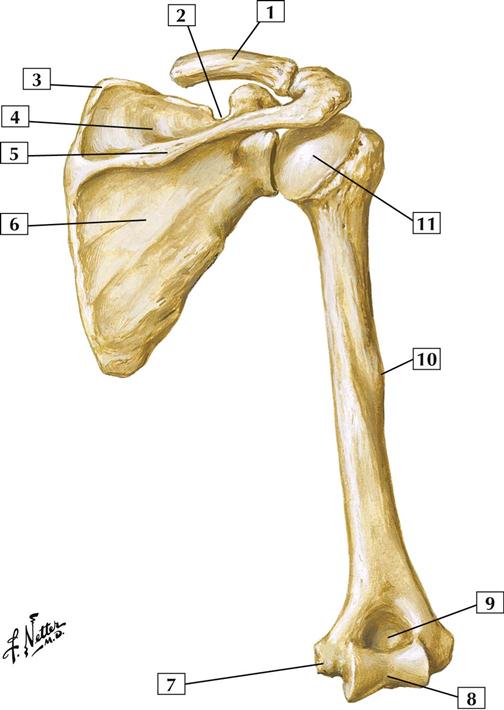
6-2 Humerus and Scapula: Posterior View
Comment:
Posteriorly, the scapula displays a prominent spine that separates the supraspinous and infraspinous fossae.
The clavicle is the 1st bone to ossify but the last bone to fuse and is formed by intramembranous ossification. It is one of the most commonly fractured bones.
Midshaft on the humerus is the deltoid tuberosity, the insertion point for the deltoid muscle.
Distally, the depression above the trochlea is called the olecranon fossa, which accommodates the olecranon of the ulna when the elbow is extended fully.
Atlas Plate 406
See also Plate 183
6-3 Shoulder (Glenohumeral) Joint: Anterior View
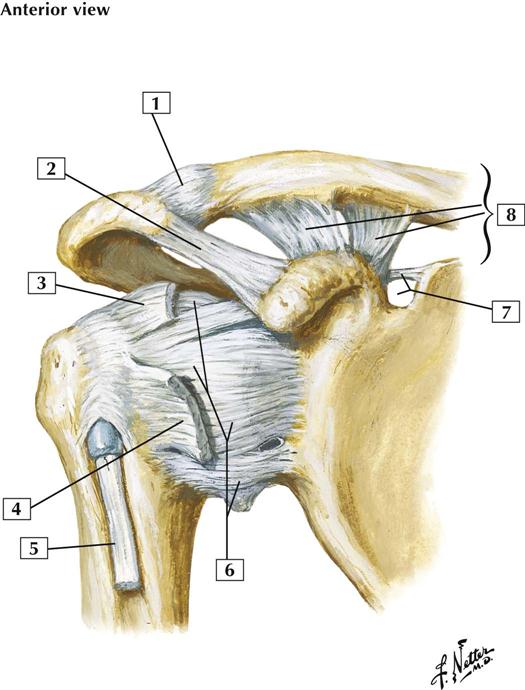
Comment:
The shoulder is a multiaxial synovial ball-and-socket (spheroidal) joint. Movements include abduction and adduction, flexion and extension, and rotation and circumduction. The shallow glenoid cavity of the scapula permits extensive movement at the shoulder but also makes this joint vulnerable to dislocation. The 4 tendons of the rotator cuff muscles help stabilize the joint.
Also shown is the acromioclavicular joint, a synovial plane joint between the acromion and clavicle. This joint permits gliding movement as the arm is raised and the scapula rotates.
Atlas Plate 408
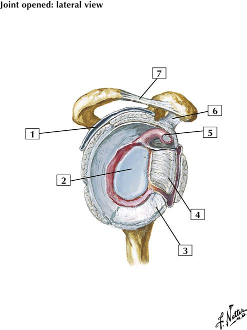
6-4 Shoulder (Glenohumeral) Joint: Lateral View
Comment:
The glenoid cavity is deepened by the presence of the glenoid labrum (lip). The joint is stabilized by a capsule, ligaments, and the 4 tendons of the rotator cuff muscles. The 4 tendons of the rotator cuff muscles reinforce the joint posteriorly, superiorly, and midanteriorly (subscapularis tendon). Most shoulder dislocations occur anteriorly, where there is less support.
Blood is supplied to the shoulder by branches of the suprascapular, humeral circumflex, and scapular circumflex arteries.
Atlas Plate 408
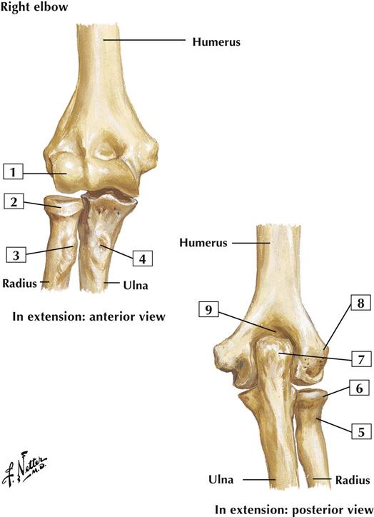
6-5 Bones of Elbow: In Extension
Comment:
The elbow bones include the humerus and the 2 bones of the forearm: the radius and ulna. The ulna lies more medially in the forearm and is the longer of the 2 bones. The point of the elbow that can be easily felt is the olecranon, located posteriorly and proximally on the ulna.
Atlas Plate 422
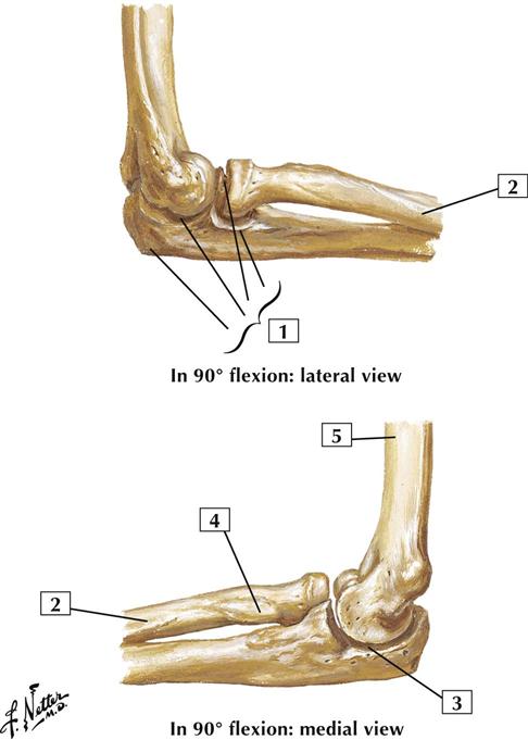
6-6 Bones of Elbow: In 90° Flexion
Comment:
The bones of the elbow include the humerus and the 2 bones of the forearm: the radius and ulna. The ulna lies more medially in the forearm and is the longer of the 2 bones. The point of the elbow that can be easily felt is the olecranon, located posteriorly and proximally on the ulna.
Atlas Plate 422
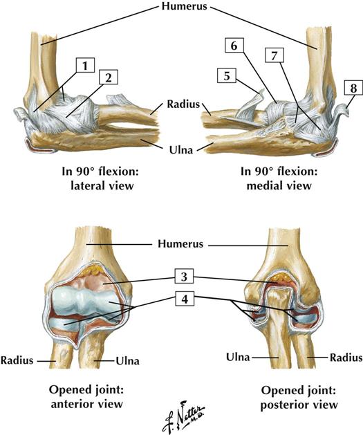
6-7 Ligaments of Elbow
Comment:
The elbow joint forms a uniaxial synovial hinge (ginglymus) joint that includes the humeroradial joint (between the capitulum of the humerus and the head of the radius) and the humero-ulnar joint (between the trochlea of the humerus and the trochlear notch of the ulna). The joint also includes a proximal uniaxial radio-ulnar synovial (pivot) joint that participates in supination and pronation (rotation). Movements about the elbow include flexion and extension.
The joint is stabilized by the laterally placed radial collateral ligament and medially placed triangular ulnar collateral ligament. The anular ligament holds the head of the radius in place.
Blood is supplied to the elbow by branches of the brachial artery and recurrent collateral branches of the radial and ulnar arteries.
Atlas Plate 424
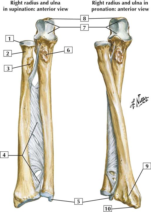
6-8 Bones of Forearm
Comment:
The bones of the forearm include the medially placed and longer ulna and the laterally placed radius.
Along the length of the forearm, the radius and ulna are connected by the interosseous membrane, which contributes to the radio-ulnar joint, a fibrous (syndesmosis) joint. The interosseous membrane divides the forearm into anterior and posterior muscular compartments.
Distally, the radius and the ulna display styloid processes.
Atlas Plate 425
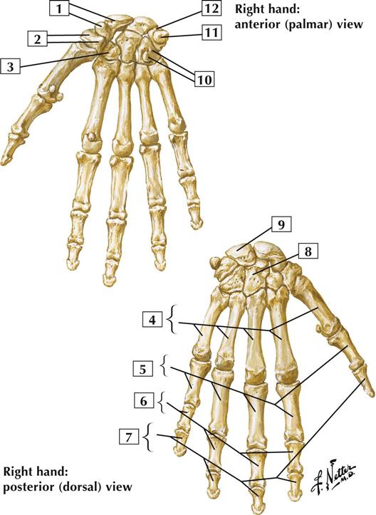
6-9 Bones of Wrist and Hand
Comment:
Bones of the wrist and hand include the 8 carpal bones; 5 metacarpal bones (1 for each digit); and, for digits 2 through 5, proximal, middle, and distal phalanges. The 1st digit, or thumb, has only a proximal phalanx and a distal phalanx.
The scaphoid, lunate, and triquetrum articulate with the distal radius to form the radiocarpal wrist joint.
Atlas Plate 443
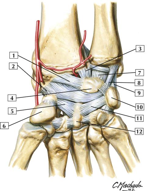
6-10 Ligaments of Wrist: Palmar View
Comment:
The wrist, or radiocarpal joint, is an ellipsoid biaxial synovial joint formed by the distal end of the radius (an articular disc) and the scaphoid, lunate, and triquetrum carpal bones. This joint is reinforced by radial and ulnar collateral ligaments and by dorsal and palmar (volar) radiocarpal ligaments. The joint permits flexion, extension, abduction, adduction, and circumduction.
Anatomists often simply lump these ligaments into a palmar radiocarpal ligament (long and short radiolunate and radioscaphocapitate ligaments [1-3 in the list above]), a palmar ulnocarpal ligament (ulnolunate, ulnocapitate, and ulnotriquetral ligaments), and various intercarpal and metacarpal ligaments.
The carpometacarpal joint of the thumb is a biaxial saddle (sellar) joint (with trapezium). It provides flexion and extension, abduction and adduction, and circumduction. The other 4 carpometacarpal joints are plane synovial joints that permit gliding movements.
Atlas Plate 441
6-11 Ligaments of Wrist: Posterior View
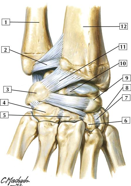
1. Ulna
2. Dorsal radio-ulnar ligament
3. Triquetrum
4. Hamate
5. Capitate
6. Trapezoid
8. Trapezium
9. Scaphoid
11. Dorsal radiocarpal ligament
12. Radius
Comment:
Proximal to the wrist lies the distal radio-ulnar joint, which is a uniaxial synovial pivot (trochoid) joint between the ulna and the ulnar notch of the radius. It allows for pronation and supination (rotation).
The wrist, or radiocarpal joint, is an ellipsoidal biaxial synovial joint formed by the distal end of the radius (an articular disc) and the scaphoid, lunate, and triquetrum carpal bones. Movements at the wrist include flexion, extension, abduction, adduction, and circumduction.
Anatomists often simplify this arrangement, designating these ligaments as a dorsal radiocarpal ligament, dorsal carpometacarpal ligaments, and intercarpal ligaments.
Between the proximal and distal rows of carpal bones lies the midcarpal (intercarpal) joints, synovial plane joints. These joints permit some gliding and sliding movements.
Atlas Plate 442
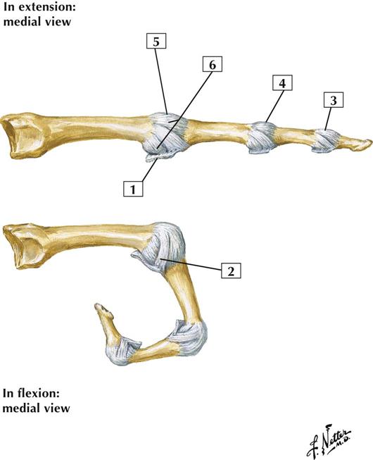
6-12 Metacarpophalangeal and Interphalangeal Ligaments: Medial Views
Comment:
The metacarpophalangeal joints are biaxial condyloid synovial joints that participate in flexion and extension, abduction and adduction, and circumduction. The capsule is supported by the collateral and palmar (volar) ligaments. The collateral ligaments are tight in flexion and loose in extension.
The interphalangeal joints (proximal interphalangeal and distal interphalangeal) are uniaxial synovial hinge joints that participate in flexion and extension. Ligaments similar to the metacarpophalangeal joints reinforce these joints. The palmar ligaments prevent hyperextension.
Atlas Plate 445
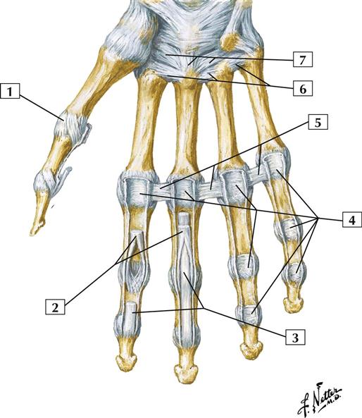
6-13 Metacarpophalangeal and Interphalangeal Ligaments: Anterior View
Comment:
The metacarpophalangeal joints are biaxial condyloid synovial joints that participate in flexion and extension, abduction and adduction, and circumduction. These joints are reinforced by the palmar (volar) ligaments and 2 collateral ligaments on either side.
The interphalangeal joints for digits 2 through 5 include a proximal interphalangeal joint and a distal interphalangeal joint. These joints are uniaxial synovial hinge joints that are reinforced by palmar ligaments and 2 collateral ligaments. They permit flexion and extension. The palmar ligaments prevent hyperextension.
Atlas Plate 445
Muscles
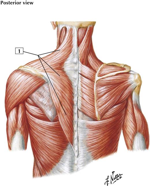
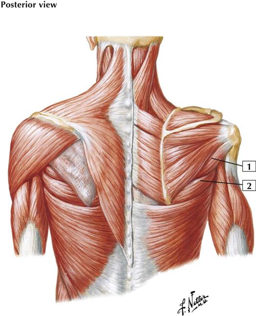
6-14 Shoulder Muscles
Origin (proximal):
External occipital protuberance and medial third of the superior nuchal line of the occipital bone, ligamentum nuchae, and spinous processes of the 7th cervical vertebra and all 12 thoracic vertebrae.
Insertion (distal):
Superior fibers insert into the posterior border of the lateral third of the clavicle. Middle fibers insert into the medial margin of the acromion and posterior border of the scapular spine. Inferior fibers converge to end in an aponeurosis inserted into the scapular spine.
Action:
The upper and lower fibers act primarily to rotate the scapula for full abduction of the upper extremity. The upper fibers, acting alone, elevate the shoulder and brace the shoulder girdle when a weight is being carried by the shoulder or hand. Central fibers run horizontally and retract the shoulder. Lower fibers draw the scapula downward. When both muscles act together, the scapula can be adducted and the head drawn directly backward.
Innervation:
Motor supply is from the accessory nerve (CN XI). Proprioceptive fibers are from the 3rd and 4th cervical nerves.
Comment:
The trapezius, in contrast to the other shoulder muscles, does not receive nerve fibers from the brachial plexus.
Atlas Plate 409
See also Plates 29, 171, 185
6-15 Shoulder Muscles
Origin (proximal):
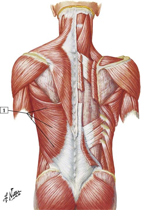
Arises from a broad aponeurosis of the posterior layer of the thoracolumbar fascia, the spinous processes of the lower 6 thoracic vertebrae, and fleshy digitations of the caudal most 3 or 4 ribs. The muscle also may attach to the iliac crest.
Insertion (distal):
The fibers converge as the muscle curves around the lower border of the teres major and twists on itself. They end as a tendon that inserts into the intertubercular groove of the humerus.
Action:
Extends, adducts, and medially rotates the humerus (arm).
Innervation:
Thoracodorsal nerve (C6-8).
Comment:
With the upper extremity fixed, the latissimus dorsi elevates the trunk when the arms are stretched above the head, as when reaching up while climbing.
The origin of the muscle from the thoracic vertebrae and lower ribs may vary.
The blood supply is by the thoracodorsal artery, a branch of the subscapular artery (which arises from the axillary artery).
Atlas Plate 171
6-16 Shoulder Muscles
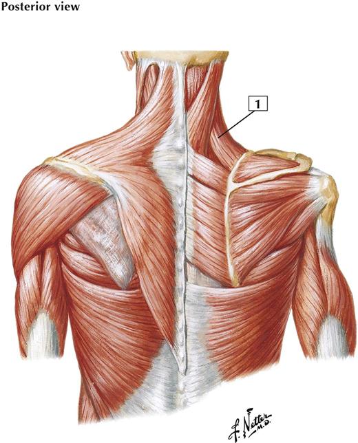
Origin (proximal):
Arises from the transverse processes of the first 4 cervical vertebrae.
Insertion (distal):
Inserts into the superior portion of the medial (vertebral) border of the scapula.
Action:
Elevates the superior angle of the scapula and tends to draw it medially. Also rotates the scapula so that the glenoid cavity is tilted inferiorly. When the scapula is held in a fixed position, the levator scapulae bends the neck laterally and rotates it slightly toward the same side.
Innervation:
By the 3rd and 4th cervical nerves from the cervical plexus and by a branch from the dorsal scapular nerve (C5) to the muscle’s lower fibers.
Comment:
Contraction of the levator scapulae helps shrug the shoulders. The blood supply to the muscle comes largely from the transverse cervical artery of the thyrocervical trunk.
Atlas Plate 409
See also Plates 29, 171, 185
6-17 Shoulder Muscles
Origin (proximal):
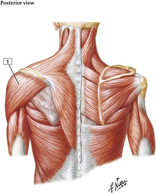
Arises from the lateral third of the clavicle, the superior surface of the acromion, and the spine of the scapula.
Insertion (distal):
The fibers converge in a thick tendon that is attached to the deltoid tuberosity on the lateral aspect of the shaft of the humerus.
Action:
The principal function is abduction of the arm at the shoulder in a movement initiated together with the supraspinatus muscle. The clavicular portion of the muscle rotates the arm medially and helps the pectoralis major flex the arm at the shoulder. The spinous portion rotates the arm laterally and helps the latissimus dorsi extend the arm at the shoulder.
Innervation:
Axillary nerve (C5 and C6).
Comment:
The deltoid is a thick, triangular muscle with coarse fibers. It covers the shoulder joint anteriorly, posteriorly, and laterally. The multipennate central portion of the muscle is most active in abduction.
The blood supply is largely via the thoraco-acromial artery and also via the anterior and posterior humeral circumflex arteries, which arise from the axillary artery.
Atlas Plate 409
See also Plates 29, 171, 185
6-18 Shoulder Muscles
Origin (proximal):
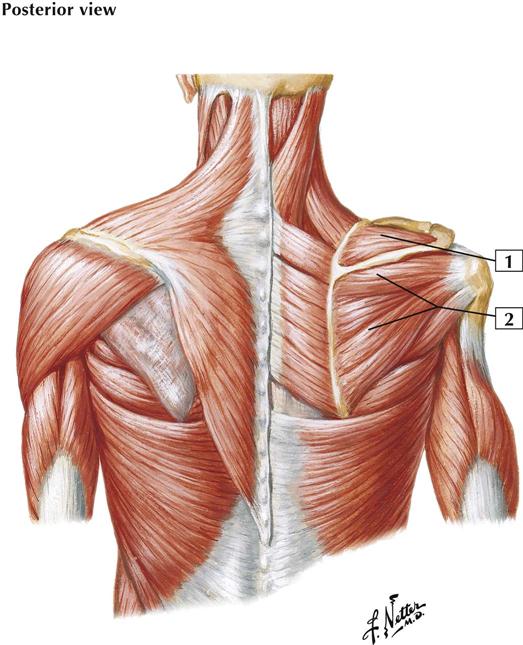
The supraspinatus muscle occupies the supraspinous fossa, originating from the medial two-thirds and arising from the strong supraspinatus fascia. The infraspinatus muscle occupies most of the infraspinous fossa; it arises from the medial two-thirds and from the infraspinatus fascia.
Insertion (distal):
Fibers of the supraspinatus converge to form a tendon that inserts into the superior facet on the greater tubercle of the humerus. The infraspinatus fibers also converge to form a tendon, which inserts into the middle facet on the greater tubercle of the humerus. The tendons of the 2 muscles adhere to each other.
Action:
The supraspinatus strengthens the shoulder joint by drawing the humerus toward the glenoid fossa. With help from the deltoid, it initiates abduction at the shoulder and is a lateral rotator of the humerus (arm). The infraspinatus strengthens the shoulder joint by bracing the head of the humerus in the glenoid fossa. It is also a lateral rotator of the humerus.
Innervation:
Both by the suprascapular nerve (C5 and C6).
Atlas Plate 409
See also Plates 29, 171, 185
6-19 Shoulder Muscles
Origin (proximal):
The teres minor originates from the lateral border of the scapula. The teres major arises from the dorsal surface of the inferior angle of the scapula.
Insertion (distal):
The teres minor inserts into the inferior facet on the greater tubercle of the humerus. The teres major inserts into the medial lip of the intertubercular groove of the humerus.
Action:
The teres minor rotates the arm laterally and weakly adducts the arm at the shoulder. Similar to the other 3 rotator cuff muscles, it draws the humerus toward the glenoid fossa, strengthening the shoulder joint. The teres major helps extend the arm from the flexed position, and it adducts and medially rotates the arm at the shoulder.
Innervation:
The teres minor is supplied by the axillary nerve (C5 and C6), whereas the teres major is innervated by the lower subscapular nerve (C6 and C7).
Comment:
The teres minor is 1 of the 4 rotator cuff muscles, and it helps stabilize the shoulder joint. Often, it is inseparable from the infraspinatus muscle.
Atlas Plate 409
See also Plates 29, 171, 185
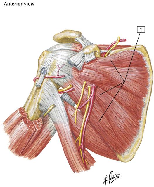
6-20 Scapulohumeral Dissection
Origin (proximal):
Arises from the medial two-thirds of the subscapular fossa and from the lower two-thirds of the lateral border of the scapula.
Insertion (distal):
The fibers converge in a tendon that is inserted into the lesser tubercle of the humerus and the anterior portion of the shoulder joint capsule.
Action:
Stay updated, free articles. Join our Telegram channel

Full access? Get Clinical Tree


