Head and Neck
Cards 1-1 to 1-84
Bones and Joints
1-1 Skull: Anterior View
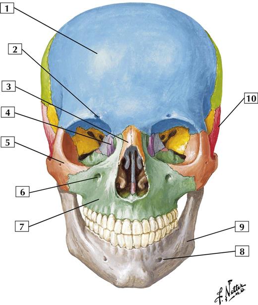
1. Frontal bone
2. Supra-orbital notch (foramen)
3. Nasal bone
7. Maxilla
9. Mandible
10. Temporal bone
Comment:
The skull bones are fused together at immovable, fibrous joints, such as the sutures.
The 2 general classes of skull bones are cranial bones (8 bones), which enclose the brain, and facial bones (14 bones). The 8 cranial bones are the frontal, occipital, ethmoidal, and sphenoidal bones, a pair of temporal bones, and a pair of parietal bones.
Associated bones of the skull include the auditory ossicles (3 in each middle ear cavity) and the unpaired hyoid bone. The skull and associated bones constitute 29 different bones (the 32 adult teeth are part of the mandible and maxilla and are not counted separately).
Atlas Plate 4
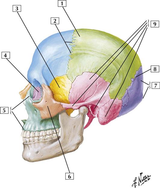
1-2 Skull: Lateral View
Comment:
This lateral view shows many bones of the cranium and some of the sutures of the skull, the immovable fibrous joints between adjacent skull bones. The coronal suture lies between the frontal bone and the paired parietal bones. The lambdoid suture lies between the paired parietal bones and the occipital bone.
The pterion is the site of union of the frontal, parietal, sphenoidal, and temporal bones. A blow to the head or a skull fracture in this region is dangerous because the bone at this site is thin, and the middle meningeal artery, supplying the dural covering of the brain, lies just deep to this area. The asterion is the site of union of the temporal, parietal, and occipital bones.
Atlas Plate 6
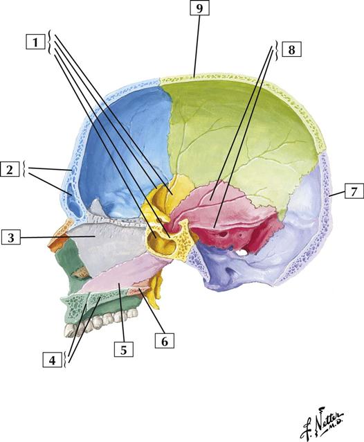
1-3 Skull: Midsagittal Section
Comment:
Note the interior of the cranium and the nasal septum. The 8 cranial bones enclosing the brain include the unpaired frontal, occipital, ethmoidal, and sphenoidal bones and the paired temporal and parietal bones. The 14 facial bones include the paired lacrimal, nasal, palatine, inferior turbinate (not shown), maxillary, and zygomatic (not shown) bones and the unpaired vomer and mandible (not shown).
The nasal septum is formed by the perpendicular plate of the ethmoidal bone, the vomer, and the palatine bones and septal cartilages.
The petrous portion of the temporal bone contains the middle and inner ear cavities and the vestibular system.
Atlas Plate 8
1-4 Lateral Wall of Nasal Cavity
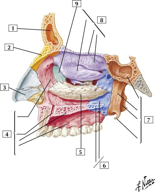
Comment:
The lateral wall of the nasal cavity prominently displays the superior and middle conchae (called turbinates when covered with mucosa) of the ethmoidal bone and the inferior concha. Portions of other bones, including the nasal bone, maxilla, lacrimal bone, palatine bone, and sphenoidal bone, contribute to the lateral wall.
The palatine processes of the maxillae and the horizontal plates of the palatine bones make up the hard palate.
Atlas Plate 37
See also Plate 8
1-5 Cranial Base: Inferior View
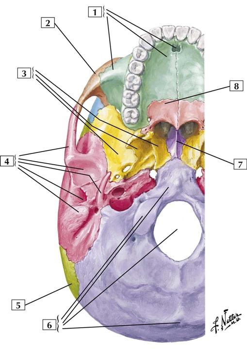
Comment:
Cranial bones and facial bones contribute to the base of the skull. Key processes and foramina associated with these bones can be seen in this inferior view.
The largest foramen of the skull is the foramen magnum, the site where the spinal cord and brainstem (medulla oblongata) are continuous.
Atlas Plate 10
1-6 Foramina of Cranial Base: Superior View
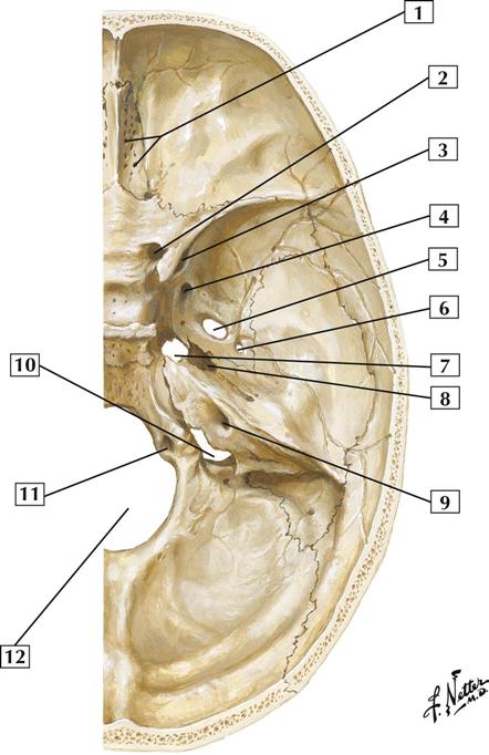
1. Foramina of cribriform plate (Olfactory nerve bundles)
2. Optic canal (Optic nerve [CN II]; Ophthalmic artery)
4. Foramen rotundum (Maxillary nerve [CN V2])
6. Foramen spinosum (Middle meningeal artery and vein; Meningeal branch of mandibular nerve)
8. Carotid canal (Internal carotid artery; Internal carotid nerve plexus)
11. Hypoglossal canal (Hypoglossal nerve [CN XII])
Comment:
Key structures passing through each foramen are noted in parentheses.
Atlas Plate 13
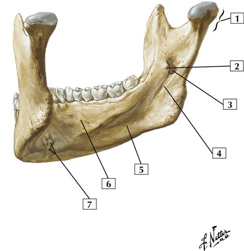
1-7 Mandible: Anterolateral Superior View
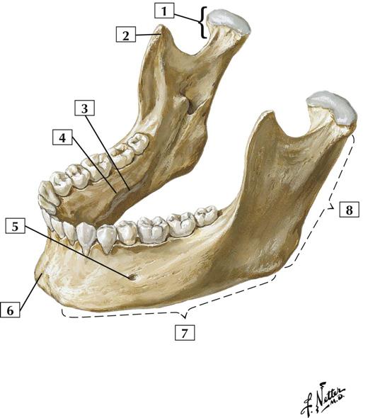
Comment:
The mandible, or lower jaw, contains the mandibular teeth and the mandibular foramen. The inferior alveolar neurovascular bundle passes through the mandibular foramen; it innervates the mandibular teeth and supplies them with blood. The nerve ends as a cutaneous branch that exits the mental foramen (mental nerve).
The condylar process of the mandible articulates with the temporal bone, forming the temporomandibular joint.
Because of its vulnerable location, the mandible is the 2nd most commonly fractured facial bone (the nasal bone is 1st). The most common sites of fracture are the cuspid (canine tooth) area and the 3rd molar area.
Atlas Plate 17
1-8 Mandible: Left Posterior View
Comment:
The inferior alveolar neurovascular bundle enters the mandibular foramen and courses through the bony mandible to supply the mandibular teeth and gums.
Depressions, or fossae, on the medial side of the mandible mark the locations of the submandibular and sublingual salivary glands.
Atlas Plate 17
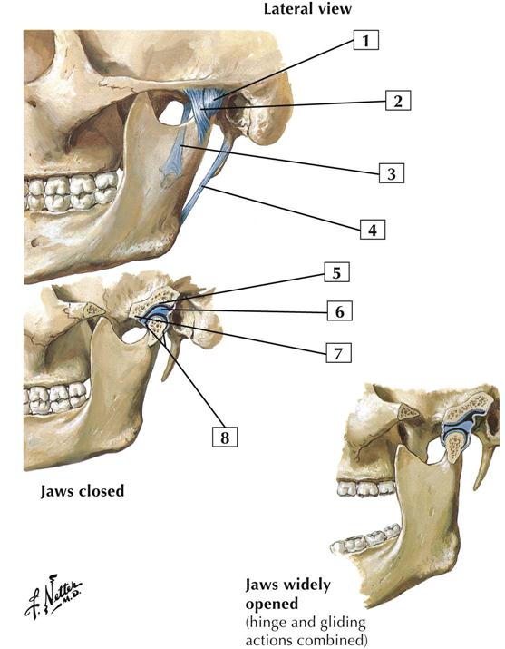
1-9 Temporomandibular Joint
Comment:
The temporomandibular joint is the synovial joint between the mandibular fossa and the articular tubercle of the temporal bone and head of the mandible. The joint’s 2 synovial cavities are separated by an articular disc of fibrocartilage.
This unique joint combines an upper uniaxial gliding joint, for forward gliding (protrusion) and backward gliding (retraction) movements and some side-to-side motion, with a lower uniaxial hinge joint, below the articular disc, for closing (elevation of) and opening (depression of) the jaw.
This joint contains an articular capsule and is reinforced by the lateral and sphenomandibular ligaments.
Atlas Plate 18
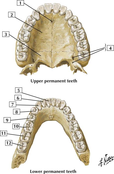
1-10 Teeth
Comment:
Humans have 2 sets of teeth: the deciduous teeth, which total 20, and the permanent teeth (shown in this illustration), which total 32 (16 maxillary and 16 mandibular teeth).
Permanent teeth in each quadrant of the jaw (mandible and maxilla) include 2 incisors, 1 canine, 2 premolars, and 3 molars. The 3rd molars are often referred to as the wisdom teeth.
The maxillary teeth are innervated by the posterior, middle, and anterior alveolar branches of the maxillary nerve. The mandibular teeth are innervated by the inferior alveolar branch of the mandibular nerve.
Atlas Plate 62
1-11 Tooth
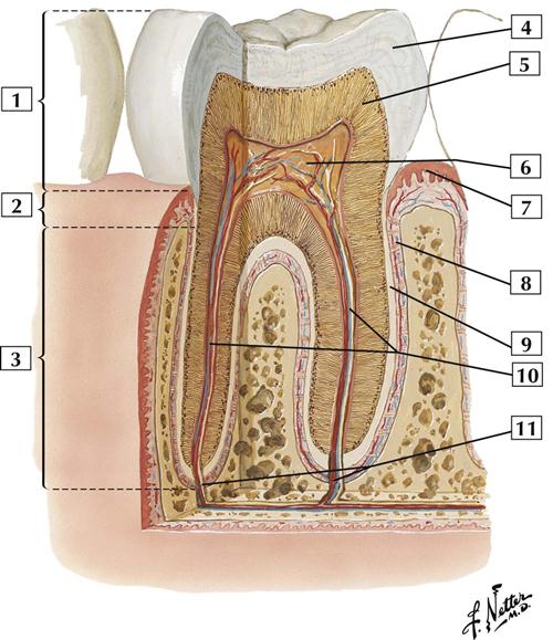
1. Crown
2. Neck
3. Root
4. Enamel (Substantia adamantina)
5. Dentine and dentinal tubules (Substantia eburnea)
6. Dental pulp containing vessels and nerves
7. Gingival (gum) epithelium (stratified)
8. Periodontium (Alveolar periosteum)
10. Root (central) canals containing vessels and nerves
11. Apical foramina
Comment:
Each tooth is composed of an enamel-covered crown, dentine, and pulp. The pulp fills a central cavity and is continuous with the root canal. Blood vessels, nerves, and lymphatics enter the pulp through an apical foramen.
The crown projects above the gum, or gingival surface. The narrow portion between the crown and root is called the neck. The root is embedded in the alveolar bone of the maxilla or mandible and is covered by cement, which is connected to the alveolar bone by the periodontal ligament.
Atlas Plate 63
1-12 Cervical Vertebrae: Atlas and Axis
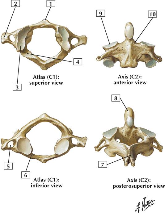
Comment:
The 1st cervical vertebra is the atlas. It is named after the Greek god Atlas, who is often depicted with the world on his shoulders. The atlas has no body or spine but is made of anterior and posterior arches. The transverse processes contain a foramen that transmits the vertebral vessels.
The 2nd cervical vertebra is the axis. Its most characteristic feature is the dens (odontoid process). The dens articulates with the anterior arch of the atlas, providing a pivot about which the atlas and head can rotate (side-to-side action of the head, as in indicating “no”).
Atlas Plate 19
See also Plates 15, 153
1-13 External Craniocervical Ligaments
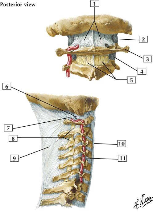
Comment:
The atlanto-occipital joint, on each side, is covered with an articular capsule and posteriorly reinforced by the posterior atlanto-occipital membrane.
The ligamentum nuchae is a strong median fibrous septum. It is an extension of the thickened supraspinous ligaments that arise from the spinous process of C7 and extend to the external occipital protuberance.
Atlas Plate 22
1-14 Internal Craniocervical Ligaments
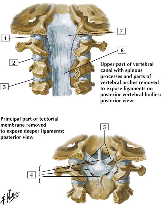
Comment:
The atlanto-occipital joint is a biaxial condyloid synovial joint between the atlas and the occipital condyles. It permits flexion and extension, as when the head is nodded up and down, and some lateral bending.
The atlanto-axial joints are uniaxial synovial joints. They consist of plane joints associated with the articular facets and a median pivot joint between the dens of the axis and the anterior arch of the atlas. The atlanto-axial joint permits the atlas and head to be rotated as a single unit, as when the head is turned from side to side.
These joints are reinforced by ligaments, especially the cruciate and alar ligaments. The alar ligaments limit rotation.
Atlas Plate 23
1-15 Cartilages of Larynx
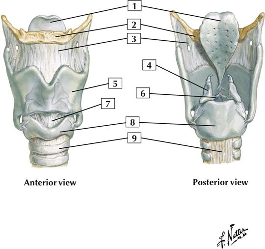
Comment:
The cartilages of the larynx include the thyroid cartilage, cricoid cartilage, epiglottis, and the paired arytenoid, corniculate, and cuneiform cartilages.
Not shown in the illustration are the cuneiform cartilages. These paired elastic cartilages lie in the ary-epiglottic folds and have no articulations with other cartilages or bones.
The thyroid cartilage possesses the anteriorly placed laryngeal prominence, or Adam’s apple.
The thyrohyoid membrane has an opening through which the internal branch of the superior laryngeal nerve enters the larynx to provide sensory innervation above the vocal folds.
Atlas Plate 79
1-16 Auditory Ossicles
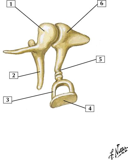
Comment:
The 3 auditory ossicles reside in the middle ear, or tympanic cavity. They amplify sonic vibrations from the tympanic membrane and transmit them to the inner ear.
The 3 ear ossicles are the malleus (hammer), incus (anvil), and stapes (stirrup). The handle of the malleus is fused with the medial aspect of the tympanic membrane; the head articulates with the incus. The incus articulates with the stapes, whose footplate is attached to the oval window.
Atlas Plate 95
Muscles
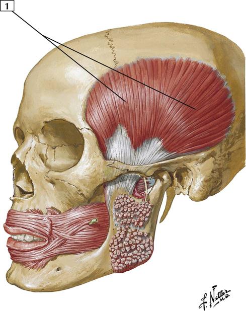
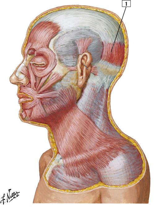
1-17 Muscles of Facial Expression: Lateral View
Origin:
This muscle has no bony origin, and its fibers arise and are continuous with 2 other anterior facial muscles, the procerus and the corrugator supercilii.
Insertion:
The fibers are directed upward. They join the galea aponeurotica anterior to the coronal suture.
Action:
Elevates the eyebrows and wrinkles the forehead, as when a person looks surprised.
Innervation:
Terminal branches of the facial nerve; temporal branch.
Comment:
The epicranius muscle consists largely of the frontal and occipital bellies and an intervening galea aponeurotica (aponeurosis).
As a muscle of facial expression, this cutaneous muscle lies within the layers of the superficial fascia. These muscles vary from person to person, and they often blend together.
Atlas Plate 25
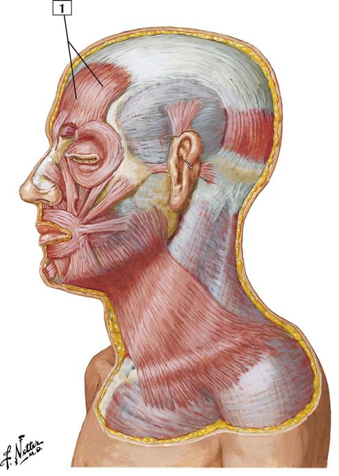
1-18 Muscles of Facial Expression: Lateral View
Origin:
Arises from the lateral two-thirds of the superior nuchal line of the occipital bone and the mastoid process of the temporal bone.
Insertion:
Inserts into the epicranial aponeurosis.
Action:
The occipital and frontal bellies of the epicranial muscle act together to draw back the scalp. This action raises the eyebrows and wrinkles the forehead.
Innervation:
Terminal branches of the facial nerve; temporal branch.
Comment:
The extensive aponeurosis called the galea aponeurotica connects the frontal belly and occipital belly of the epicranial muscle.
As a muscle of facial expression, this cutaneous muscle lies within the layers of the superficial fascia. These muscles vary from person to person, and they often blend together.
Atlas Plate 25
1-19 Muscles of Facial Expression: Lateral View
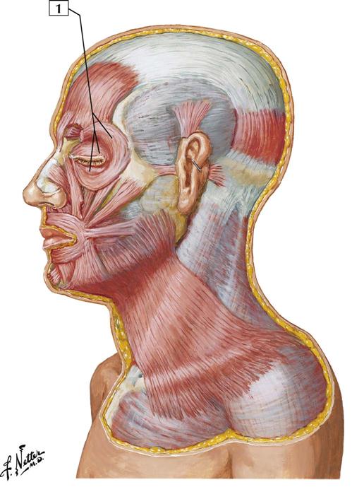
Origin:
Arises from the nasal portion of the frontal bone, the frontal process of the maxilla, the lacrimal bone, and the medial palpebral ligament.
Insertion:
Attaches to the skin of the eyelids, surrounds the bony orbit, and inserts into the superior and inferior tarsi medial to the lacrimal puncta.
Action:
This muscle is a sphincter that closes the eyelids. Its palpebral portion closes the lids gently, as in blinking. The orbital portion closes the eyelids more forcibly.
Innervation:
Terminal branches of the facial nerve; primarily the zygomatic branch.
Comment:
The orbicularis oculi has 3 parts: an orbital part, which is thicker and surrounds the orbital margin; a palpebral part, which is thin and lies in the eyelids; and a lacrimal part.
As a muscle of facial expression, this cutaneous muscle lies within the layers of the superficial fascia.
Atlas Plate 25
1-20 Muscles of Facial Expression: Lateral View
Origin:
Fibers arise near the median plane of the maxilla above and from the mandible below.
Insertion:
Fibers insert into the skin of the lips and into the mucous membrane beneath the lip.
Action:
This muscle acts primarily to close the lips. Its deep and oblique fibers pull the lips toward the teeth and alveolar arches. When all of its fibers act together, they can protrude the lips.
Innervation:
Terminal branches of the facial nerve; primarily the mandibular branch.
Comment:
A major portion of this muscle is derived from the buccinator and blends with other facial muscles around the oral cavity. This muscle is especially important in speech because it alters the shape of the mouth.
As a muscle of facial expression, this cutaneous muscle lies within the layers of the superficial fascia.
Atlas Plate 25
1-21 Muscles of Facial Expression: Lateral View
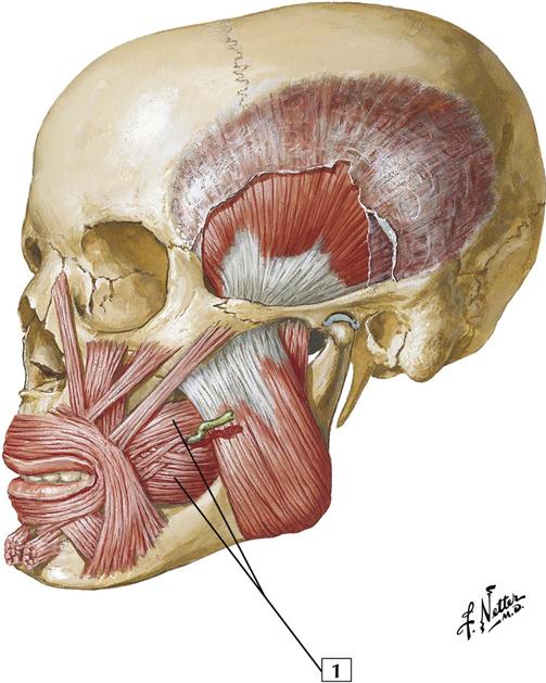
Origin:
Arises from the mandible, pterygomandibular raphe, and alveolar processes of the maxilla and mandible.
Insertion:
Attaches to the angle of the mouth.
Action:
Contraction of this muscle presses the cheek against the molar teeth and aids in chewing. This muscle also can expel air from the mouth, as when a musician plays a woodwind or brass instrument.
Innervation:
Terminal branches of the facial nerve; buccal branch.
Comment:
By pressing the cheek against the teeth, the buccinator holds food between the molars. When the muscle contracts too forcefully during chewing, the teeth bite the cheek.
The term buccinator is Latin for “trumpet player.” This muscle may be well developed in a trumpet player. The buccinator is a muscle of facial expression.
Fibers of this muscle blend with other muscles around the mouth.
Atlas Plate 48
See also Plate 25
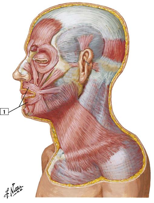
1-22 Muscles of Facial Expression: Lateral View

Origin:
Arises from the superficial fascia covering the superior portions of the pectoralis major and deltoid muscles.
Insertion:
Ascends over the clavicle and is directed medially to insert into the mandible below the oblique line. Other portions of the muscle insert into the skin and subcutaneous tissue of the lower portion of the face.
Action:
Draws the lower lip and corner of the mouth inferolaterally and partially opens the mouth, as during an expression of surprise. When all the fibers act together, the skin over the clavicle and lower neck is wrinkled and drawn upward toward the mandible.
Innervation:
Terminal branches of the facial nerve; cervical branch.
Comment:
As a muscle of facial expression, this cutaneous muscle lies within the layers of the superficial fascia.
Atlas Plate 25
1-23 Muscles of Facial Expression: Lateral View
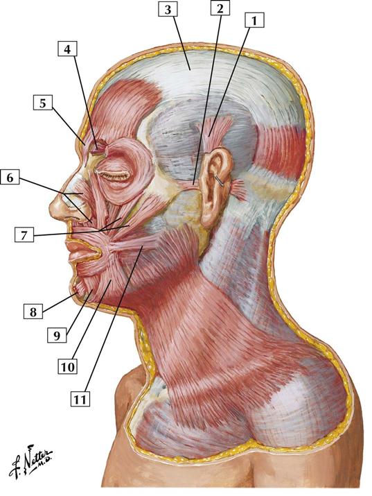
1. Auricularis superior muscle
2. Auricularis anterior muscle
4. Corrugator supercilii muscle (Frontalis and Orbicularis oculi, partially cut away)
6. Nasalis muscle (Transverse part; Alar part)
7. Zygomaticus minor and major muscles
9. Depressor labii inferioris muscle
10. Depressor anguli oris muscle
11. Risorius muscle
Comment:
This lateral view shows additional muscles of facial expression. The muscles around the eyes, ears, nose, and mouth blend with muscles of the lips, chin, and cheek. All are innervated by terminal branches of the facial nerve.
As muscles of facial expression, these cutaneous muscles lie within the layers of the superficial fascia. They vary from person to person, and they often blend together.
All of the muscles of facial expression are derived embryologically from the 2nd pharyngeal (branchial) arch and are innervated by the facial nerve (CN VII).
Atlas Plate 25
1-24 Extrinsic Eye Muscles
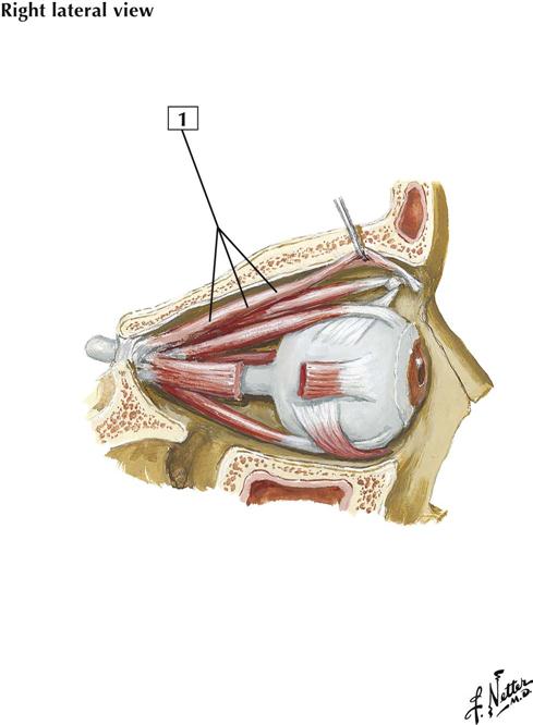
Origin:
Arises from the lesser wing of the sphenoidal bone, anterior and superior to the optic canal.
Insertion:
Attaches to the skin and tarsal plate of the upper eyelid.
Action:
Raises the upper eyelid.
Innervation:
Oculomotor nerve (CN III). At the distal end of this muscle, near its attachment to the tarsal plate, is a small amount of smooth muscle called the superior tarsal muscle. The fibers of the superior tarsal muscle are supplied by postganglionic sympathetic fibers of the autonomic nervous system.
Comment:
Because of the dual nature of this muscle (it is skeletal and has a small smooth muscle component), drooping of the upper eyelid can result from a nerve lesion affecting the oculomotor nerve or the sympathetic fibers. This drooping is called ptosis.
Atlas Plate 86
1-25 Extrinsic Eye Muscles
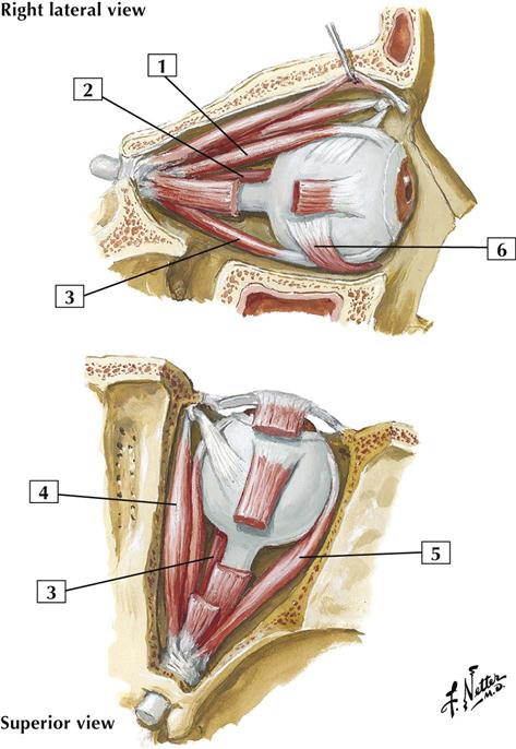
Origin:
The 4 rectus muscles and the superior oblique arise from a common tendinous ring (anulus of Zinn) on the body of the sphenoidal bone. The inferior oblique arises from the floor of the orbit.
Insertion:
The 4 rectus muscles insert into the sclera, just posterior to the cornea. The superior oblique muscle passes forward, and its tendon passes through a fibrous ring (trochlea) and inserts into the sclera deep to the superior rectus muscle. The inferior oblique inserts into the sclera deep to the lateral rectus muscle.
Actions:
In clinical testing, when the eye is abducted, the superior rectus elevates the globe and the inferior rectus depresses it. When the eye is adducted, the superior oblique depresses the globe and the inferior oblique elevates it. The medial rectus is a pure adductor, whereas the lateral rectus is a pure abductor. The anatomic actions differ from the actions tested for clinical evaluation of the muscles.
Innervation:
The lateral rectus is innervated by the abducens nerve (CN VI); the superior oblique is innervated by the trochlear nerve (CN IV). All the other rectus muscles and the inferior oblique are innervated by the oculomotor nerve (CN III).
Atlas Plate 86
1-26 Muscles Involved in Mastication