Lower Limb
Cards 7-1 to 7-72
Bones and Joints
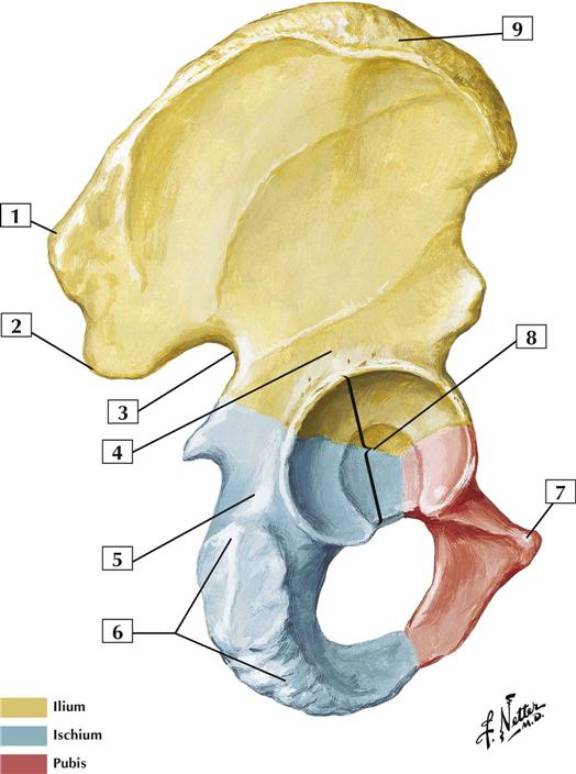
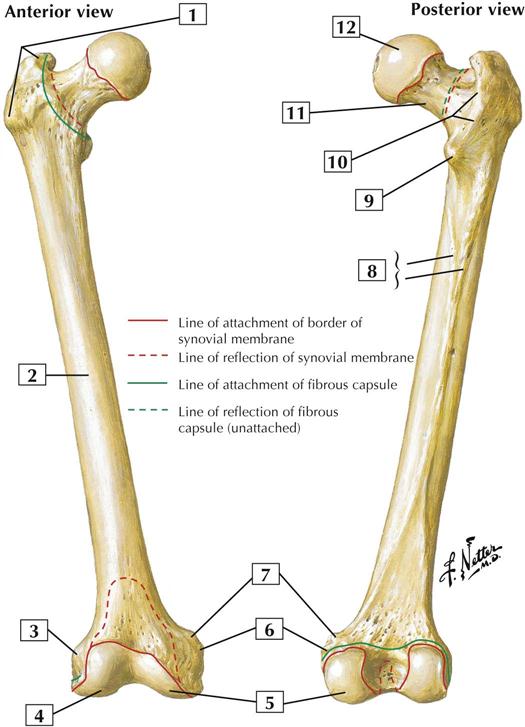
7-1 Hip (Coxal) Bone: Lateral View
Comment:
The hip bone consists of 3 bones: the ilium, ischium, and pubis. Before puberty, these bones are joined by cartilage, but they begin to fuse by midadolescence and are completely fused in adulthood. All 3 fused bones contribute to the acetabulum, the cup-like cavity for articulation of the head of the femur.
The fused hip bone articulates with the femur (thigh bone) and with the vertebral column (spine). Specifically, the ilium articulates with the sacrum in a plane synovial joint that allows for little movement, in contrast to the shoulder joint, and provides great stability. This stability is important for standing, walking, and running on 2 legs (bipedalism).
Atlas Plate 473
See also Plates 243, 333, 334
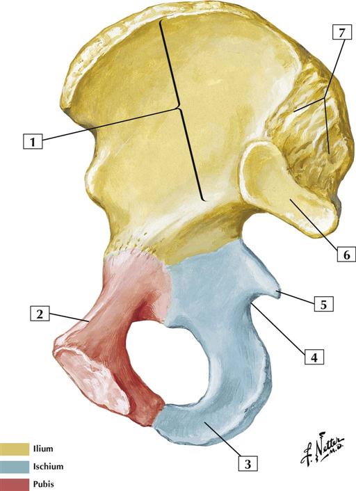
7-2 Hip (Coxal) Bone: Medial View
Comment:
The hip bone consists of 3 bones: the ilium, ischium, and pubis. Before puberty, these bones are joined by cartilage, but they begin to fuse by midadolescence and are completely fused in adulthood.
Anteriorly, the 2 pubic bones articulate with one another at the pubic symphysis. A fibrocartilage disc separates the 2 bones, and this joint allows some movement.
Atlas Plate 473
See also Plates 243, 333, 334
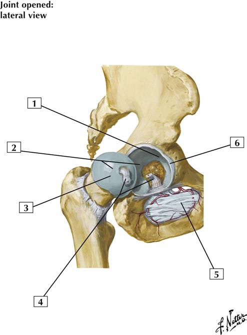
7-3 Hip Joint: Lateral View
Comment:
The hip joint is a multiaxial ball-and-socket synovial joint between the acetabulum and the head of the femur. The acetabular labrum deepens the acetabular cavity even farther, and the fibrous joint capsule is reinforced by 3 ligaments. Within the acetabulum, the ligament of the head of the femur attaches to the femoral head and provides a pathway for a small artery derived from the obturator artery.
The hip participates in abduction and adduction, flexion and extension, and rotation and circumduction.
The ligament of the head of the femur contains the acetabular branch (artery of the round ligament of the femoral head) that arises from the obturator artery.
Blood is supplied to the hip by branches of the medial and lateral femoral circumflex arteries, the gluteal arteries, and the obturator artery.
Atlas Plate 474
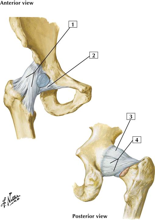
7-4 Hip Joint: Anterior and Posterior Views
Comment:
The hip joint is a multiaxial ball-and-socket synovial joint between the acetabulum and the head of the femur. The acetabular labrum deepens the acetabular cavity even farther, and the fibrous joint capsule is reinforced by 3 ligaments.
The iliofemoral ligament is the most important ligament reinforcing the hip joint. This ligament forms an inverted Y ligament (of Bigelow) that limits hyperextension and lateral rotation. The pubofemoral ligament limits extension and abduction, whereas the ischiofemoral ligament limits extension and medial rotation. If one notices where these ligaments attach, one can understand how they limit movement in a certain direction.
The hip participates in abduction and adduction, flexion and extension, and rotation and circumduction.
Atlas Plate 474
7-5 Femur
Comment:
The femur, or thigh bone, is the longest bone in the body. When a person is standing, the femur transmits the weight of the body from the hip to the tibia.
The head of the femur articulates with the coxal (hip) bone at the acetabulum. The femoral neck is a common fracture site. The greater trochanter is the point of the hip and an attachment site for several of the gluteal muscles (abductors of the thigh at the hip). The lesser trochanter is an attachment site for the iliopsoas tendon, a strong flexor of the thigh at the hip.
Atlas Plate 476
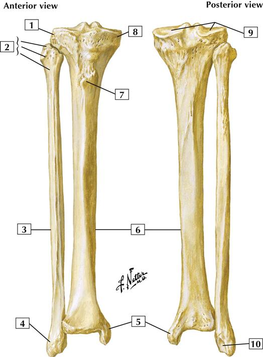
7-6 Tibia and Fibula
Comment:
The tibia articulates with the condyles of the femur and is the weight-bearing bone of the leg.
The smaller fibula lies posterolateral to the tibia. It exists largely for muscle attachment.
The tibial tuberosity is the insertion site for the patellar ligament (tendon of attachment for the quadriceps muscles of the anterior thigh that extend the leg at the knee joint).
The proximal tibiofibular joint is a plane synovial joint that permits limited gliding movement. The distal tibiofibular joint is a fibrous joint (syndesmosis), which allows almost no movement.
Atlas Plate 500
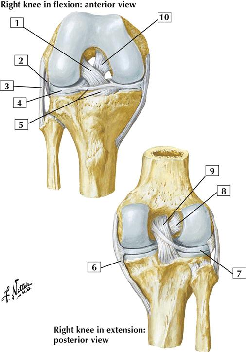
7-7 Knee: Cruciate and Collateral Ligaments
Comment:
The knee is the largest and most complicated joint in the body. It is a biaxial condylar synovial joint between the condyles of the femur and tibia. It also includes a saddle joint between the femur and patella.
The knee participates in flexion and extension. When flexed, it also participates in some gliding and rotation movements. When the knee extends fully, the femur rotates slightly and medially on the tibia, pulling each of the ligaments taut and stabilizing the joint.
The menisci, the cruciate ligaments, and the transverse ligament are intracapsular ligaments. The transverse ligament binds and stabilizes the menisci.
Most of the blood supply to the knee is from genicular branches of the popliteal artery.
Atlas Plate 496
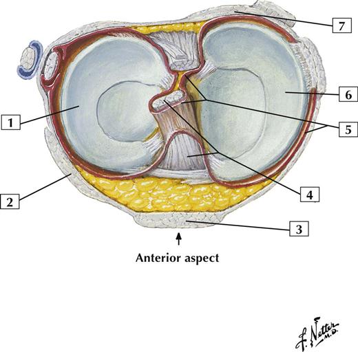
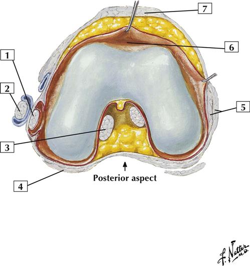
7-8 Knee: Interior (Superior View)
Comment:
The knee is surrounded by a thin, fibrous capsule that is stabilized by the surrounding muscle attachments and intracapsular and extracapsular ligaments. Intracapsular ligaments include the anterior and posterior cruciate ligaments, medial and lateral menisci, and transverse ligament. Extracapsular ligaments include the medial and lateral collateral ligaments, patellar ligament, and arcuate and oblique popliteal ligaments.
Of the 2 cruciate ligaments, the anterior is the weaker and is most taut when the knee is fully extended, preventing hyperextension. It is usually torn in hyperextension with the tibia medially (internally) rotated. The posterior cruciate tightens most during flexion of the knee, preventing excessive anterior displacement of the femur on the tibia or excessive posterior displacement of the tibia on the femur. Both cruciate ligaments maintain some level of tautness during movements of the knee.
The tibial collateral ligament limits extension and abduction of the leg and is attached to the medial meniscus. The fibular collateral ligament limits extension and adduction of the leg.
Atlas Plate 495
7-9 Knee: Interior (Inferior View)
Comment:
The knee is surrounded by a thin, fibrous capsule that is stabilized by the surrounding muscle attachments and intracapsular and extracapsular ligaments. Intracapsular ligaments include the anterior and posterior cruciate ligaments, medial and lateral menisci, and transverse ligament. Extracapsular ligaments include the medial and lateral collateral ligaments, patellar ligament, and arcuate and oblique popliteal ligaments.
Of the 2 cruciate ligaments, the anterior is the weaker and is most taut when the knee is fully extended, preventing hyperextension. The posterior cruciate tightens most during flexion of the knee, preventing excessive anterior displacement of the femur on the tibia or excessive posterior displacement of the tibia on the femur.
Atlas Plate 495
7-10 Bones of Foot
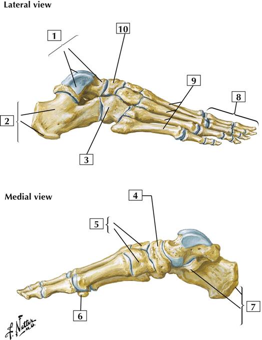
Comment:
The bones of the foot include the 7 tarsal bones, of which only the talus articulates with the leg bones. Five metatarsal bones articulate proximally with the tarsals and distally with the phalanges. Similar to the thumb, the 1st toe (big toe) has only 2 phalanges. Toes 2 through 5 have a proximal, middle, and distal phalanx.
The trochlea of the talus (ankle bone) articulates with the tibia and fibula, and the head of the talus articulates with the navicular bone. The calcaneus (heel bone) articulates with the talus superiorly and the cuboid anteriorly.
Atlas Plate 512
7-11 Ligaments and Tendons of Ankle: Lateral View
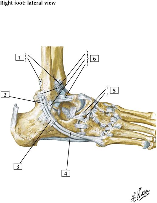
Comment:
The ankle (talocrural) joint is a hinge-type (ginglymus) uniaxial synovial joint between the tibia and fibula and the trochlea of the talus. This joint permits dorsiflexion (extension) and plantarflexion. Its thin, fibrous capsule is reinforced by the medial (deltoid) ligament, which has 4 parts, and the lateral collateral ligament, which has 3 parts.
Of the tarsal joints, the talocalcaneal (subtalar) joint is a plane synovial joint between the talus and calcaneus. It permits inversion and eversion of the foot.
The talocalcaneonavicular joint is a partial ball-and-socket synovial joint between the head of the talus and the calcaneus and navicular bones (along with the calcaneocuboid joint, it forms the transverse tarsal joint). It is supported by the spring ligament and is important in gliding and rotational movements of the foot.
Atlas Plate 514
7-12 Ligaments and Tendons of Ankle: Medial View
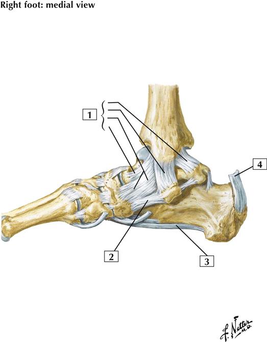
Comment:
The ankle (talocrural) joint is a hinge-type (ginglymus) uniaxial synovial joint between the tibia and fibula and the trochlea of the talus. This joint permits dorsiflexion (extension) and plantarflexion. Its thin, fibrous capsule is reinforced by the medial (deltoid) ligament, which has 4 parts, and the lateral collateral ligament, which has 3 parts.
The medial (deltoid) ligament has 4 parts and limits eversion of the foot. This ligament helps maintain the medial long arch of the foot, whereas the plantar calcaneonavicular (spring) ligament provides strong plantar support for the head of the talus (which maintains the arch of the foot).
Atlas Plate 514
7-13 Ligaments and Tendons of Foot: Plantar View
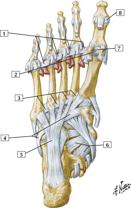
Comment:
The tarsometatarsal joints are plane synovial joints. They consist of articular capsules and are strengthened by plantar, dorsal, and interosseous ligaments. They permit gliding and sliding movements.
The metatarsophalangeal joints are multiaxial condyloid synovial joints surrounded by articular capsules and strengthened by plantar and collateral ligaments. They permit flexion and extension, some abduction and adduction, and circumduction. The plantar (plate) ligaments are part of the weight-bearing surface of the foot.
The interphalangeal joints are uniaxial hinge-type (ginglymus) synovial joints that also are enclosed by capsules and strengthened by plantar and collateral ligaments. They permit flexion and extension.
Atlas Plate 515
Muscles
7-14 Muscles of Lower Limb
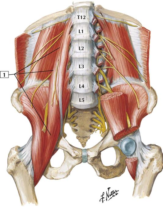
Origin (proximal):
Arises from the transverse processes of all 5 lumbar vertebrae and from the sides of the T12-L5 vertebrae and the intervertebral discs between them.
Insertion (distal):
Tapers inferiorly, crossing in front of the sacrum and sacro-iliac joint to join with the iliacus muscle and to insert on the lesser trochanter of the femur.
Action:
With the iliacus, the psoas major flexes the thigh at the hip and is an important flexor of the trunk at the hip. Acting alone, it laterally flexes the trunk ipsilaterally.
Innervation:
Ventral rami of lumbar nerves L1-3.
Comment:
The psoas major and iliacus muscles are commonly referred to as the iliopsoas muscle because they act in unison. Their action is especially important in flexing the trunk against gravity, as when a person does sit-ups with the legs straight (hips extended).
About half the population has a smaller muscle, the psoas minor, on the anterior surface of the psoas major.
Atlas Plate 483
See also Plate 258
7-15 Muscles of Lower Limb
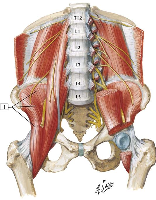
Origin (proximal):
The fan-shaped iliacus arises from the inner surface of the wing of the ilium (iliac fossa).
Insertion (distal):
The iliacus fibers blend with those of the psoas major and insert on the lesser trochanter of the femur.
Action:
The iliacus acts in unison with the psoas major muscle. The 2 muscles are often called the iliopsoas muscle. The iliopsoas flexes the thigh at the hip and is an important flexor of the trunk.
Innervation:
Femoral nerve (L2, L3, and L4).
Comment:
The iliacus is innervated by branches derived from the femoral nerve as this larger nerve descends to pass into the thigh.
Atlas Plate 483
See also Plate 258
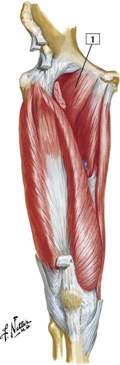
7-16 Muscles of Hip and Thigh: Lateral View

Origin (proximal):
Arises from the anterior superior iliac spine and the anterior portion of the iliac crest.
Insertion (distal):
As its name suggests, this muscle inserts into the iliotibial tract. This strong tendinous tract inserts on the lateral condyle of the tibia.
Action:
This muscle flexes, abducts, and medially rotates the thigh at the hip. With the assistance of the gluteus maximus, this muscle stabilizes the hip joint. The tensor fasciae latae also stabilizes the extended knee.
Innervation:
Superior gluteal nerve (L4 and L5).
Comment:
The chief action of the tensor fasciae latae is hip flexion. The muscle also acts with the gluteus maximus to control anteroposterior tilting of the pelvis when 1 leg supports all of the body’s weight. Stabilization of the hip occurs because the muscle holds the femoral head in the acetabulum. The tensor fasciae latae also stabilizes the knee in extension.
Atlas Plate 481
7-17 Muscles of Thigh: Anterior View
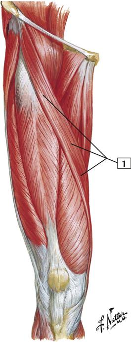
Origin (proximal):
Arises from the anterior superior iliac spine.
Insertion (distal):
Inserts on the superior portion of the medial surface of the tibial shaft, close to the insertions of the gracilis and semitendinosus muscles.
Action:
The sartorius muscle crosses the hip and knee joints. Consequently, it is a flexor, abductor, and lateral rotator of the thigh at the hip joint. It is also a flexor of the leg at the knee joint. Along with other muscles originating from the pelvis, it helps to balance the pelvis.
Innervation:
Femoral nerve (L2 and L3).
Comment:
Sartorius is Latin for “tailor.” By sitting cross-legged in a tailor’s position, one can appreciate the function of the sartorius muscle.
Atlas Plate 479
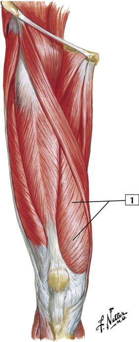
7-18 Muscles of Thigh: Anterior View
Origin (proximal):
Arises by 2 separate heads, a straight head from the anterior inferior iliac spine and a reflected head from the ilium just superior to the acetabulum.
Insertion (distal):
The tendons of origin combine to form a fusiform muscle belly that inserts into the quadriceps tendon. This tendon inserts into the base of the patella and, by extension of the patellar ligament, into the tibial tuberosity.
Action:
This muscle acts on the knee through the patellar ligament and is an extensor of the leg at the knee joint. Because it also crosses the hip joint, it helps the iliopsoas flex the thigh at the hip.
Innervation:
Femoral nerve (L2, L3, and L4).
Comment:
The rectus femoris and the 3 vastus muscles form the quadriceps femoris complex. These muscles are powerful extensors of the knee. Of the 4 quadriceps muscles, only the rectus femoris crosses the hip and the knee joints.
Atlas Plate 479
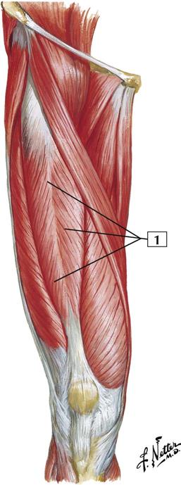
7-19 Muscles of Thigh: Anterior View
Origin (proximal):
Arises from the posterior aspect of the femur, beginning at the greater trochanter and continuing inferiorly along the lateral lip of the linea aspera of the femur.
Insertion (distal):
Most of the muscle inserts into the lateral patella and the tendon of the rectus femoris to form the quadriceps tendon. The patellar ligament inserts into the tibial tuberosity.
Action:
Extension of the leg at the knee.
Innervation:
Femoral nerve (L2, L3, and L4).
Comment:
The vastus lateralis is 1 of the 4 muscles making up the quadriceps femoris extensor complex of the knee. It covers essentially the entire lateral portion of the thigh.
Atlas Plate 479
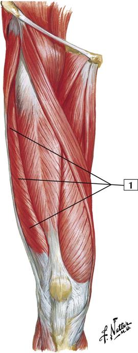
7-20 Muscles of Thigh: Anterior View
Origin (proximal):
Arises from the anterior and lateral aspects of the femoral shaft and the lateral intermuscular septum.
Insertion (distal):
Inserts into the posterior surface of the upper border of the patella and forms part of the quadriceps tendon. The patellar tendon inserts into the tibial tuberosity.
Action:
Extension of the leg at the knee joint.
Innervation:
Femoral nerve (L2, L3, and L4).
Comment:
The vastus intermedius is 1 of the 4 muscles of the quadriceps femoris group that makes up the extensor complex of the knee. Tapping the patellar tendon of this extensor complex elicits the knee jerk reflex and tests spinal cord segments L3 and L4.
Atlas Plate 479
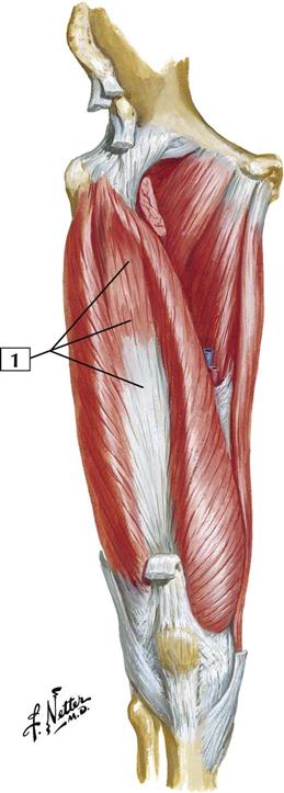
7-21 Muscles of Thigh: Anterior View
Origin (proximal):
Arises from the intertrochanteric line and medial lip of the linea aspera of the femur and from the medial intermuscular septum.
Insertion (distal):
Inserts into the medial border of the quadriceps tendon, but some of its inferior fibers insert directly into the medial side of the patella. The patellar tendon inserts into the tibial tuberosity.
Action:
Extension of the leg at the knee joint.
Innervation:
Femoral nerve (L2, L3, and L4).
Comment:
The vastus medialis is 1 of the 4 muscles of the quadriceps femoris complex that extends the knee. Similar to the vastus lateralis, the vastus medialis contributes some aponeurotic fibers to the knee joint capsule.
Atlas Plate 479
7-22 Muscles of Thigh: Anterior View
Origin (proximal):
Arises from the pecten of the pubic bone.
Insertion (distal):
Inserts on the pectineal line of the shaft of the femur just inferior to the lesser trochanter.
Action:
Adducts and flexes the thigh at the hip joint and assists with medial rotation of the thigh.
Innervation:
Femoral nerve (L2 and L3) and occasionally a branch from the obturator nerve.
Comment:
The pectineus muscle is medial to the iliopsoas and forms a portion of the floor of the femoral triangle. The muscle is usually flat and quadrangular.
Stay updated, free articles. Join our Telegram channel

Full access? Get Clinical Tree


