Unicentric Plasma Cell Variant Castleman Disease
Pei Lin, MD
Key Facts
Terminology
Histologically distinctive reaction pattern in lymph node characterized by
Marked interfollicular plasmacytosis
Regressive (hyaline-vascular) changes in small subset of follicles
Defined in this chapter as HHV8(-) and not associated with either multicentric CD or POEMS syndrome
Clinical Issues
˜ 10-20% of localized/unicentric cases of CD
Broad age range; median 3rd-4th decade
Peripheral lymph nodes are most commonly affected
˜ 10-20% of patients have systemic symptoms &/or laboratory abnormalities
Subset of cases reported with these abnormalities may be unrecognized multicentric CD
Usually cured by surgical excision
Microscopic Pathology
Preserved lymph node architecture
Marked plasmacytosis in interfollicular areas
No cytologic atypia
Germinal centers are hyperplastic and usually subset are atretic with changes resembling CD-HV
Ancillary Tests
No evidence of HHV8 infection
No evidence of monoclonality
Top Differential Diagnoses
HHV8(+) multicentric CD
Autoimmune diseases
Marginal zone B-cell lymphoma
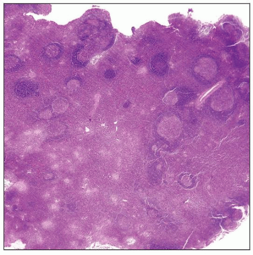
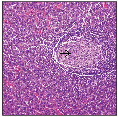
 Lymph node involved by unicentric Castleman disease, plasma cell variant (CD-PV). Multiple follicles are present and the interfollicular zones are expanded by plasma cells. |
TERMINOLOGY
Abbreviations
Castleman disease, plasma cell variant (CD-PV)
Synonyms
Unicentric Castleman disease, plasma cell variant
Angiofollicular lymph node hyperplasia
Giant lymph node hyperplasia
Angiomatous lymphoid hamartoma
Benign giant lymphoma
Definitions
Histologically distinctive reaction pattern in lymph node characterized by
Marked interfollicular plasmacytosis
Regressive (hyaline-vascular) changes in small subset of follicles in subset of cases
ETIOLOGY/PATHOGENESIS
Unknown
Data supporting role for dysregulation of interleukin-6 (IL-6) in pathogenesis
Lymphocytes in CD-PV express IL-6
B cells express IL-6 receptor (CD126)
Autocrine or paracrine mechanisms may be involved
In mice, forced expression of IL-6 in bone marrow cells causes syndrome that resembles, in part, CD-PV
Immune dysregulation also may be involved
As defined in this chapter, there is no evidence of human herpes virus 8 (HHV8) infection
CLINICAL ISSUES
Epidemiology
Incidence
Accounts for 10-20% of localized or unicentric cases of CD
Age
Broad range; median in 3rd-4th decade
Gender
No preference
Site
Peripheral lymph nodes most common
Mediastinal involvement less common (than hyaline vascular variant of CD)
Presentation
Most patients present with lymphadenopathy without systemic symptoms
10-20% of patients reported in literature had systemic symptoms
Fever, night sweats, weight loss, malaise
However, it seems likely that many of these patients were HHV8(+)
Therefore better classified as HHV8-associated &/or multicentric CD
Small subset of patients reported were associated with POEMS syndrome
POEMS = peripheral neuropathy, organomegaly, endocrinopathy, monoclonal M protein, skin lesions
These patients also likely to be HHV8(+)
Probably better classified as HHV8-associated &/or multicentric CD
Laboratory Tests
Many patients lack laboratory abnormalities
Subset of patients (˜ 10-20%) can have cytopenias
Anemia and thrombocytopenia
Serum IL-6 levels can be increased
Treatment
Surgical approaches
Usually curative by excision
Prognosis
Good
Small subset of patients may evolve to multicentric CD
Possibly were cases of multicentric CD at time of initial biopsy
IMAGE FINDINGS
Radiographic Findings
Lymphadenopathy
Often multiple lymph nodes in an anatomic group are large
PET scan shows increased FDG uptake
MACROSCOPIC FEATURES
Size
Usually lymphadenopathy is of modest size
MICROSCOPIC PATHOLOGY
Histologic Features
Less well defined than HV-CD
Preserved overall lymph node architecture
Marked plasmacytosis in interfollicular areas
Some plasma cells can be binucleated
Vascularity in interfollicular areas can be prominent
Sinuses usually patent
Widely spaced lymphoid follicles
Lymphoid follicles contain hyperplastic germinal centers, but small subset of germinal centers often show regressive changes
Resemble follicles seen in CD-HV
Others have used term “mixed or transitional” type because of these follicles
Atretic follicles are usually present and part of spectrum of CD-PV
Mantle zones are usually well defined and can be expanded
Plasmablasts are absent or rare in mantle zones
Cytologic Features
Plasma cells are cytologically normal without atypia
Lymphocytes show range in cytologic appearance
Difficult to establish specific diagnosis of CD-PV by fine needle aspiration
ANCILLARY TESTS
Immunohistochemistry
Interfollicular plasma cells express polytypic immunoglobulin light chains
Follicles composed of polytypic B cells and T cells
Germinal centers are Bcl-2(-)
Atretic follicles show increased follicular dendritic cells
CD21(+), CD23(+), CD35(+)
Rare cases are reported with monoclonal plasma cells
These cases most likely HHV8(+) &/or multicentric CD


