Tuberculous Epididymo-orchitis
Steven S. Shen, MD, PhD
Jae Y. Ro, MD, PhD
Key Facts
Terminology
Infection of testis and epididymis due to Mycobacterium tuberculosis
Etiology/Pathogenesis
Mycobacterium tuberculosis
Most tuberculous epididymo-orchitis are associated with other genitourinary tract involvement
Clinical Issues
Affects any age but mainly adults (> 72% are older than 35 years)
Mild testicular enlargement and scrotal pain
Macroscopic Features
Epididymis is almost always primary site of involvement with secondary spread to testis
Irregular mass with foci of caseating necrosis
Microscopic Pathology
Multiple confluent granulomas with central caseating necrosis
Aggregates of epithelioid cells with peripheral rim of lymphocytes
Destruction of seminiferous tubules and interstitium by caseating or noncaseating granulomatous inflammation
Langhans giant cells may be present
Late stage with fibroblastic response with scar formation
Ancillary Tests
Acid-fast bacteria stain positive
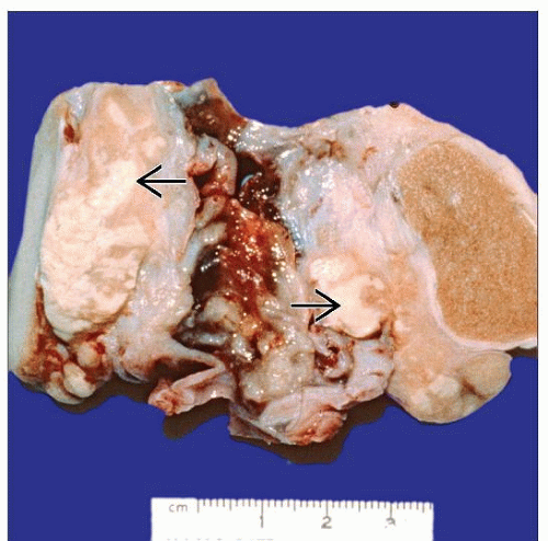 Gross photograph of a tuberculous epididymo-orchitis shows involvement of testis and epididymis by an irregular gray-white pseudotumorous mass with chalky white areas of necrosis  . . |
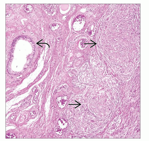 Several large epithelioid granulomas  are seen in the epididymis and surrounding paratesticular soft tissue are seen in the epididymis and surrounding paratesticular soft tissue  . Most testicular tuberculosis in adults starts with involvement of the epididymis. . Most testicular tuberculosis in adults starts with involvement of the epididymis. |
TERMINOLOGY
Definitions
Infection of testis and epididymis due to Mycobacterium tuberculosis
ETIOLOGY/PATHOGENESIS
Infectious Agents
Mycobacterium tuberculosis
Most cases of tuberculous epididymo-orchitis are associated with genitourinary tract involvement at other sites
In adults, almost all are result of tuberculous prostatitis
In children, > 1/2 of patients have advanced pulmonary tuberculosis and miliary spread
CLINICAL ISSUES
Epidemiology
Incidence
High incidence in developing countries, immigrants, and immunocompromised patients
May be late manifestation of intravesical bacillus Calmette-Guérin (BCG) therapy
Age
Affects any age but mainly adults (> 72% are older than 35 years)
Presentation
Mild testicular enlargement and scrotal pain
Associated with other constitutional symptoms of tuberculous infection
Commonly associated with tuberculosis of lung and genitourinary tract
Bilateral involvement (30%)
Formation of abscess or sinus tract (50%)
Secondary hydrocele (30%)
Treatment
Surgical resection and systemic antituberculous therapy
Prognosis
Excellent with modern antituberculous treatment
IMAGE FINDINGS
General Features
Nonspecific heterogeneous or homogeneous mass of epididymis or testis on ultrasonography
MACROSCOPIC FEATURES
General Features
Epididymis is almost always primary site of involvement with secondary spread to testis
Irregular mass with foci of caseating necrosis
When testis is involved, swollen and nodular
Late stages: Extensive cystic change due to necrosis, associated hydronephrosis
MICROSCOPIC PATHOLOGY
Histologic Features
Destruction of epididymis/tubules with caseating or noncaseating granulomatous inflammation in interstitium
Multiple confluent granulomas with central caseating necrosis
Aggregates of epithelioid cells with peripheral rim of lymphocytes
Langhans giant cells (fusion of epithelioid cells with nuclei arranged in horseshoe-shaped pattern, often pointing toward necrosis)
Schaumann (basophilic, shell-like crystals) and asteroid bodies may be present
Late stage with fibroblastic response with scar formation
Predominant Pattern/Injury Type
Infectious
Predominant Cell/Compartment Type
Histiocytes/macrophages, lymphocytes, and plasma cells
ANCILLARY TESTS
Histochemistry
Ziehl-Neelsen (acid-fast bacillus)
Reactivity: Positive
DIFFERENTIAL DIAGNOSIS
Nonspecific Granulomatous Orchitis
Lack of necrosis or well-formed granuloma
Stay updated, free articles. Join our Telegram channel

Full access? Get Clinical Tree


