Fig. 13.1 Proposed sites for injection in treatment of hemifacial spams. The corrugator, orbital orbicularis oculi, frontalis and procerus are marked. Starting doses are from 2.5 to 3.75 U onabotulinumtoxinA depending upon the severity of facial twitching.
Hemifacial spasm is more prevalent in females, commonly begins in the fifth decade and tends to have a fluctuating course. In contrast to essential blepharospasm, symptoms often continue during sleep and can provoke insomnia. Emotion and stress tend to exacerbate facial twitching. Ear clicks can occur which resolve with treatment of the HFS (Rudzinska et al., 2010). Although benign, HFS can be disabling because of social embarrassment and excessive closure of the affected eye, interfering with vision. Symptoms can progress over time and facial weakness may develop independent from botulinum neurotoxin (BoNT) therapy. Hypertension is thought to be a risk factor for the development of HFS (Oliveira et al., 1999).
Imaging studies have confirmed that the most frequent cause of HFS is vascular compression of the facial nerve at the root exit zone. The severity of compression correlates with the severity of HFS symptoms (Banik and Miller, 2004). Vascular compression generally involves the anterior inferior cerebellar artery, the posterior inferior cerebellar artery, the vertebral basilar artery and the internal auditory artery. The internal auditory artery can be tortuous or ectatic. The offending vessel is ipsilateral to the facial nerve and side of the HFS. Non-vascular origins of HFS occur less commonly and include demyelination, various tumors and space-occupying lesions in the cerebellopontine angle. Evidence suggests that there may be a genetic susceptibility toward vascular anomaly resulting in HFS (Miwa et al., 2002; Lagalla et al., 2010).
Hemifacial spasm must be distinguished from other conditions involving the facial musculature, including blepharospasm, facial myokymia, oromandibular dystonia, facial tic, hemimasticatory spasm, post-Bell’s palsy synkinesis and focal seizures. Blepharospasm usually occurs bilaterally at onset and concerns only the eyelids (with the exception that it can be part of involvement of other facial muscles in Meige’s syndrome). Blepharospasm is a form of dystonia that causes involuntary closure of the eyes by muscle spasm, or without spasms in a form called apraxia of eyelid opening. Bright light can exacerbate the condition, which subsides during sleep. Blepharospasm may uncommonly coexist with HFS, complicating the diagnosis. One study found blepharospasm to occur following the onset of HFS (Tan et al., 2004). Facial myokymia, a fine rippling movement of the facial muscles, is associated with an abnormality of the brainstem such as that seen in multiple sclerosis. Oromandibular dystonia, another form of dystonia involving only the lower facial muscles, subsides during sleep, similar to other dystonias. Facial tics tend to be multifocal and not unilateral, have more complex movements and are usually associated with premonitory sensations and mild voluntary suppression. Hemimasticatory spasm affects jaw closure, with painful muscle contractions. Facial synkinesiae are caused by a misdirected axonal sprouting after facial nerve lesions. Periocular muscle activation would be associated with perioral movements and vice versa. They may be mistaken for HFS; orbicular synkinesis can be treated similar to HFS with BoNT injections (Roggenkamper et al., 1994). Finally, focal seizures, including epilepsia partialis continua, may be erroneously diagnosed as HFS (Wang and Jankovic, 1998).
Diagnostic tests for HFS include a brain MRI with attention to the cerebellopontine angle, with and without contrast, which will detect any space-occupying lesion requiring neurosurgical intervention. Magentic resonance angiography of the intracranial vessels may help to define the site of vascular compression. Electromyography can help with the differential diagnosis in difficult cases, and electroencephalography may be able to detect epileptiform discharges characteristic of a focal seizure.
Treatment
Treatment of HFS has included medications, microvascular decompression and BoNT injections. Medications such as baclofen, clonazepam, carbamazepine and phenytoin may provide mild improvement at the expense of side effects. Microvascular decompression (Janetta’s operation) involves placing surgical gauze inbetween the facial nerve and the compressing blood vessel. Success rates from microvascular decompression vary from 88 to 97%, and a small percentage of patients may experience recurrence of HFS following surgery (Miller and Miller, 2012). Surgical complications include hearing loss and facial weakness, in addition to the accepted surgical risk of intracranial hemorrhage, stroke and even death.
Injections of BoNT are the preferred treatment of HFS. They are successful in over 90% of patients. Patients with HFS tend to require a lower dose, with a longer duration of effect compared with that in those with blepharospasm (Cannon et al., 2010). Injections of BoNT provide relief from symptoms for a period of approximately 12 weeks, and repeated injections have been found to be effective for many years. They can provide relief from symptoms without the adverse effects of neurosurgery. Serotype A BoNT, in the forms of onabotulinumtoxinA, abobotulinumtoxinA and incobotulinumtoxinA, and serotype B (rimabotulinumtoxinB) have all been used in the treatment of HFS. Use of EMG guidance during injection is not necessary.
Side effects of BoNT injections tend to be those associated with other facial injections: erythema and ecchymosis of the region injected, dry eyes, mouth droop, ptosis, lid edema and facial muscle weakness (Elston, 1986; Yoshimura et al., 1992; Park et al., 1993). Ptosis and facial muscle weakness are transient and will resolve within 1 to 4 weeks. Ptosis could be caused by local diffusion of the BoNT to affect the levator palpabrea (Brin et al., 1987).
The beneficial effect of BoNT injections occurs within 3 days to 2 weeks, with a peak effect at approximately 2–3 weeks. The effects are transient, with a mean duration of improvement of approximately 2.8 months (Yoshimura et al., 1992). There is a high variability of duration of the beneficial effect among patients.
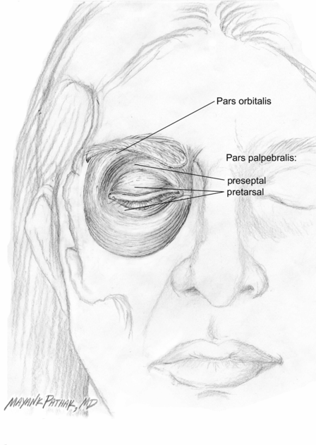
The muscles injected to treat HFS may be the orbicularis oculi, corrugator, frontalis, risorius, buccinator and depressor anguli oris. The orbicularis oculi is composed of two parts: the pars palpebralis, which ordinarily closes the eyelid, and the pars orbitalis, which squeezes the eye shut with stronger contractions. The pars palpebralis also has two parts: a preseptal and a pretarsal region (Fig. 13.2).

Fig 13.2 The orbicularis oculi and its parts..
Stay updated, free articles. Join our Telegram channel

Full access? Get Clinical Tree


