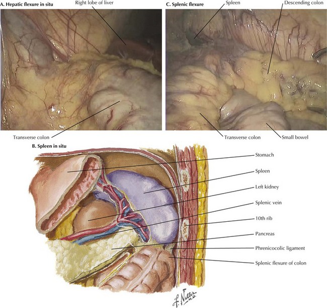
Transverse Colectomy

Chapter 23


Transverse Colectomy


Surgical Principles


Transverse colectomy requires detailed knowledge of the vascular supply of the colon, the anatomy of the hepatic and splenic flexures, and the relationship of the omentum to the colon and stomach. The goal of the operation is to resect the transverse colon and create a tension-free, well-vascularized bowel anastomosis. The procedure usually involves the following steps:


The procedure is somewhat unique in that steps 1 to 4 can be performed in any order desired by the surgeon. This is particularly important for the distal colonic segment, which will be perfused retrograde from the inferior mesenteric artery through the marginal artery of Drummond.


Anatomy for Transverse Colectomy


Hepatic Flexure


The hepatic (or right) flexure is the anatomic name for the bend in the colon as it transitions from the ascending colon to the transverse colon. The hepatic flexure usually contains only small, unnamed vessels, although in some patients there are larger vessels that require ligation. Certain disease states, such as portal hypertension with retroperitoneal collateralization, can cause the hepatic flexure to become quite vascular.


The intraoperative photograph demonstrates the hepatic flexure in situ (Fig, 23-1, A). The liver is cephalad and the right kidney is posterior. The ascending colon is mobilized from its lateral attachments at the white line of Toldt. As the ascending colon is mobilized medially, the dissection is complete when the duodenum is identified and preserved posteriorly with the retroperitoneum. Aggressive traction near the end of the mobilization may cause avulsion injury to the middle colic vein, which results in difficult-to-control hemorrhage.


An alternative and more common strategy for mobilization of the hepatic flexure is to begin by dividing the lateral attachments of the ascending colon along the white line of Toldt. The dissection then proceeds distally around the hepatic flexure at the ascending-transverse junction and continues medially.


Splenic Flexure


The splenic (or left) flexure of the colon is the bend in the bowel where the distal transverse colon transitions to the descending colon. To mobilize the splenic flexure, the avascular lateral attachments of the descending colon to the retroperitoneum must be divided along the white line of Toldt. The splenocolic (lienocolic) ligament is the superior extension of this and forms connective bands connecting the apex of the splenic flexure to the inferior aspect of the splenic capsule. The anatomic relations of the splenic flexure in situ are shown in Figure 23-1, B and C.


The dissection continues from distal to proximal on the descending colon in the proper plane along the white line of Toldt, as appropriate medial traction is applied. Aggressive downward traction on the flexure can cause avulsion injury to the splenic capsule, resulting in dangerous bleeding that may rarely require splenectomy. Variation in the splenic flexure redundancy, angle, and location can greatly affect the difficulty in mobilization. A low-lying, nonredundant flexure is much easier to mobilize than a high, redundant colon that is closely adherent to the spleen.


For difficult splenic flexures in open surgery, the incision must be of adequate size to provide optimal lighting and retraction. In laparoscopic surgery, extralong instruments, additional working ports, and changes in patient position may be necessary.


Dorsally, attachments to the kidney and filmy adhesions to the tail of the pancreas must be freed to supply adequate mobilization of the splenic flexure. The renocolic ligament is connective tissue posteriorly adherent from the colon to the kidney and must be divided to complete the dissection.

Stay updated, free articles. Join our Telegram channel

Aug 12, 2016 | Posted by in ANATOMY | Comments Off on Transverse Colectomy

Full access? Get Clinical Tree

Get Clinical Tree app for offline access