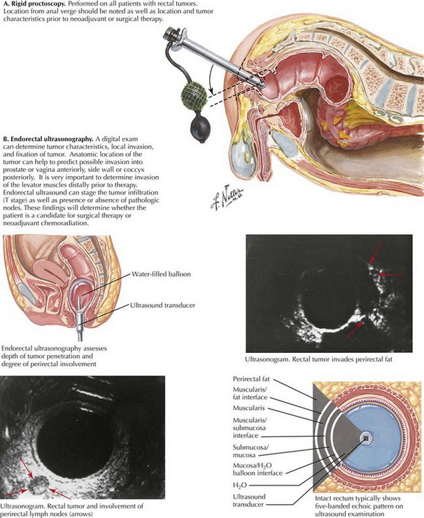
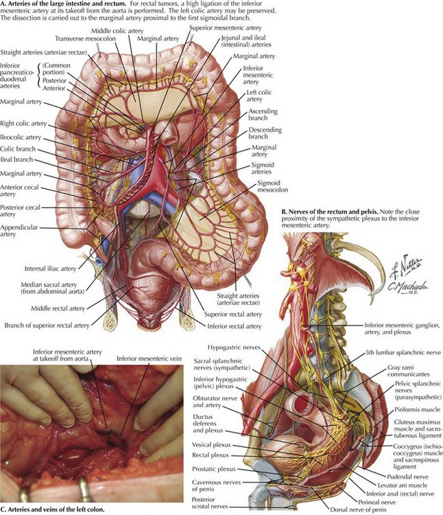
Abdominoperineal Resection

Chapter 25


Abdominoperineal Resection


Principles of Preoperative Evaluation


The patient is screened with a full colonoscopy. Digital rectal examination and proctoscopy are performed to confirm tumor location and to assess feasibility of a sphincter-sparing approach (Fig. 25-1, A). Digital vaginal examination and vaginoscopy are performed with the proctoscope to assess for local invasion. CT scanning of the chest, abdomen, and pelvis is done to survey for metastatic disease. Endorectal ultrasound is used for staging to assess the need for preoperative chemoradiation (Fig. 25-1, B).


Pelvic magnetic resonance imaging (MRI) is increasingly used, providing a more complete and less operator-dependent picture of the extent of the tumor in the pelvis. MRI can provide extremely useful information on circumferential mesorectal margins or frank involvement of the pelvic side wall, sacrum, or anterior organs. MRI is particularly useful in men with anteriorly based tumors, because it can determine whether local involvement of the prostate, seminal vesicles, or bladder exists, indicating a need for exenteration.


Patients staged with clinical stage II or stage III tumors are usually treated with preoperative chemoradiation. Long-course therapy is routinely used, and surgery is typically performed 8 weeks after radiation therapy. The patient is reassessed with proctoscopy and the response to chemoradiation is noted. Some patients not thought to be candidates for a low anterior resection may be determined to be suitable for sphincter-sparing procedures when assessed after neoadjuvant therapy. Caution should be used in determining the extent of resection necessary. For patients with sphincter involvement or adjacent organ involvement before neoadjuvant therapy, the surgeon should excise the clinically involved tissue en bloc. Microscopic deposits are frequently seen in deep specimens despite clear mucosa.


Anatomic Approach to Left Colon Mobilization


The left colon is mobilized just medial to the line of Toldt, preserving the fascia of the mesocolon. This approach allows a bloodless mobilization of the descending colon to the midline. The left gonadal and ureter are easily identified and protected throughout the dissection because they lie posterior to Toldt’s fascia, which is kept intact over the retroperitoneum. If difficult to find, dissection either proximally toward the kidney or distally into the pelvis can assist in identifying the ureter.


The mobilization is extended to the root of the mesentery, and the inferior mesenteric artery is identified at its takeoff from the aorta (Fig. 25-2, A). Branches of the sympathetic nerves, which lie deep to the IMA, are protected by keeping close to the fascia of the mesocolon as it wraps around the IMA, if necessary sweeping nerve branches dorsally and away from the vessel (Fig. 25-2, B). The IMA is isolated, clamped, and ligated. The left colic artery and the inferior mesenteric vein are divided and ligated at the level of the IMA (Fig. 25-2, C). The mesentery is divided perpendicularly to the level of the marginal artery, just proximal to the 1st sigmoidal branch. Unlike in low anterior resection, where extra length is needed for a tension-free colorectal anastomosis, mobilization of the splenic flexure is not required unless the patient is morbidly obese and extra length is needed for stoma construction.


Stay updated, free articles. Join our Telegram channel

Aug 12, 2016 | Posted by in ANATOMY | Comments Off on Abdominoperineal Resection

Full access? Get Clinical Tree

Get Clinical Tree app for offline access