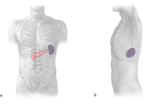
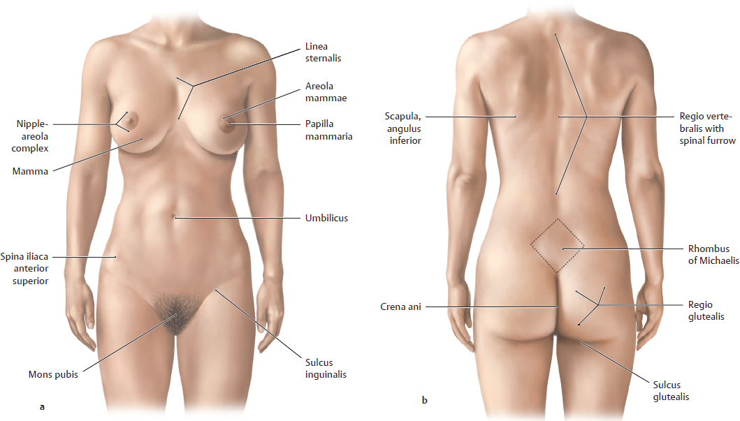
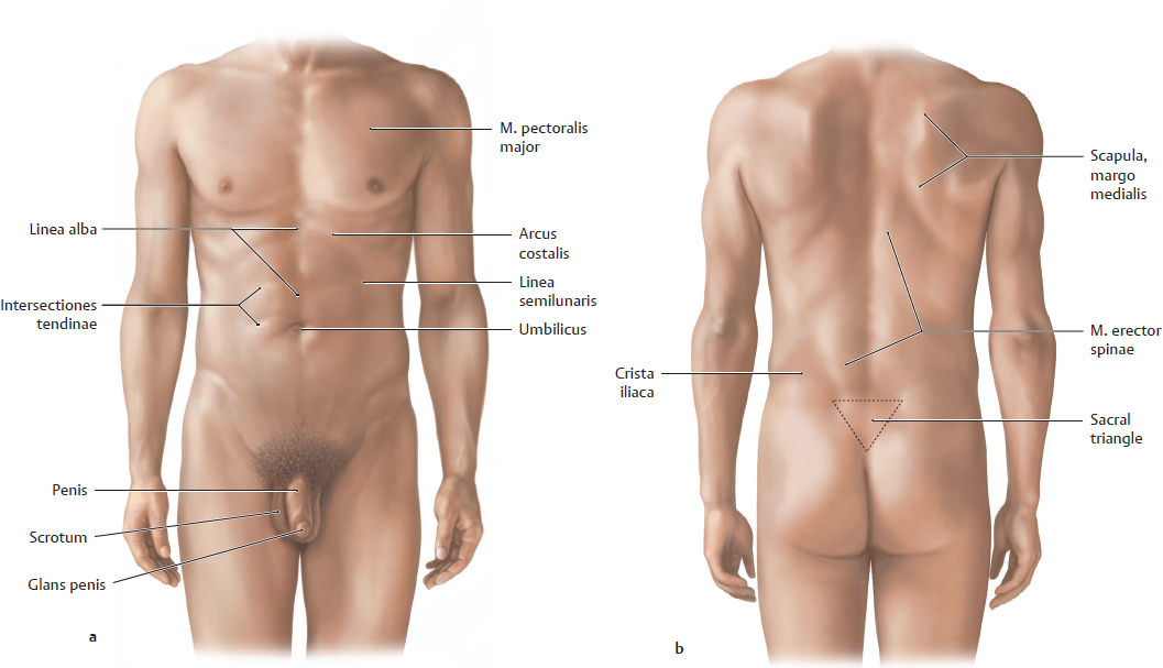
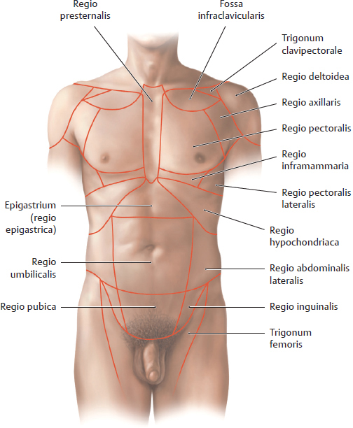
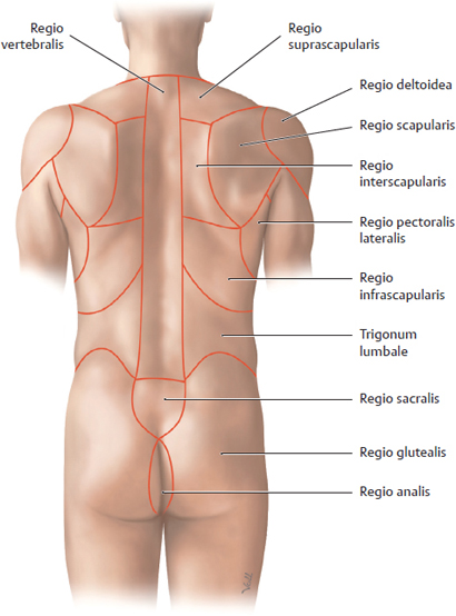
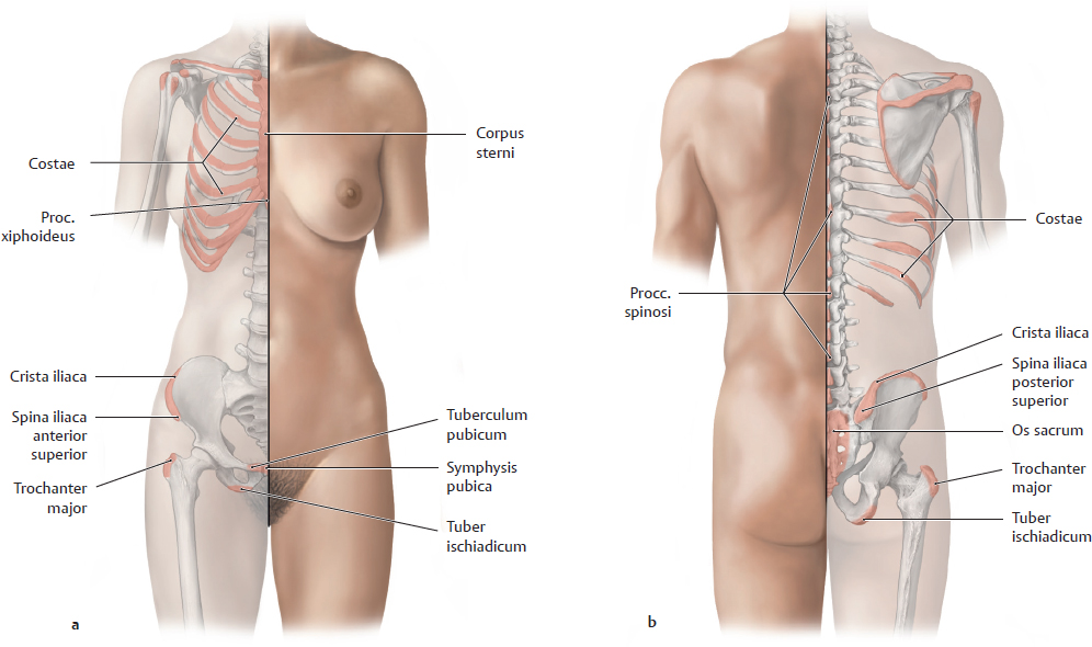
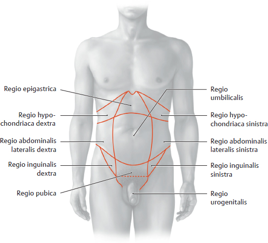
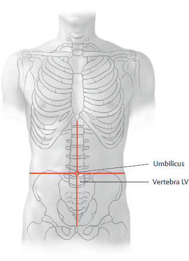
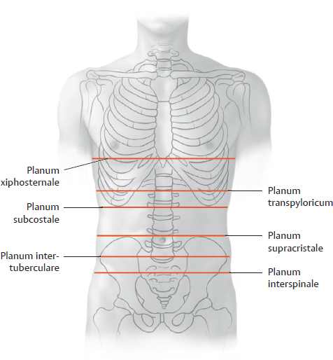
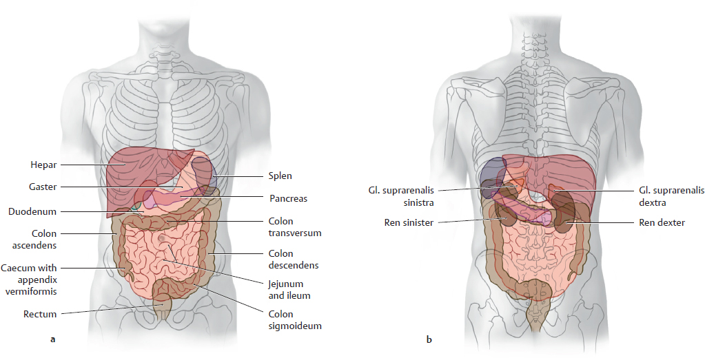
21. Topographical Anatomy A Surface anatomy of the female a Anterior view; b Posterior view. B Surface anatomy of the male a Anterior view; b Posterior view. C Thoracic and abdominal regions Anterior view. D Back and gluteal regions Posterior view. E Body surface contours and palpable bony landmarks of the trunk a Anterior view; b Posterior view. A Regions of the anterior trunk wall Three levels can be identified on the abdominal wall from above downward: the epigastrium, mesogastrium, and hypogastrium. Each level consists of three regions: • The median region of the epigastrium is the regio epigastrica. It is flanked laterally by the right and left regiones hypochondriacae. • The median region of the mesogastrium is the regio umbilicalis, which is bounded laterally by the right and left regiones abdominales laterales. • The median region of the hypogastrium is the regio pubica, which is flanked by the right and left regiones inguinales. The regio pubica is bounded inferiorly by the regiones urogenitales. The levels of the abdomen are defined by horizontal planes that are determined by palpable bony landmarks (see C). B Quadrants of the anterior trunk wall The quadrants of the anterior trunk wall are centered on the umbilicus, which lies at the level of the corpora vertebrarum L 3–4 and are named right and left upper and lower quadrant (RUQ, LUQ, RLQ, LLQ). C Horizontal (transverse) planes in the anterior trunk wall The anterior trunk wall is divided transversely by the following imaginary planes of section: • Planum xiphosternale: passes through the synchondrosis between the processus xiphoideus and corpus sterni. • Planum transpyloricum: plane midway between the incisura jugularis of the sternum and the superior border of the symphysis pubica. Located at the level of the L 1 vertebra, it divides the anterior trunk wall into upper and lower halves. The pylorus of the gaster is generally located slightly below this plane. • Planum subcostale: passes through the lowest points of the arcus costalis of the tenth rib at the level of the corpus vertebrae L 2. It marks the boundary between the epigastrium and mesogastrium (see A). • Planum supracristale: usually passes through the corpus vertebrae L 4, connecting the highest points on the cristae iliacae. • Planum intertuberculare: connects the tubercula iliaca and passes through the corpus vertebrae L 5. The planum intertuberculare marks the boundary between the mesogastrium and hypogastrium. • Planum interspinale: connects the two spinae iliacae anteriores superiores. Note: The three upper planes are variable in their location, which depends on the position and shape of the cavea thoracis. The key variables are respiratory position, age, sex, and constitutional type. D Projection of the abdominal and pelvic organs onto the trunk wall a Anterior trunk wall; b Posterior trunk wall. The surface projection of organs on the trunk wall depends on body posture, age, constitutional type, sex, nutritional state, and respiratory position. Note the overlap of the cavitates abdominis and thoracica: Perforating injuries of the cavitas abdominis that involve the hepar, for example, may also involve the cavitas pleuralis (“multicavity injury”). The projections of individual organs are shown in E. E Projection of anatomical structures in the abdomen and pelvis onto the columna vertebralis The spinal notation refers to corpora vertebrarum.
21.1 Surface Anatomy, Topographic Regions, and Palpable Bony Landmarks
21.2 Location of the Abdominal and Pelvic Organs and their Projection onto the Trunk Wall
T 7 | Superior border of the hepar |
T 12 | Hiatus aorticus |
L 1 | • Planum transpyloricum (generally the pylorus is at or below this plane) • Fundus vesicae biliaris • Hilum renale • Pars superior duodeni • Collum pancreatis • Origin of the truncus coeliacus • Origin of the a. mesenterica superior • Attachment of the mesocolon transversum • Hilum splenicum |
L 1/2 | • Origin of the aa. renales |
L 2 | Flexura duodenojejunalis |
L 3 | Origin of the a. mesenterica inferior |
L 3/4 | Umbilicus |
L 4 | Bifurcatio aortae |
L 5 | Origin of the v. cava inferior from the vv. iliacae communes |
S 3 | Upper (cranial) border of the rectum |
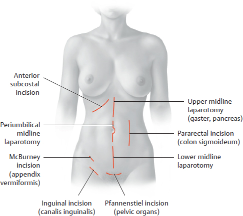
F Placement of surgical skin incisions in the anterior abdominal wall
Note: The periumbilical midline incision passes around the left side of the umbilicus to avoid cutting the remnant of the v. umbilicalis on the right side (the ligamentum teres hepatis, see p. 245). This v. umbilicalis remnant is generally but not always obliterated, and injury to the vessel, if it is still patent, may cause significant bleeding.
The McBurney incision is also called the gridiron incision because it changes direction in different planes of the trunk wall. The muscles of the trunk wall can be divided less traumatically by tailoring the direction of the cut to the prevailing fiber direction of the various muscle layers.
21.3 Topography of the Opened Cavitas Peritonealis (Supracolic Part and Infracolic Part)
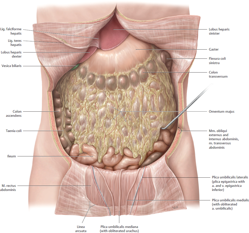
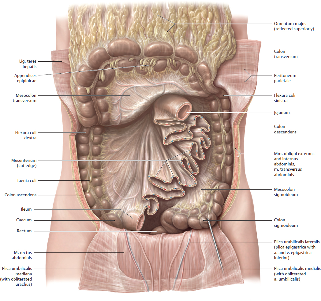
A The omentum majus in situ
Anterior view. The layers of the abdominal wall have been opened and retracted to display the omentum majus in its normal anatomical position. The omentum majus is draped over the loops of intestinum tenue, which are visible only at the inferior border of the omentum. The omentum majus is an apron-like fold of peritoneum suspended from the curvatura major of the gaster and covering the anterior surface of the colon transversum. It develops from the embryonic mesogastrium dorsalis, which becomes greatly enlarged to form a peritoneal sac suspended from the curvatura major (see p. 361). The omentum majus is relatively mobile, and is subject to considerable variation. Not infrequently, adhesions form between the omentum majus and the peritoneal covering of organs, especially as a result of local inflammation. While these adhesions help contain the spread of inflammation, they also limit the mobility of the organ to which the omentum is adherent. Peritoneal adhesions may undergo fibrotic changes over time, forming tough bands of scar tissue that may cause extrinsic narrowing and obstruction of organs such as the intestinum tenue. In many cases the omentum majus also assumes importance as a lymphoid organ through the secondary acquisition of nodi lymphoidei. The omentum minus is described on p. 356.
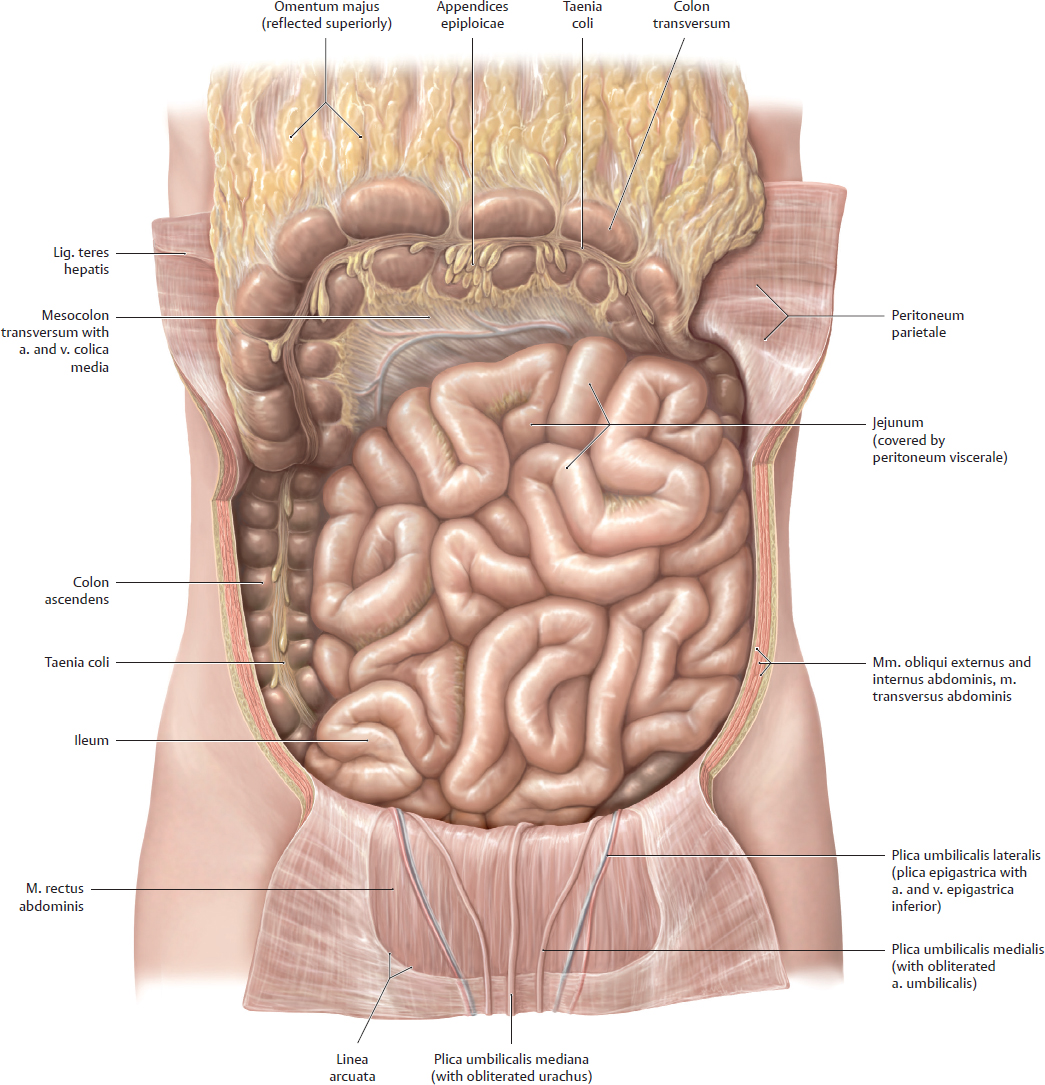
B Dissection with the omentum majus reflected superiorly and the intestinum tenue in situ
Anterior view. The omentum majus has been reflected superiorly, carrying with it the colon transversum, to demonstrate how the intraperitoneal part of the intestinum tenue is framed by the colon segments. The mesocolon transversum divides the cavitas peritonealis into a supracolic part and an infracolic part (see B, p. 200).
The large epithelial surface area of the peritoneum is important clinically:
• With bacterial infection (caused by external trauma or the seepage of septic material from an inflamed appendix), pathogenic microorganisms can easily spread within the cavitas peritonealis, where bacterial toxins are readily absorbed and carried into the bloodstream. As a result, bacterial peritonitis (inflammation of the peritoneum) generally constitutes a very serious and life-threatening condition.
• Localized inflammations may result in peritoneal adhesions and scar tissue bands (see A).
• The large surface area can be utilized for peritoneal dialysis in patients with renal failure: A dialysis solution instilled into the cavitas peritonealis can absorb waste products from the blood through the peritoneum, allowing them to be removed from the body.
21.4 Drainage Spaces and Recesses within the Cavitas Peritonealis
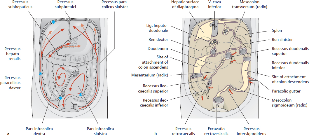
A Drainage spaces and recesses within the cavitas peritonealis
a Anterior view with the omentum majus and intestinum tenue removed; preferred metastatic sites (see blue stars);
b Posterior wall of the cavitas peritonealis, anterior view. The mesenteric roots and sites of organ attachment create partially bounded spaces (recesses or sulci). Peritoneal fluid released by the peritoneal epithelium (transudate) can flow freely within these spaces.
B Upper abdomen
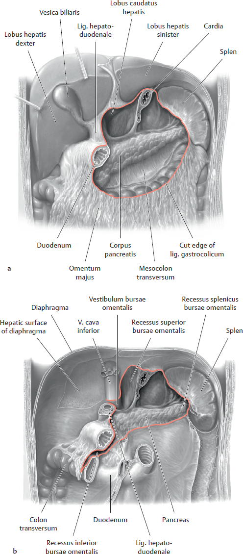
Anterior view of the upper abdomen; the hepar has been lifted to better display the recessus subhepaticus. The arrow points to the foramen omentale, the opening into and out of the bursa omentalis behind the omentum minus.
C Recesses in the posterior wall of the cavitas peritonealis
Anterior view of a male abdomen and pelvis. Because the peritoneum extends between organs, it forms recesses and sulci (see also A). In a sense, the bursa omentalis may be considered the largest recess in the cavitas peritonealis (see p. 360).
Note: The individual recesses are located between an organ and the wall of the cavitas peritonealis or between organs. Freely mobile loops of the intestinum tenue may become entrapped in these recesses (“internal hernia”) hindering the passage of the intestinal contents and potentially causing a life-threatening bowel obstruction (“mechanical ileus”).
21.5 Overview of the Mesenteria
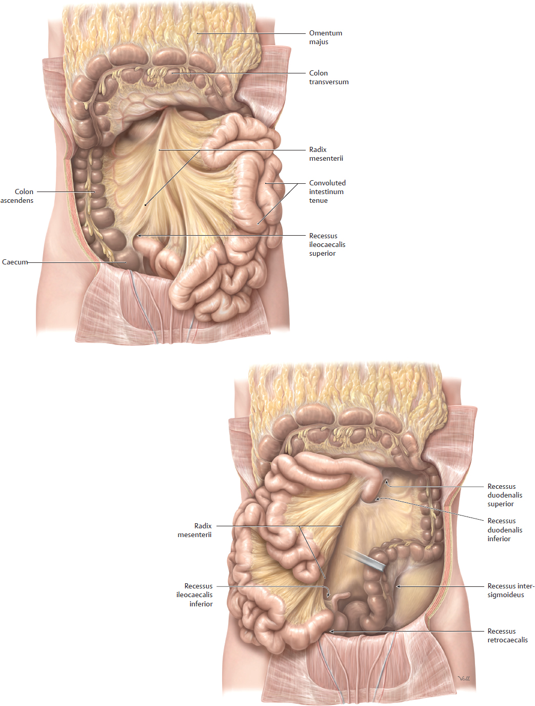
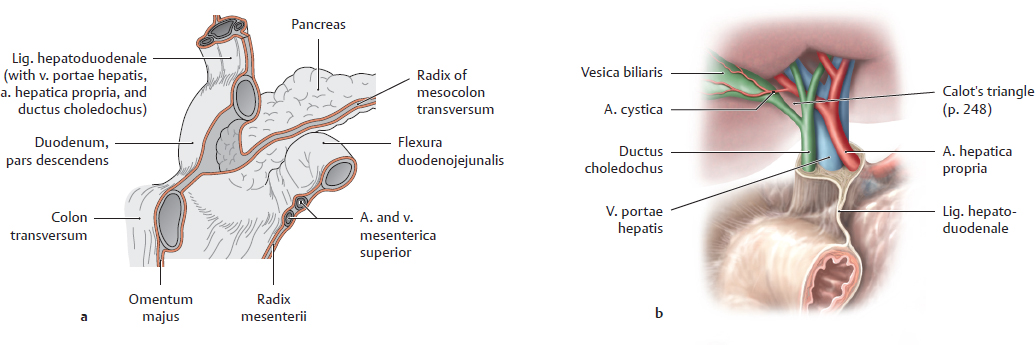
A Overview of the mesenteria with the omentum majus reflected superiorly and the intestinum tenue removed
Anterior view. The colon transversum and omentum majus have been reflected superiorly and the intraperitoneal intestinum tenue has been removed, leaving short stumps of jejunum and ileum. Three principal mesenteria are distinguishable in relation to the intestina tenue and crassum (the formation of the mesenteria is described on p. 32):
• The mesenterium of the intestinum tenue (the mesenterium proper)
• The mesocolon transversum
• The mesocolon sigmoideum (called also the mesosigmoid)
The origins of the mesenterium are shown in B. Smaller mesenteria are found on the appendix vermiformis (mesoappendix vermiformis) and rarely the upper part of the rectum (mesorectum, see C).
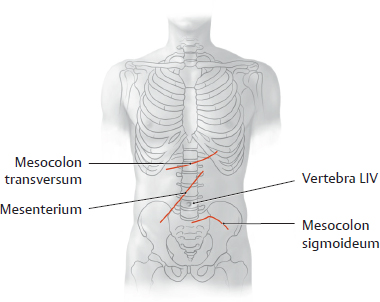
B Projection of the mesenteric roots onto the skeleton
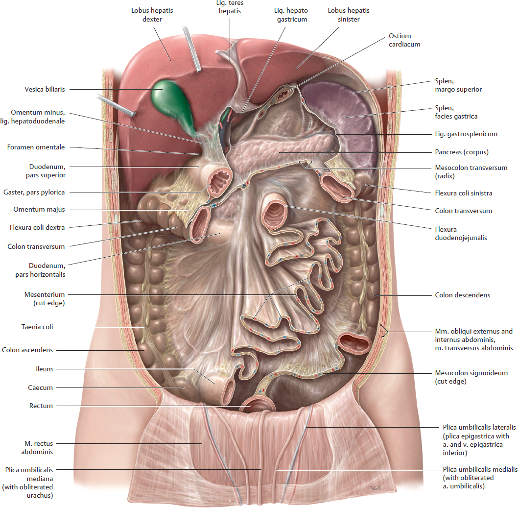
C Overview of the mesenteria* with the omentum majus removed
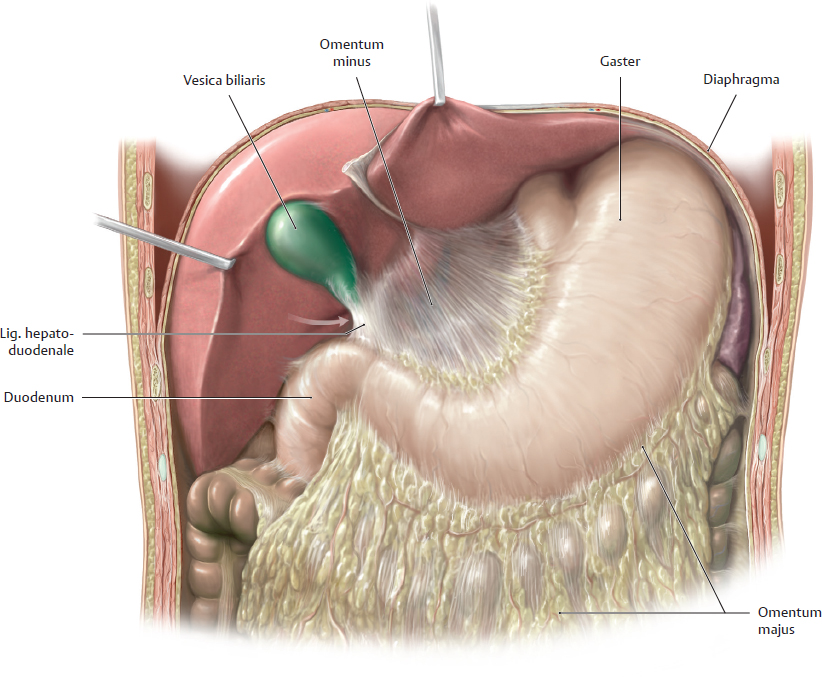
Anterior view. The mesenteria have been exposed by removing the gaster, jejunum, and ileum, leaving short stumps of intestinum tenue. The hepar has been reflected superiorly to display one part of the omentum minus: the lig. hepatoduodenale, which connects the hepar to the pylorus and duodenum. The other part of the omentum minus, the lig. hepatogastricum (peritoneal fold between the hepar and curvatura minor of the gaster), has been removed with the gaster, opening the anterior wall of the bursa omentalis. Most of the colon transversum and colon sigmoideum have been removed to display the roots of the mesocolon transversum and mesocolon sigmoideum.
Note: The cola ascendens and descendens become attached to the posterior wall of the cavitas peritonealis during the fourth month of embryonic development. The mesenteria of the cola ascendens and descendens become fused to the posterior wall of the cavitas peritonealis. The mesocolon transversum crosses over the duodenum, whose mesenteriumalso fuses to the posterior wall of the mesocolon transversum during embryonic development (see p. 36). The mesocolon transversum necessarily passes over this “retroperitoneal portion” of the duodenum because of its attachment to the posterior wall of the cavitas peritonealis. Developmentally, almost all of the mesenteria are mesenteria dorsalia. Only upper abdominal organs like the gaster and hepar have mesenteria ventralia.
21.6 Topography of the Bursa Omentalis
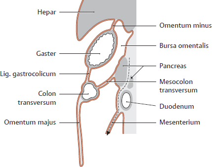
A Shape and location of the bursa omentalis in sagittal section
Left lateral view. The bursa omentalis is the largest potential space in the cavitas peritonealis. It is located behind the omentum minus and gaster.
Note: As the gaster rotates during embryonic development, the bursa omentalis comes to lie posterior to the gaster. The pancreas, which migrates into the retroperitoneum secondarily, thus forms part of the posterior wall of the bursa, which provides a route for gaining surgical access to that organ. As the gaster rotates in the clockwise direction (viewed from the front), its curvatura minor points to the right and also superiorly, simultaneously displacing the hepar superiorly and to the right. As a result of this, the bursa omentalis comes to lie partially posterior to the hepar.
B Boundaries of the bursa omentalis
Anterior | Omentum minus, lig. gastrocolicum |
Posterior | Pancreas, aorta (pars abdominalis), truncus coeliacus, a. and v. splenica, plica gastropancreatica, left gl. suprarenalis, polus superior of left kidney |
Superior | Hepar (with lobus caudatus), recessus superior of bursa omentalis |
Inferior | Mesocolon transversum, recessus inferior of bursa omentalis |
Left | Splen, lig. gastrosplenicum, recessus splenicus of bursa omentalis |
Right | Hepar, bulbus duodeni |
C Surgical approaches to the bursa omentalis (see A)
• Through the foramen omentale (natural opening, see E)
• Between the curvatura major of the gaster and the colon transversum through the lig. gastrocolicum
• Through the mesocolon transversum after elevating the colon transversum (inferior approach)
• Between the curvatura minor of the gaster and the hepar (through the omentum minus)
• From the curvatura major of the gaster after division of the omentum majus
D Bursa omentalis, anterior view
a Boundaries of the bursa omentalis, also the shape and location of the gastric bed
b Structure of the posterior wall of the bursa omentalis
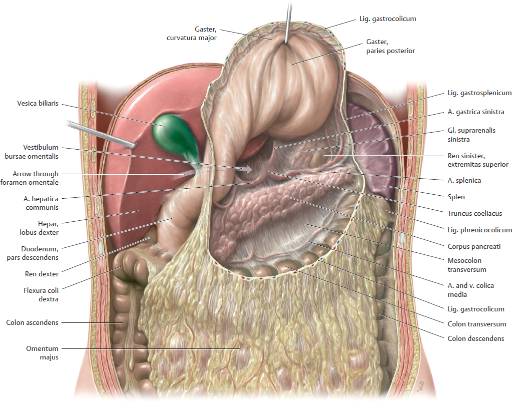
E Bursa omentalis in the upper abdomen
Anterior view. The lig. gastrocolicum has been divided, the gaster has been reflected superiorly (surgical approach), and the hepar has been retracted superolaterally. The foramen omentale (arrow) is the only natural orifice of the bursa omentalis (opens posterior to the lig. hepatoduodenale). The vestibulum of the bursa omentalis lies just past the foramen and forms the initial portion of the bursa cavity.
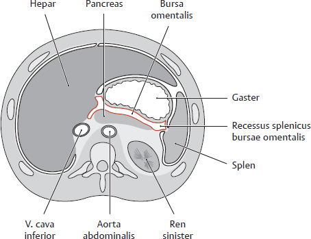
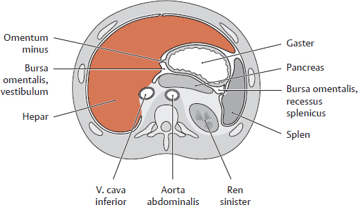
F Transverse section through the bursa omentalis
Schematic section through the abdomen at the T 12/L 1 level, viewed from below.
Note the walls and recesses that result from the formation of the bursa during the embryonic rotation of the gaster. Because the initial upper right portion of the embryonic body cavity moves posteriorly as part of the 90° rotation of the gaster, structures that were formerly posterior (splen) move to the left side while structures that were formerly anterior (hepar) move to the right side. Recesses in the bursa omentalis extend close to these organs (see B).
21.7 Topography of the Upper Abdominal Organs: Hepar, Vesica Biliaris, Duodenum, and Pancreas
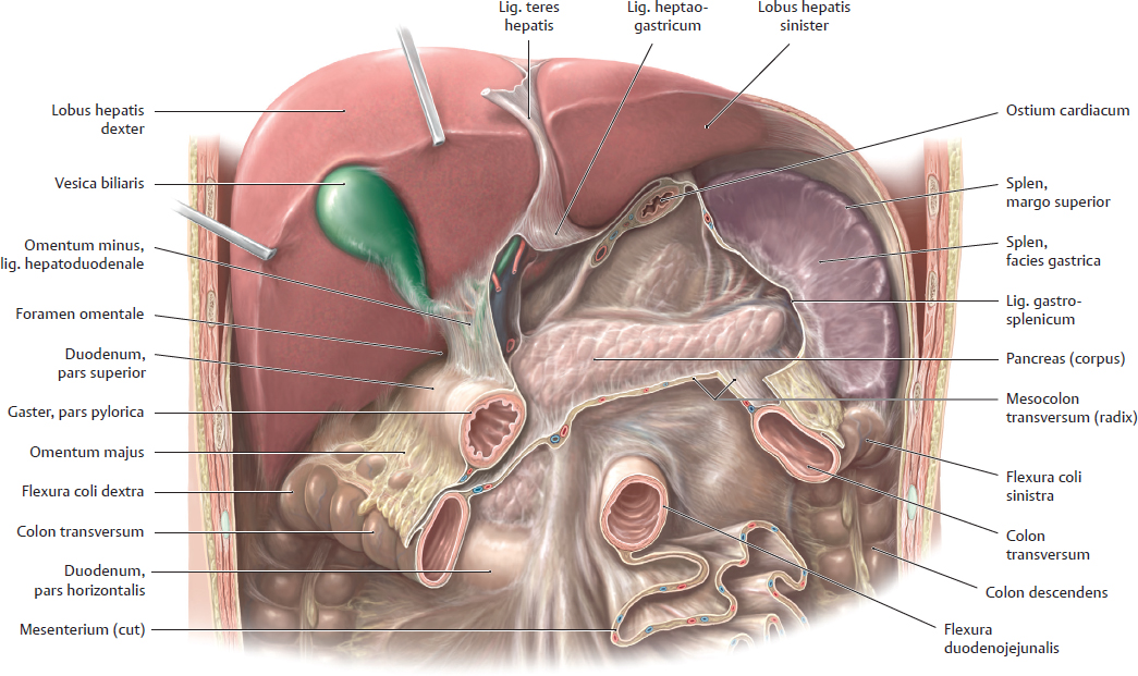
A Location of the hepar and vesica biliaris
Anterior view. The gaster and intestinum tenue have been removed, leaving a short stump of the jejunum. Most of the colon transversum has been removed. The hepar has been lifted for better exposure of parts of the omentum minus, lig. hepatoduodenale and pancreas (for the contents of the lig. hepatoduodenale see Eb).
B Position of the hepar
Transverse section through the abdomen at approximately the T 12/L 1 level, viewed from below. The hepar is intraperitoneal except for the area nuda, which is not visible here. The lobus hepatis sinister extends into the LUQ, where it is anterior to the gaster. The peritoneal fold between the hepar and the curvatura minor of the gaster (omentum minus) can be seen. Portions of the hepar form the right boundary of the bursa omentalis.
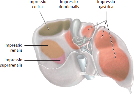
C Areas of contact with other organs
View of the visceral surface of the hepar.
Note: Impressions from organs that are in direct contact with the hepar are visible only on a liver that has been hardened in place by a chemical preservative (“fixation”). An unfixed hepar from a cadaver that has not been chemically preserved is so soft that generally it will not show organ impressions. Diseases of the hepar may easily spread to other organs, and vice versa, at areas of contact with adjacent organs (extensive owing to the size and topography of the hepar).
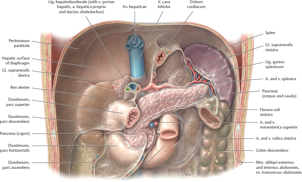
D Location of duodenum and pancreas
Anterior view. The hepar, gaster and intestinum tenue have been removed, leaving the duodenum and a very small stump of the jejunum. The cola ascendens and transversum have been removed to expose the right kidney, pancreas, and duodenal loop. The secondarily retroperitoneal colon descendens is left in situ. The pancreas and duodenum are also secondarily retroperitoneal (for peritoneal relationships see p. 201).
Both kidneys and gll. suprarenales, located in the spatium retroperitoneale, are visible through the peritoneum parietale. The kidneys and gll. suprarenales are primarily retroperitoneal. The intraperitoneal splen is located in the left upper quadrant in a compartment called the splenic niche.
Note: The root of the (intraperitoneal) colon transversum crosses anterior to the duodenum and pancreas.
E Peritoneal relationships of the duodenum and pancreas; contents of the ligamentum hepatoduodenale
a Peritoneal relationships of the duodenum and pancreas, anterior view. The root of the mesocolon transversum crosses over the pars descendens of the duodenum and the pancreas.
b Contents of the ligamentum hepatoduodenale. The lig. hepatoduodenale is part of the omentum minus and connects the hepar with the pylorus and pars superior of the duodenum. It contains the v. portae hepatis, the a. hepatica propria, and the ductus choledochus.
21.8 Topography of the Upper Abdominal Organs: Gaster and Splen
A Location of the gaster and splen
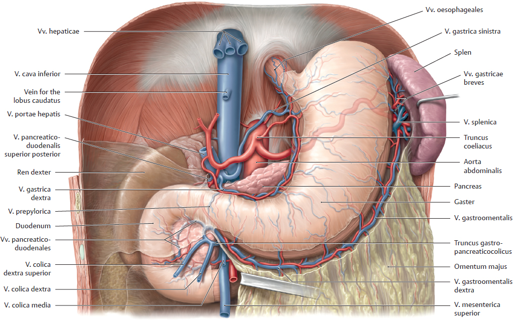
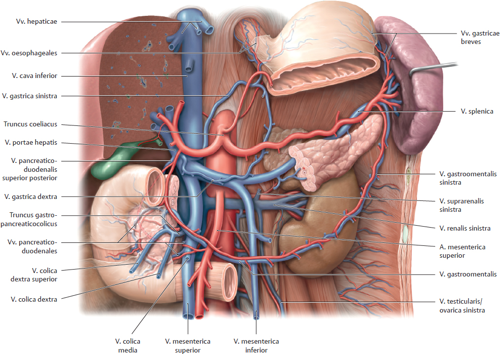
Anterior view. The hepar and omentum minus have been removed, the omentum majus has been opened and retracted to the left, and the gaster pulled slightly downward for better exposure. At several sites, the peritoneum has been removed or windowed for better exposure of the opening of the vv. hepaticae into the v. cava inferior and the opening of the vv. gastricae into the v. portae hepatis at the margin of the lig. hepatoduodenale (here completely opened). The splen is retracted from its “niche” and lies close to the fundus and curvatura major of the gaster. The intraperitoneal gaster covers most of the retroperitoneal pancreas. The omentum majus, a remnant of the mesogastrium dorsale, is suspended from the curvatura major of the gaster. The gaster is partially shown transparent to display the a. splenica that extends behind the gaster from the truncus coeliacus to the splen.
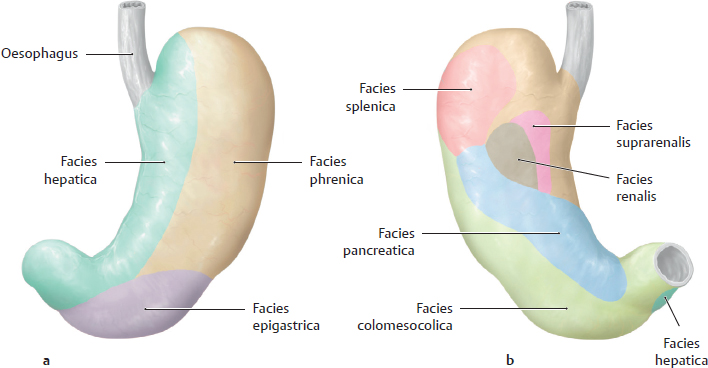
B Areas of contact with adjacent organs
a, b Anterior and posterior views of the parietes gastrici. Because the gaster is intraperitoneal, it is very mobile relative to adjacent organs. But since the gaster is in close contact with other organs, lesions that penetrate the paries gastri (ulcers, malignant tumors) may spread to nearby organs or may cause adhesions to develop between the gaster and adjacent organs.
C Location of the pancreas, splen, and major vessels
Anterior view. The gaster has been partially removed and pulled slightly downward, and most of the intestina have been removed leaving only the duodenum. The splen has been lifted from its bed and retracted anterolaterally toward the fundus gastricus. Part of the corpus pancreatis has been resected. Most of the peritoneum has been removed, and retroperitoneal fat and connective tissue have been cleared away.
The secondarily retroperitoneal pancreas crosses over the polus superior of the left kidney that also lies in the spatium retroperitoneale. The diagram shows that the displayed retroperitoneal organs are not arranged in a coronal plane but oriented anterior to posterior. The most anterior organ in the right upper quadrant is the duodenum. Posterior to the duodenum lies the pancreas that is oriented transversely in the right and left upper quadrants. The most posterior organs are the two kidneys (here only the left kidney is clearly visible, the right kidney is covered by the duodenum and the caput pancreatis).